. 2010 Jan 19;18(1):136-47.
doi: 10.1016/j.devcel.2009.11.010.

Neuronal regulation of the spatial patterning of neurogenesis

Affiliations

Neuronal regulation of the spatial patterning of neurogenesis

Rosa Gonzalez-Quevedo et al. Dev Cell. .

Abstract

Precise regulation of neurogenesis is achieved in specific regions of the vertebrate nervous system by formation of distinct neurogenic and nonneurogenic zones. We have investigated how neurogenesis becomes confined to zones adjacent to rhombomere boundaries in the zebrafish hindbrain. The nonneurogenic zone at segment centers comprises a distinct progenitor population that expresses fibroblast growth factor (fgfr) 2, erm, sox9b, and the retinoic acid degrading enzyme, cyp26b1. FGF receptor activation upregulates expression of these genes and inhibits neurogenesis in segment centers. Cyp26 activity is a key effector inhibiting neuronal differentiation, suggesting antagonistic interactions with retinoid signaling. We identify the critical FGF ligand, fgf20a, which is expressed by specific neurons located in the mantle region at the center of segments, adjacent to the nonneurogenic zone. Fgf20a mutants have ectopic neurogenesis and lack the segment center progenitor population. Our findings reveal how signaling from neurons induces formation of a nonneurogenic zone of neural progenitors.


Figures

Figure 1
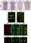
Figure 1. FGF signaling is restricted to non-neurogenic regions in the zebrafish hindbrain
Dorsal views of flat mounted embryos, anterior to the top, at the indicated stages. Following in situ hybridisation, embryos of the same batch were developed for the same amount of time. Arrowheads indicate segment centres, and arrows point at hindbrain boundaries. All scale bars, 50 µm. (A–H): time course of neurog1 (A–D) and neurod4 (E–H) expression from 22 somites to 48 h. (I–J): Higher power views showing the spatial restriction of neurogenesis marked by neurog1 (same embryo as 1D) and neurod4 (1J, same embryo as 1H). (K–N): erm expression. (O–T): Double fluorescent in situ hybridization using probes for erm and dld (O–Q), and fgfr2 and neurog1 (R–T). Images shown are a merge of confocal stacks through the hindbrain at 36 hpf.
Figure 2
Figure 2. Blocking FGFR activation results in proneural gene expression and differentiating neurons in segment centres
In situ hybridisation of 40 hpf zebrafish to detect proneural gene expression (neurog1, dld, dla; A–F and A’–F’) or differentiating neurons (neurod4; G–H and G’–H’) in either wild type (wt) or dominant negative fgfr1 embryos (Tg(hsp70l:dnfgfr1-EGFP)). Heat shock was started at the 22 somite stage. Black arrowheads indicate segment centres and red arrowheads indicate ectopic neurogenesis. Scale bar for A–H, 50 µm. (A’–H’): higher power view of images in A–H. Scale bar, 25 µm. Punctate line: midline. See also Figure S1.
Figure 3
Figure 3. FGF signaling maintains a Sox9b expressing population in segment centres
(A–L): In situ hybridisation for erm, fgfr2, neurog1 or neurod4 (red) followed by immunostaining with anti-Sox9 antibody (green). Images shown are a projection of confocal stacks. Scale bar, 50 µm. White arrowheads indicate segment centres, and yellow arrowheads indicate colocalization of Sox9b with fgfr2 and erm. The staining in segment centres is due to Sox9b, as it is lost in Sox9b morphant embryos (not shown). (M–N): Whole mount immunostaining of Sox9b in 36 hpf wild type (wt; M) or transgenic dominant-negative fgfr1 embryos (Tg(hsp70l:dnfgfr1-EGFP); N). White arrowheads indicate segment centres in wt embryos, and open arrowheads in transgenic embryos point at centres where Sox9b expression is absent. Images shown are merged confocal stacks. Scale bar, 20 µm. (O–R): In situ hybridizations of 26 hpf wild type embryos (left) or embryos expressing constitutively-active FGFR1 (Tg(hsp70:ca-fgfr1)), using erm (O–P) or sox9b (Q–R) probes. Embryos were heat shocked at 24 hpf and fixed 2 h later. Arrowheads indicate segment centres. Red arrowheads indicate upregulation of erm or sox9b expression. Scale bar, 50 µm. See also Figure S2.
Figure 4
Figure 4. Cyp26b1 is expressed in segment centres and regulated by FGF signaling
(A–D): In situ hybridisations to detect the time course of cyp26b1 expression. Images are a merge of confocal stacks of Fast Red staining. Scale bar, 50 µm. White arrowheads indicate segment centres. (D–F): In situ hybridisation of cyp26b1 followed by immunostaining with anti-Sox9 antibody. Yellow arrowheads show colocalization of cyp26b1 with Sox9b in segment centres. (G–H): In situ hybridization of 40 hpf embryos to detect cyp26b1 expression in wild type (wt; G) or dominant negative FGFR1 embryos (Tg(hsp70l:dnfgfr1-EGFP)) (H). Heat shocks were started at the 22 somite stage. Black arrowheads indicate segment centres, and open arrowheads indicate the disappearance of cyp26b1 expression from centres. Scale bar, 50 µm. (I–J): In situ hybridization of 26 hpf embryos to detect cyp26b1 in either wild type (wt; I) or constitutively active fgfr1 embryos, Tg(hsp70:ca-fgfr1) (J). Heat shocks were started at 24 hpf and embryos fixed 2 h later. Black arrowheads indicate segment centres, and red arrowheads centres in embryos with cyp26b1 upregulation. Scale bar, 50 µm.
Figure 5
Figure 5. Blocking Cyp26 activity results in premature neurogenesis
(A–F): In situ hybridization of 40 hpf embryos to detect expression of neurog1 (A–B), dla (C–D) or neurod4 (E–F) in DMSO or R115866 treated embryos. Treatments were started at 24–26 h. Black arrowhead points at r5. Scale bar, 50 µm. (A’–F’): higher power views of r4 and r5 shown in A–F (black arrowheads). Red arrowheads indicate ectopic proneural expression. Scale bar, 25 µm. (G–J). Blocking RA signaling with DEAB partially rescues loss of Cyp26. In situ hybridization of 36 hpf embryos to detect expression of neurog1 in DMSO (G), R115866 (H), DEAB (I) or R115866 + DEAB (J) treated embryos.
Figure 6
Figure 6. Fgf20a is expressed in neurons at segment centres
(A–D): Time course of fgf20a expression at 18 somites (A), 22 somites (B), 24 hpf (C) and 30 hpf (D). Embryos belong to the same batch and were developed for the same amount of time. Black arrowheads point at the centre of r5. Scale bar, 50 µm. (E–I): Merge of confocal stacks of double-stained embryos. Scale bar, 100 µm. (E) In situ hybridisation of fgf20a (red) and anti-EphA4 staining (green) to reveal r3 and r5 (white arrowheads) at 24 h. (F) In situ hybridisation of fgf20a (red) and antibody staining for Sox9b (green) in 28 hpf embryo. White arrowheads indicate segment centres. (G–I): In situ hybridisation of fgf20a (red) and antibody staining for the pan-neuronal marker HuC/D in 24 hpf embryos. White arrowheads indicate segment centres, and yellow arrowheads show colocalization of fgf20a with specific HuC/D-expressing neurons. (J–L): Double labelling of fgf20a (red) and HuC/D (green). Images show a merge of confocal stacks through r4 at 24 hpf in transverse sections. Dorsal is to the top. In r4, fgf20a expressing cells form clusters (white arrowheads) in the mantle zone and colocalize with specific HuC/D expressing neurons (yellow arrowheads). vz: ventricular zone. Scale bar, 50 µm. See also Figure S3.
Figure 7
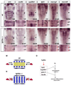
Figure 7. Fgf20a is required for inhibition of neurogenesis in segment centres
In situ hybridisations of wild type (A–F) or fgf20a homozygous embryos (G–L) raised at 25°C. (A’–L’) show higher power images of A–L. Scale bar, 50 µm for A–L; 20 µm for A’–L’. erm expression in segment centres is significantly reduced in fgf20a mutants (open arrowheads in G’). Markers of segment centres, sox9b, cyp26b1 and fgfr2 are greatly decreased in fgf20a −/− embryos (open arrowheads in H’–J’). (K–L): fgf20a mutant embryos have ectopic neurogenesis in segment centres, detected by neurog1 (K, E) and neurod4 expression (L, F). Red arrowheads indicate ectopic neurogenesis in segment centres (K’, L’). (M–N) Model of the patterning of neurogenesis by fgf20a in hindbrain segments. In wild type embryos (M), fgf20a secreted from neurons in the adjacent mantle region (red ovals) prevents neuronal differentiation (blue circles) in segment centres by maintaining a population of progenitors (yellow circles). (N) In fgf20a mutants there is ectopic neurogenesis and low level expression of segment centre markers. (O) Summary of the regulation of genes in the non-neurogenic zone of progenitors in segment centres. fgf20a upregulates a set of genes that control different aspects of maintaining an undifferentiated population.

Similar articles

Cited by

References

    1. Amoyel M, Cheng YC, Jiang YJ, Wilkinson DG. Wnt1 regulates neurogenesis and mediates lateral inhibition of boundary cell specification in the zebrafish hindbrain. Development. 2005;132:775–785. - PubMed
    1. Bae YK, Shimizu T, Hibi M. Patterning of proneuronal and interproneuronal domains by hairy- and enhancer of split-related genes in zebrafish neuroectoderm. Development. 2005;132:1375–1385. - PubMed
    1. Bally-Cuif L, Hammerschmidt M. Induction and patterning of neuronal development, and its connection to cell cycle control. Curr Opin Neurobiol. 2003;13:16–25. - PubMed
    1. Bertrand N, Castro DS, Guillemot F. Proneural genes and the specification of neural cell types. Nat Rev Neurosci. 2002;3:517–530. - PubMed
    1. Borello U, Cobos I, Long JE, Murre C, Rubenstein JL. FGF15 promotes neurogenesis and opposes FGF8 function during neocortical development. Neural Develop. 2008;3:17. - PMC - PubMed

Publication types

MeSH terms

LinkOut - more resources