

Magnetic resonance imaging of the anteroposterior position and thickness of the aging, accommodating, phakic, and pseudophakic ciliary muscle
- PMID: 20152603
- PMCID: PMC2826892
- DOI: 10.1016/j.jcrs.2009.08.029
Magnetic resonance imaging of the anteroposterior position and thickness of the aging, accommodating, phakic, and pseudophakic ciliary muscle
Abstract
Purpose: To quantify accommodative and age-related changes in the anteroposterior position and thickness of the ciliary muscle in phakic and pseudophakic eyes.
Setting: Department of Surgery/Bioengineering, UMDNJ-Robert Wood Johnson Medical School, Piscataway; Institute of Ophthalmology and Visual Science UMDNJ-New Jersey Medical School, Newark, New Jersey; MRI Research, Inc., Middleburg Heights, Ohio, USA.
Methods: Magnetic resonance images were taken of phakic and pseudophakic eyes.
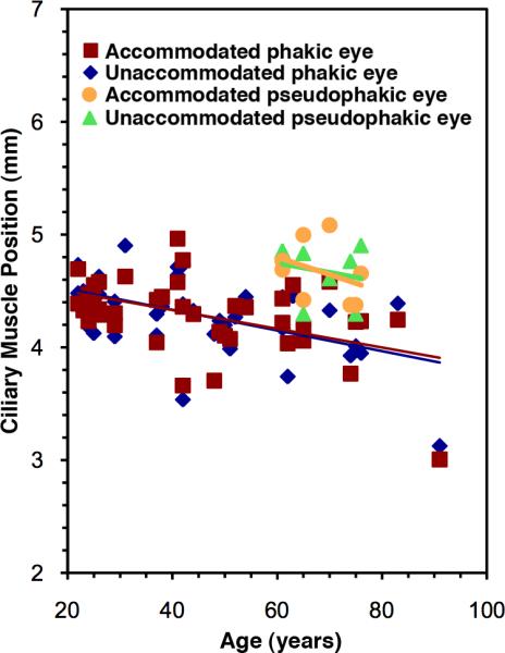
Results: The cohort comprised 32 phakic volunteers and 8 volunteers with a monocular intraocular lens (IOL) aged 22 to 91 years. No anteroposterior accommodative movement of the ciliary muscle apex occurred in either group. The muscle moved closer to the cornea with advancing age in phakic eyes; IOL implantation returned the muscle to a youthful position. An age-dependent increase in ciliary muscle anteroposterior thickness occurred that was not mitigated by IOL implantation. Muscle thickness increased with accommodation in only phakic eyes.
Conclusions: Presbyopia-correction strategies cannot rely on accommodative anterior movement of the ciliary muscle. Forces on the uvea by crystalline lens-pupillary margin contact may increase with accommodation and lens growth, producing accommodative and age-dependent increases in muscle thickness and significant age-dependent anterior muscle displacement. Intraocular lens implantation removed these forces, allowing choroidal elasticity to restore the muscle to a youthful position; however, the increase in thickness was permanent and likely due to an age-dependent increase in connective tissue. This supports the geometric theory of presbyopia development and that the mechanical forces in human accommodation and presbyopia are very different from those in the rhesus monkey model.
Copyright 2010 ASCRS and ESCRS. Published by Elsevier Inc. All rights reserved.
Figures


Similar articles
-
Magnetic resonance imaging of aging, accommodating, phakic, and pseudophakic ciliary muscle diameters.J Cataract Refract Surg. 2006 Nov;32(11):1792-8. doi: 10.1016/j.jcrs.2006.05.031. J Cataract Refract Surg. 2006. PMID: 17081859 Free PMC article.
-
Extralenticular and lenticular aspects of accommodation and presbyopia in human versus monkey eyes.Invest Ophthalmol Vis Sci. 2013 Jul 26;54(7):5035-48. doi: 10.1167/iovs.12-10846. Invest Ophthalmol Vis Sci. 2013. PMID: 23745002 Free PMC article.
-
Quantification of age-related and per diopter accommodative changes of the lens and ciliary muscle in the emmetropic human eye.Invest Ophthalmol Vis Sci. 2013 Feb 7;54(2):1095-105. doi: 10.1167/iovs.12-10619. Invest Ophthalmol Vis Sci. 2013. PMID: 23287789 Free PMC article.
-
[Deterioration of amplitude of the accommodation with age and its possible restoration in the intraocular lens implanted eye].Nippon Ganka Gakkai Zasshi. 1992 Sep;96(9):1071-8. Nippon Ganka Gakkai Zasshi. 1992. PMID: 1414696 Review. Japanese.
-
The effect of aging on the ciliary muscle and its potential relationship with presbyopia: a literature review.PeerJ. 2024 Dec 24;12:e18437. doi: 10.7717/peerj.18437. eCollection 2024. PeerJ. 2024. PMID: 39735562 Free PMC article. Review.
Cited by
-
Stretch-dependent changes in surface profiles of the human crystalline lens during accommodation: a finite element study.Clin Exp Optom. 2015 Mar;98(2):126-37. doi: 10.1111/cxo.12263. Clin Exp Optom. 2015. PMID: 25727940 Free PMC article.
-
Age- and refraction-related changes in anterior segment anatomical structures measured by swept-source anterior segment OCT.PLoS One. 2020 Oct 23;15(10):e0240110. doi: 10.1371/journal.pone.0240110. eCollection 2020. PLoS One. 2020. PMID: 33095821 Free PMC article.
-
Intraocular accommodative movements in monkeys; relationship to presbyopia.Exp Eye Res. 2022 Sep;222:109029. doi: 10.1016/j.exer.2022.109029. Epub 2022 Mar 11. Exp Eye Res. 2022. PMID: 35283107 Free PMC article.
-
A biomechanical model for evaluating the performance of accommodative intraocular lenses.J Biomech. 2022 May;136:111054. doi: 10.1016/j.jbiomech.2022.111054. Epub 2022 Mar 18. J Biomech. 2022. PMID: 35344827 Free PMC article.
-
Long-term visual outcomes of laser anterior ciliary excision.Am J Ophthalmol Case Rep. 2018 Feb 4;10:38-47. doi: 10.1016/j.ajoc.2018.01.033. eCollection 2018 Jun. Am J Ophthalmol Case Rep. 2018. PMID: 29780911 Free PMC article.
References
-
- Thornton SP. Lens implantation with restored accommodation. Curr Canadian Ophthalmic Prac. 1986;4:60, 62, 82.
-
- Findl O, Leydolt C. Meta-analysis of accommodating intraocular lenses. J Cataract Refract Surg. 2007;33:522–527. - PubMed
-
- Strenk SA, Strenk LM, Koretz JF. The mechanism of presbyopia. Prog Retin Eye Res. 2005;24:379–393. - PubMed
-
- Baikoff G, Lutun E, Wei J, Ferraz C. Anterior chamber optical coherence tomography study of human natural accommodation in a 19-year-old albino. J Cataract Refract Surg. 2004;30:696–701. - PubMed
Publication types
MeSH terms
Grants and funding
LinkOut - more resources
Full Text Sources
Other Literature Sources
Medical
Miscellaneous
