Computer-aided diagnosis of lung nodules on CT scans: ROC study of its effect on radiologists' performance
- PMID: 20152726
- PMCID: PMC3767437
- DOI: 10.1016/j.acra.2009.10.016
Computer-aided diagnosis of lung nodules on CT scans: ROC study of its effect on radiologists' performance
Abstract
Rationale and objectives: The aim of this study was to evaluate the effect of computer-aided diagnosis (CAD) on radiologists' estimates of the likelihood of malignancy of lung nodules on computed tomographic (CT) imaging.
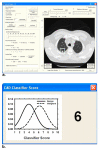
Methods and materials: A total of 256 lung nodules (124 malignant, 132 benign) were retrospectively collected from the thoracic CT scans of 152 patients. An automated CAD system was developed to characterize and provide malignancy ratings for lung nodules on CT volumetric images. An observer study was conducted using receiver-operating characteristic analysis to evaluate the effect of CAD on radiologists' characterization of lung nodules. Six fellowship-trained thoracic radiologists served as readers. The readers rated the likelihood of malignancy on a scale of 0% to 100% and recommended appropriate action first without CAD and then with CAD. The observer ratings were analyzed using the Dorfman-Berbaum-Metz multireader, multicase method.
Results: The CAD system achieved a test area under the receiver-operating characteristic curve (A(z)) of 0.857 +/- 0.023 using the perimeter, two nodule radii measures, two texture features, and two gradient field features. All six radiologists obtained improved performance with CAD. The average A(z) of the radiologists improved significantly (P < .01) from 0.833 (range, 0.817-0.847) to 0.853 (range, 0.834-0.887).
Conclusion: CAD has the potential to increase radiologists' accuracy in assessing the likelihood of malignancy of lung nodules on CT imaging.
Figures


References
-
- Sone S, Takashima S, Li F, et al. Mass screening for lung cancer with mobile spiral computed tomography scanner. Lancet. 1998;351:1242–1245. - PubMed
-
- Henschke CI, McCauley DI, Yankelevitz DF, et al. Early lung cancer action project: overall design and findings from baseline screening. Lancet. 1999;354:99–105. - PubMed
-
- Nawa T, Nakagawa T, Kusano S, et al. Lung cancer screening using low-dose spiral CT: results of baseline and 1-year follow-up studies. Chest. 2002;122:15–20. - PubMed
-
- Diederich S, Wormanns D, Semik M, et al. Screening for early lung cancer with low-dose spiral CT: Prevalence in 817 asymptomatic smokers. Radiology. 2002;222:773–781. - PubMed
-
- Henschke CI, Yankelevitz DF, Libby DM, et al. Survival of patients with stage I lung cancer detected on CT screening. N Engl J Med. 2006;355:1763–1771. - PubMed
Publication types
MeSH terms
Grants and funding
LinkOut - more resources
Full Text Sources
Other Literature Sources
Medical
Miscellaneous

