. 2010 Apr 15;340(2):504-17.
doi: 10.1016/j.ydbio.2010.02.008. Epub 2010 Feb 10.

Tenectin is a novel alphaPS2betaPS integrin ligand required for wing morphogenesis and male genital looping in Drosophila

Affiliations

Tenectin is a novel alphaPS2betaPS integrin ligand required for wing morphogenesis and male genital looping in Drosophila

Stéphane Fraichard et al. Dev Biol. .

Abstract

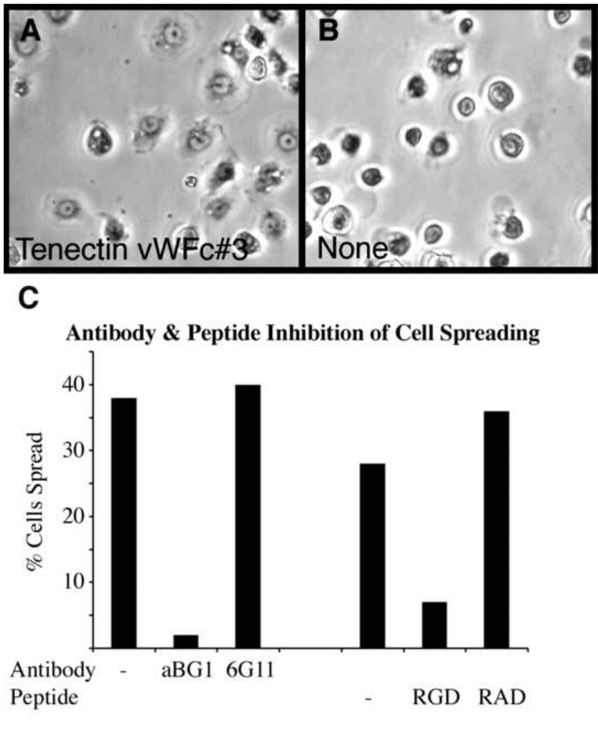
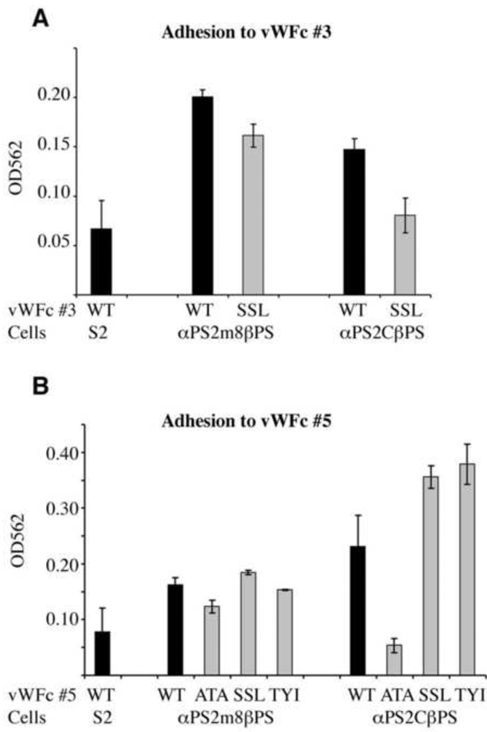
Morphogenesis of the adult structures of holometabolous insects is regulated by ecdysteroids and juvenile hormones and involves cell-cell interactions mediated in part by the cell surface integrin receptors and their extracellular matrix (ECM) ligands. These adhesion molecules and their regulation by hormones are not well characterized. We describe the gene structure of a newly described ECM molecule, tenectin, and demonstrate that it is a hormonally regulated ECM protein required for proper morphogenesis of the adult wing and male genitalia. Tenectin's function as a new ligand of the PS2 integrins is demonstrated by both genetic interactions in the fly and by cell spreading and cell adhesion assays in cultured cells. Its interaction with the PS2 integrins is dependent on RGD and RGD-like motifs. Tenectin's function in looping morphogenesis in the development of the male genitalia led to experiments that demonstrate a role for PS integrins in the execution of left-right asymmetry.


Figures

Fig. 1
Fig. 1
tenectin gene organization. (A) Schematic of the tenectin gene showing the start and stop codons and intron splice sites for the two RNAs transcribed from 2 promoters (Pr. 1 and Pr. 2). The two transcripts contain identical translated sequences. The positions of the P{EPgy2}CG31422EY16369 element and sequence used for making the inverted repeat RNAi construct (IR1) are marked. For comparison, the flybase gene structure, with errors in intron/exon positions, is presented at the top of the figure (tnc-PA). (B) Schematic of the deduced protein showing the signal peptide, the RGD motifs, von Willebrand Factor type-c (VWC) domains and internal repeats. (C) Sequence alignments of VWC domains in tenectin and tenebrin. Consensus cysteines are also shown.
Fig. 2
Fig. 2
Developmental profile of tenectin expression. Northern blot hybridization of RNA isolated from staged late third instar larvae, prepupae, pupae and unstaged adults. The probe used for hybridization was prepared from the common fifth exon. The blot was reprobed to detect an early gene (E74) and a prepupal gene (β-Ftz-F1). Previously identified peaks in ecdysone titer are shown (Richards, 1981; Handler, 1982; Warren et al., 2006). Developmental times are shown on top, in hours after puparium formation (APF). Hybridization to detect rp49 mRNA (O’Connell and Rosbash, 1984) was used as a control for loading and transfer. This experiment was performed twice with very similar results (data not shown).
Fig. 3
Fig. 3
Stimulation of tenectin transcription by 20E. (A) tenectin RNA levels are shown for mass isolated third instar larval imaginal discs maintained in culture without added hormone (black bars) or treated with 5 × 10⁻⁶ M 20E (grey bars) for 0, 3 or 6 hours. (B) tenectin mRNA levels in larval organs cultured for 6 hours in the absence (C) or presence of 20E alone, 20E and cycloheximide (20E+Cy), or cycloheximide (Cy) alone. Total RNA was analyzed by Northern blot hybridization and tenectin mRNA was quantified by volume integration densitometry. Hybridization to detect rp49 mRNA (O’Connell and Rosbash, 1984) was used as a control for loading and transfer. Error bars are s.e.m. and asterisks indicate significant differences from control using Student’s t test (P<0.01).
Fig. 4
Fig. 4
tenectin is expressed in brain and imaginal discs during post-embryonic development. tenectin nucleic acid or antibody probes were hybridized to brain and discs dissected from late third instar larvae (A–I). Brain (A) and eye-antennal disc (B) stained for tenectin transcript (green) and tenectin protein (red: A) or elav protein (red: B). Cells of the optic lobes corresponding to the lamina region expressing tenectin transcripts are indicated (arrowhead in A). Leg (C), male genital (D) and wing (E) discs probed for tenectin transcript. Leg (F), male genital (G) wing (H) and eye-antennal discs (I) probed for tenectin protein. Prepupal wing disc, transversal cut, stained for tenectin protein (J) showing its localization to the apposed surfaces of the dorsal and ventral wing layers (arrowhead). Regions of the male genital discs corresponding to different abdominal segment cells (Casares et al., 1997) are indicated (D and G). n: notum; wh: wing hinge.
Fig. 5
Fig. 5
tenectin mRNA is reduced in flies expressing tnc–IRs. tenectin transcript levels were quantified by real time PCR in staged third instar larvae heterozygous for tnc-IR1a or tnc-IR1b and these levels were set as 100% for each line (tnc-IR). Levels of both transcripts produced from the tenectin gene were measured and compared with the levels in flies carrying one copy of tnc-IR1a (or tnc-IR1b) and one copy of the Act-GAL4 driver.
Fig. 6
Fig. 6
Rotation of genitalia and spermiduct looping in tnc–IR and mysb13 males. (A) Image of a wild type male external genitalia (posterior view with dorsal upwards), showing the position of the anus and penis. The direction and extent of genitalia rotation is schematized by a looping arrow (bottom left). (B and C) Images of a representative tnc–IR and mysb13 males showing genitalia malrotation. (D) Schematic representation (Ádám et al., 2003) and (E) dissected wild type male abdomen showing the rightward (when viewed from the posterior) looping of the spermiduct. (F) Dissected tnc–IR male abdomen with under-rotation phenotype. sp, spermiduct; g, gut; p, penis.
Fig. 7
Fig. 7
Tnc-IRs knockdowns exhibit wing defects. Table (A) shows the different classes of wing defects exhibited by tenectin RNAi lines. Dorsal views of adult wings (B–F) either wild type (B and E) or double heterozygous for tnc-IR and Act-GAL4 (C, D and F). Lateral views of wild type (G) or transheterozygous for tnc-IR and Act-GAL4 (H). tenectin mutants exhibit wing defects: blistered wings (C), malformed wings (F), wing expansion failure (H), and wing margin nicking (D).
Fig. 8
Fig. 8
Tenectin supports PS2 integrin-mediated cell spreading. (A) Phase contrast microscopy of cells transformed to express PS2m8 integrins cells spreading on tenectin VWC#3. (B) In the absence of ligand the cells remain round and unspread. The parental S2/M3 cells, which do not express PS2 integrins, show no spreading on tenectin VWC#3 (not shown). (C) The anti-βPS integrin function blocking antibody aBG1 inhibits cells spreading on tenectin VWC#3 while the control anti-βPS integrin CF.6G11 has no effect. Both antibodies were purified and used at a concentration of 15 µg/ml. 1.5 mg/ml of an integrin inhibitory peptide GRGDSP (RGD) inhibits cell spreading on tenectin VWC#3 while the same concentration of a control peptide GRADSP (RAD) has no effect.
Fig. 9
Fig. 9
Tenectin VFC#3 andVFC#5 support PS2-mediated cell adhesion. Untransformed (S2) cells or the same expressing PS2m8 (αPS2m8βPS) and PS2c (αPS2cβPS) integrins were allowed to adhere to tenectin VWC#3 (A) or VFC#5 (B). The tenectin fusion proteins used were either wild type (WT) or the same whose potential integrin binding motifs had been mutated; RGD or RSD>SSL (SSL in A and B respectively); RDD>ATA (ATA); RYE>TYI (TYI). Adhesion was defined by the number of cells remaining attached after washing, 20 minutes after settling on the plate. The number of cells was determined by staining the cells with crystal violet and dye levels were determined using a microplate reader. To obtain tenectin dependent adhesion values, background adhesion observed in wells coated with BSA was subtracted. Three wells were scored for each ligand and the values are the mean ± s.e.m. of these three values. Differences between S2 cells and PS2m8 cells on VWF#3, between both PS2m8 cells and PS2c cells on VWF#3 wild type verses RGD>SSL, and PS2c cells on wild type VWF#5 verses RDD>ATA were significant (P< 0.05). Consistent, but less dramatic, were the differences between PS2c cells and S2 cells on VWF#3 (P=0.06), PS2c cells and S2 cells on VWF#5 (P=0.10), PS2m8 cells on wild type VWF#5 verses RDD>ATA (P=0.09), PS2m8 cells and S2 cells on VWF#5 (P=0.13).

Similar articles

Cited by

References

    1. Abbott MK, Lengyel JA. Embryonic head involution and rotation of male terminalia require the Drosophila locus head involution defective. Genetics. 1991;129:783–789. - PMC - PubMed
    1. Ádám G, Perrimon N, Noselli S. The retinoic-like juvenile hormone controls the looping of left-right asymmetric organs in Drosophila. Development. 2003;130:2397–2406. - PubMed
    1. Andres AJ, Fletcher JC, Karim FD, Thummel CS. Molecular analysis of the initiation of insect metamorphosis: a comparative study of Drosophila ecdysteroid-regulated transcription. Dev. Biol. 1993;160:388–404. - PubMed
    1. Bellen HJ, Levis RW, Liao G, He Y, Carlson JW, Tsang G, Evans-Holm M, Hiesinger PR, Schulze KL, Rubin GM, Hoskins RA, Spradling AC. The BDGP gene disruption project: single transposon insertions associated with 40% of Drosophila genes. Genetics. 2004;167:761–781. - PMC - PubMed
    1. Bloor JW, Brown NH. Genetic analysis of the Drosophila alphaPS2 integrin subunit reveals discrete adhesive, morphogenetic and sarcomeric functions. Genetics. 1998;148:1127–1142. - PMC - PubMed

Publication types

MeSH terms