. 2010 Apr 15;340(2):518-27.
doi: 10.1016/j.ydbio.2010.02.007. Epub 2010 Feb 10.

Midline signaling regulates kidney positioning but not nephrogenesis through Shh

Affiliations

Midline signaling regulates kidney positioning but not nephrogenesis through Shh

Piyush Tripathi et al. Dev Biol. .

Abstract

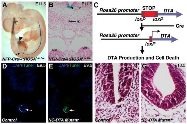
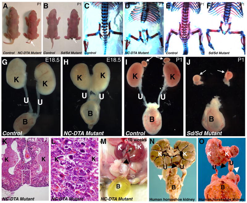
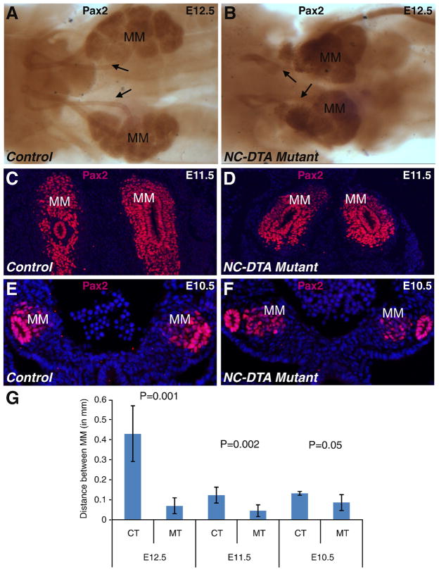
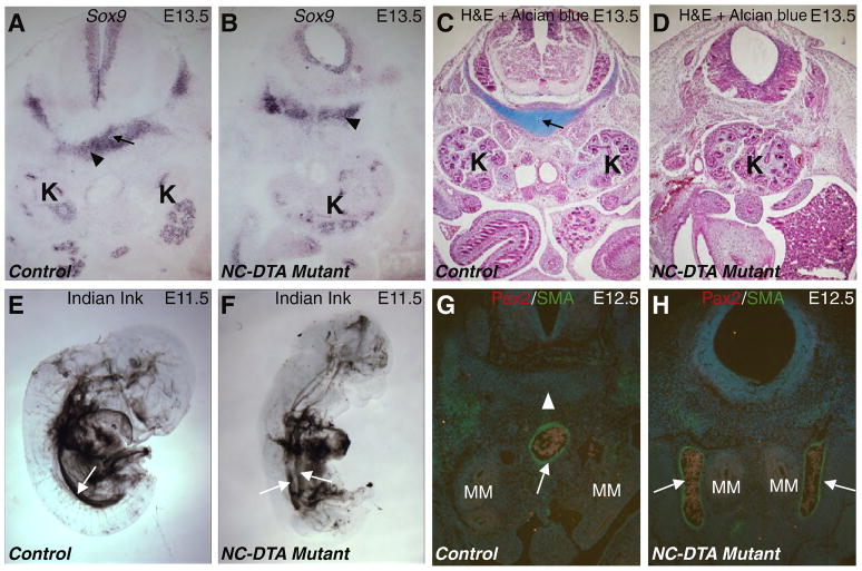
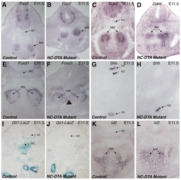
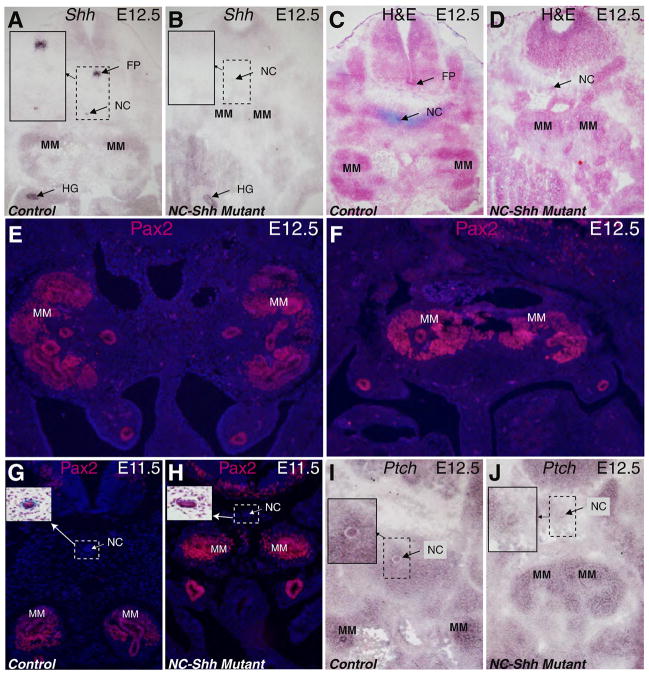
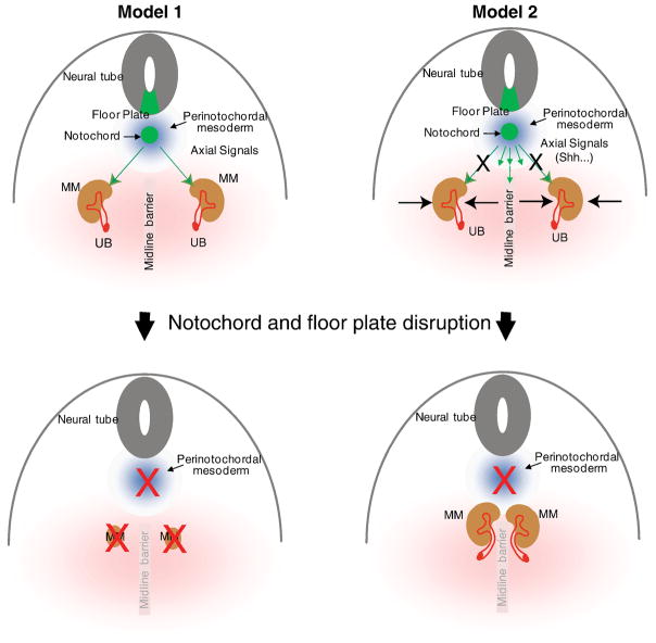
The role of axial structures, especially the notochord, in metanephric kidney development has not been directly examined. Here, we showed that disruption of the notochord and floor plate by diphtheria toxin (DTA)-mediated cell ablation did not disrupt nephrogenesis, but resulted in kidney fusions, resembling horseshoe kidneys in humans. Axial disruptions led to more medially positioned metanephric mesenchyme (MM) in midgestation. However, neither axial disruption nor the ensuing positional shift of the MM affected the formation of nephrons and other structures within the kidney. Response to Shh signaling was greatly reduced in midline cell populations in the mutants. To further ascertain the molecular mechanism underlying these abnormalities, we specifically inactivated Shh in the notochord and floor plate. We found that depleting the axial source of Shh was sufficient to cause kidney fusion, even in the presence of the notochord. These results suggested that the notochord is dispensable for nephrogenesis but required for the correct positioning of the metanephric kidney. Axial Shh signal appears to be critical in conferring the effects of axial structures on kidney positioning along the mediolateral axis. These studies also provide insights into the pathogenesis of horseshoe kidneys and how congenital kidney defects can be caused by signals outside the renal primordia.


Figures

Fig. 1
Fig. 1. Transgenic ablation of the notochord and floor plate by diphtheria toxin (DTA)
A. The ROSALacZ reporter revealed the expression of NFP-Cre in notochord and floor plate (arrow). The weak expression was also observed in part of the hindgut (arrowhead). B, A transverse section at the level of hind limb bud (shown by dashed line in Fig. 1A) showed specific X Gal staining in the notochord and floor plate. C, when combined with a ROSADTA allele, NFP-Cre induces the production of DTA, translational arrests, and apoptosis in Cre-positive cells. TUNEL assay on transverse sections of E9.5 control (D) and mutant (E) embryos at the level of the MM. Notochords are outlined by dotted circles. Apoptotic cells were seen only in the notochord of the mutant (E). F and G, transverse sections of the control (F) and mutant (G) embryos revealed notochord degeneration in the mutants at E10.5. Arrow in D–F points to the notochord. FP: floor plate; NC: notochord.
Fig. 2
Fig. 2. DTA mediated notochord and floor plate ablation results in kidney fusion but not renal agenesis
A, Newborn control and NC-DTA mutant mice. B, Newborn wild-type and Danforth Short tail homozygous (Sd/Sd) mutants. Both NC-DTA mutants and Sd/Sd mutants have short tails (arrows in A and B). CF, Skeletal preparations from newborn control (C) and NC-DTA mutant mice (D). In NC-DTA mutants, vertebral column is severely defective below the thoracic level, similar to skeletal defects in Sd/Sd mutants (E–F). Urinary systems from control mice (G and I), a NC-DTA mutant (H) and a Sd/Sd mutant (J). The NC-DTA mutants have fused kidneys resembling horseshoe kidneys (H), different from Sd/Sd mutants that have bilateral renal agenesis (J). White arrows indicate adrenal glands. K, H&E stained paraffin section of the fused kidneys. The area within the rectangle is shown in L at a higher magnification. Cortices from left and right kidneys are connected. M, a NC-DTA mutant with fused and hydronephrotic kidney. White arrow points to hydronephrotic kidney. Black arrow points to expanded pelvic area. N–O, Horseshoe kidneys in human fetus. Black triangle points to the fused lower poles of the left and right kidneys and white triangle points to hydroureter. K: kidney, U: ureter, B: bladder.
Fig. 3
Fig. 3. Disruption of the notochord and floor plate affects the mediolateral positioning of the MM
A–B, Pax2 immunostaining outlined the MM, WD, and UB at E12.5 in the control sample (A) and the mutant sample (B). C–F, Anti-Pax2 antibody stained transverse sections of E11.5 control (C) and mutant (D) and E10.5 control (E) and mutant (F) embryos. DAPI staining highlights the nuclei. G, Metric data measuring the distance between MM tissues from both sides. The MM tissues are closer to each other in the mutants (two-tailed t-test). Distance between the left and right MM cell masses (in millimeters) at E12.5: CT 0.431667±0.14 (n=4), MT 0.07±0.04 (n=4), P=0.001; at E11.5: CT 0.123333±0.04 (n=4), MT 0.045±0.03 (n=4), P=0.002; at E10.5: CT 0.132±0.01 (n=4), MT 0.086±0.04 (n=5), P=0.05. MM: metanephric mesenchyme; CT: control; MT: mutant. Arrow: UB/ureter.
Fig. 4
Fig. 4. Disruption of the notochord and floor plate affects chondrogenesis of perinotochordal cells and midline vascular development
A–B, Sox9 RNA in situ hybridization on transverse sections at the MM level in control (A) and mutant (B) E13.5 embryos. Black arrow in A points to the notochord that is absence in the mutant (B). Black triangles point to the perinotochordal cells positive for Sox9 in both control and mutant samples. C–D, Transverse sections at the MM level in control (C) and mutant (D) E13.5 embryos stained with H&E and Alcian blue (for cartilage). Black arrow in C points to the notochord that is absence in the mutant (D). E–F, India ink injection into the left ventricle of E11.5 control (E) and mutant (F) embryos. The white arrows point to the dorsal aorta. At this time, the control dorsal aorta does not have major branches below the aortic arches (E). However, the mutant aorta is still bifurcated for most of the trunk region (F). G–H, transverse sections at the MM level from control (G) and NC-DTA mutant (H) E12.5 embryos. White arrows point to the αSMA staining (Green) in the aortic wall. White triangle points to the notochord (absent in the mutant). MM is highlighted by Pax2 staining in red. MM: metanephric mesenchyme; K: kidney.
Fig. 5
Fig. 5. Axial disruption affects the mediolateral positioning of the MM, but has little impact on the nephrogenic program inside the kidneys
Pax2 RNA in situ hybridization outlines the MM from the control (A) and mutant (B) at E11.5. C–D, expression patterns of Gdnf in MM are similar between control and mutant at E11.5. Foxd1 expression in the outer portion of the MM and other mesodermal cells, including cells that contribute to the formation of the renal capsule, is apparent in both the control (E) and the mutant (F) samples. Arrowhead marks a medial expansion of its expression domain in mutant. Shh is normally expressed in the floor plate, notochord, and the hindgut (G). In the mutant, the expression in the floor plate and notochord is missing but the expression in the hindgut remains (H). Expression of Gli1lacZ is seen around notochord in the control (I) but there is a drastic reduction of Gli1lacZ positive cells in the midline cell populations in the mutant sample. (J). The expression pattern of Id2 is similar in control and mutants (K–L). FP: floor plate; HG: hindgut; MM: metanephric mesenchyme; NC: notochord; NT: Neural tube.
Fig. 6
Fig. 6. Inactivation of Shh in the notochord and floor plate also leads to kidney fusion but not renal agenesis
Shh expression is detected in the notochord, floor plate and the hindgut on the transverse section at the MM level in the E12.5 control embryo (A). Shh expression in the notochord and floor plate is absent in the littermate mutant (B), though the notochord is still present at this stage. Insets show the enlarged images of the rectangular areas. H&E and Alcian blue (for cartilage) stained transverse sections from control (C) and mutant (D) embryos at E12.5 showed that the notochord persists in NC-Shh mutants with the MM tissues fused. Pax2 staining on E12.5 cross sections shows the separation of the left and right MM tissues in the control (E) and the fused MM tissues in the mutant (F). Pax2 staining on E11.5 control (G) and mutant (H) littermates revealed that MM tissues are closer to each other in the mutants. Insets show notochord images from H&E stained adjacent sections. Compared to the control (I) at E12.5, Ptch1 expression in the mutant sample (J) is drastically decreased in perinotochordal mesoderm at the MM level (Insets show the enlarged images of the rectangular areas.). FP: floor plate; HG: hindgut; MM: metanephric mesenchyme; NC: notochord.
Fig. 7
Fig. 7. Models for axial regulation of metanephric kidney development
The finding of renal fusion in the NC-DTA mutants contradicts the hypothesis that degeneration of the notochord leads to renal agenesis (Model 1). In a new model (Model 2), signals from the notochord and floor plate, such as Shh, do not directly affect nephrogenesis. Instead, these signals affect the development of the midline structures and the positioning of the MM. See text for details. UB: ureteric bud; MM: metanephric mesenchyme.

Similar articles

Cited by

References

    1. Aoki Y, Mori S, Kitajima K, Yokoyama O, Kanamaru H, Okada K, Yokota Y. Id2 haploinsufficiency in mice leads to congenital hydronephrosis resembling that in humans. Genes Cells. 2004;9:1287–96. - PubMed
    1. Bai CB, Auerbach W, Lee JS, Stephen D, Joyner AL. Gli2, but not Gli1, is required for initial Shh signaling and ectopic activation of the Shh pathway. Development. 2002;129:4753–61. - PubMed
    1. Barak H, Rosenfelder L, Schultheiss TM, Reshef R. Cell fate specification along the anterior-posterior axis of the intermediate mesoderm. Dev Dyn. 2005;232:901–14. - PubMed
    1. Chang CP, McDill BW, Neilson JR, Joist HE, Epstein JA, Crabtree GR, Chen F. Calcineurin is required in urinary tract mesenchyme for the development of the pyeloureteral peristaltic machinery. J Clin Invest. 2004;113:1051–8. - PMC - PubMed
    1. Chen F. Genetic and developmental basis for urinary tract obstruction. Pediatr Nephrol. 2009;24:1621–32. - PMC - PubMed

Publication types

MeSH terms

LinkOut - more resources