. 2010 Jul;171(1):82-7.
doi: 10.1016/j.jsb.2010.02.005. Epub 2010 Feb 10.

Fluorescence Detection of Heavy Atom Labeling (FD-HAL): a rapid method for identifying covalently modified cysteine residues by phasing atoms

Affiliations

Fluorescence Detection of Heavy Atom Labeling (FD-HAL): a rapid method for identifying covalently modified cysteine residues by phasing atoms

Vincent Chaptal et al. J Struct Biol. 2010 Jul.

Abstract

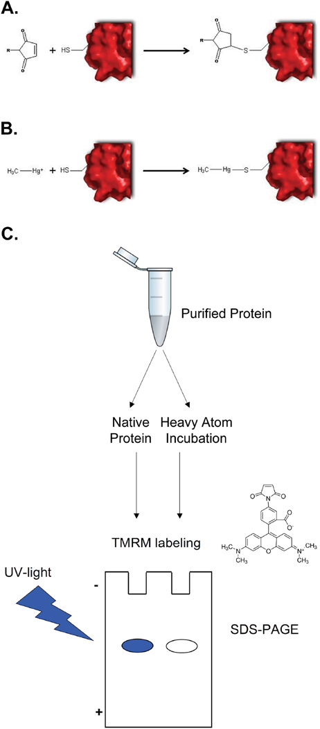
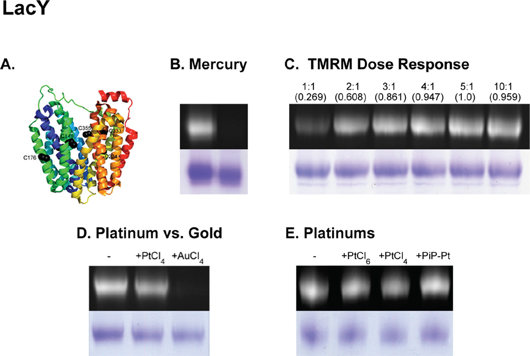
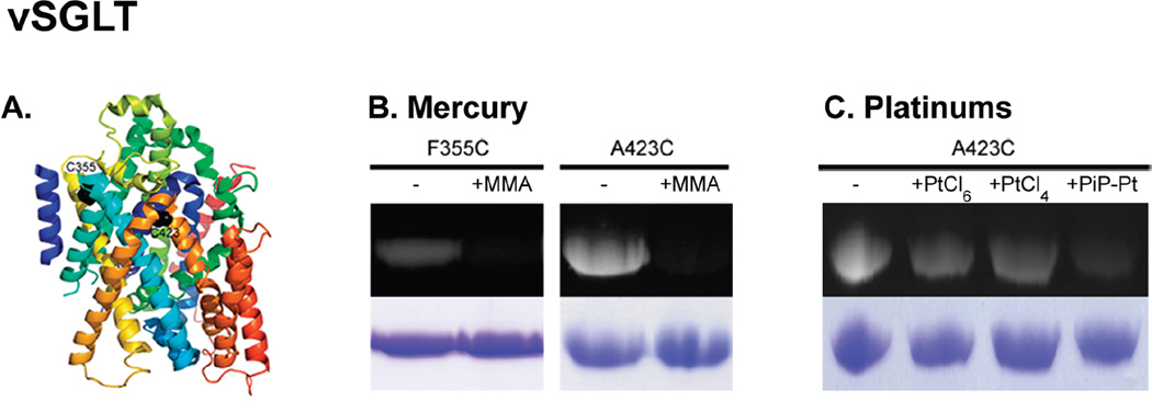
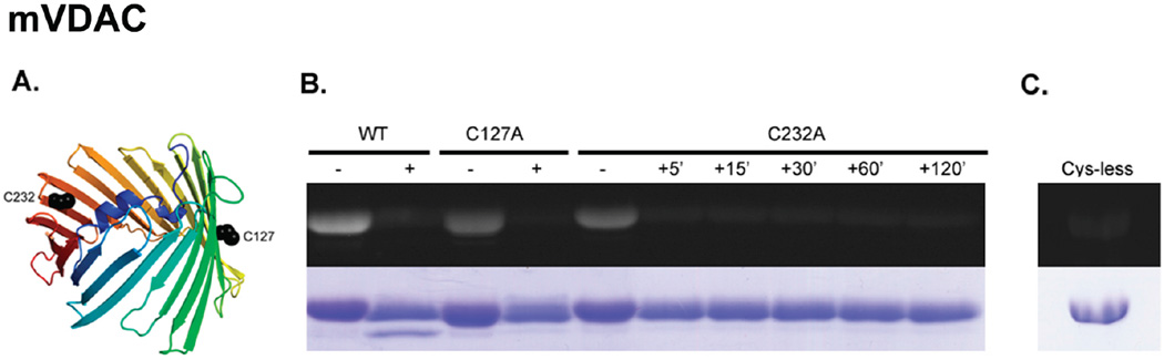
Membrane protein crystallography frequently stalls at the phase determination stage due to poor crystal diffraction and the inability to identify heavy atom derivatization prior to data collection. Thus, a majority of time, effort and resources are invested preparing potential derivatized crystals for synchrotron data collection and analysis without knowledge of heavy atom labeling. To remove this uncertainty, we introduce Fluorescence Detection of Heavy Atom Labeling (FD-HAL) using tetramethylrhodamine-5-maleimide (a fluorescent maleimide compound) to monitor in-gel cysteine residue accessibility and ascertain covalent modification by mercury, platinum and gold compounds. We have tested this technique on three integral membrane proteins (LacY, vSGLT and mVDAC1) and can quickly assess the optimal concentrations, time and heavy atom compound to derivatize free cysteine residues in order to facilitate crystal phasing. This, in conjunction with cysteine scanning for incorporating heavy atoms at strategic positions, is a useful tool that will considerably assist in phasing membrane protein structures.


Figures

Figure 1
Figure 1. FD-HAL technique
(A) Alkylation of cysteine by a maleimide compound. (B) Derivatization of cysteine by a HA (Methyl Mercury Acetate used as an example) [27]. (C) Schematic flow-diagram of the Fluorescence Detection of HA Labeling (FD-HAL) procedure. A purified protein sample is labeled by HAs and subsequently incubated with TMRM. The test sample is run on a SDS-PAGE and the TMRM binding fluorescence is monitored under UV-light. As a control, TMRM incubation (in the absence of HA labeling) is performed in a similar manner. The comparison of fluorescence intensity between the native vs. derivatized protein determines the degree of HA labeling. A decrease in fluorescence indicates the cysteine(s) have been derivatized by HAs and are no longer available for TMRM labeling.
Figure 2
Figure 2. FD-HAL on LacY
(A) Crystal structures of C154G LacY (PDB code: 1PV7) represented as a rainbow cartoon with cysteine residues shown as black spheres. (B) MMA labeling. (C) Dose response of TMRM incubation on native LacY. The numbers on top indicate the molar ratio of TMRM to LacY and number in parentheses are the Relative Fluorescence Intensities quantified using ImageJ software [28]. The results are averaged over 3 different SDS-PAGE and normalized to the (5:1) molar ratio. (D) K2PtCl4 and K2AuCl4 labeling. (E) K2PtCl6, K2PtCl4 and PiP-Pt labeling. (B)-(F) Top panels: UV fluorescence of TMRM. Lower panels: Coomassie Brilliant Blue staining as loading controls. The protein concentration was measured to assure equivalent protein sample loads. (-) indicates protein not labeled with HA.
Figure 3
Figure 3. FD-HAL on vSGLT
(A) Crystal structure of vSGLT (PDB code: 3DH4) represented as a rainbow cartoon with cysteine residues shown as black spheres. (B) MMA labeling of the two single cysteine mutants F355C and A423C. To be able to correctly visualize the TMRM fluorescence signal for the F355C mutant, 20 µg of protein was run on the SDS-PAGE. (C) K2PtCl6, K2PtCl4 and PiP-Pt labeling of the A423C vSGLT single cysteine mutant.
Figure 4
Figure 4. FD-HAL on mVDAC1
(A) Crystal structure of mVDAC1 (PDB code: 3EMN) represented as a rainbow cartoon with cysteine residues shown as black spheres. (B) MMA labeling of wild type, and the two single cysteine C127A and C232A mVDAC1 mutants. A time-course incubation with MMA was carried out on C232A-mVDAC1, over 5, 15, 30, 60 and 120 minutes before washing away excess MMA. (C) Basal TMRM fluorescence observed by incubating TMRM on a cysteine-free mVDAC1 mutant.

Similar articles

Cited by

References

    1. White SH. Biophysical dissection of membrane proteins. Nature. 2009 May 21;459(7245):344–346. - PubMed
    1. Boggon TJ, Shapiro L. Screening for phasing atoms in protein crystallography. Structure. 2000;Vol 8(No 7):R143–R149. - PubMed
    1. Cohen SL, Padovan JC, Chait BT. Mass spectrometric analysis of mercury incorporation into proteins for X-ray diffraction phase determination. Anal Chem. 2000 Feb 1;72(3):574–579. - PubMed
    1. Sun PD, Hammer CH. Mass-spectrometry assisted heavy-atom derivative screening of human Fc gamma RIII crystals. Acta Crystallogr D Biol Crystallogr. 2000 Feb;56(Pt 2):161–168. - PubMed
    1. Wittig I, Schägger H. Features and applications of blue-native and clear-native electrophoresis. Proteomics. 2008 Oct;8(19):3974–3990. - PubMed

Publication types

LinkOut - more resources