. 2010 Feb 26;32(2):253-65.
doi: 10.1016/j.immuni.2010.01.010. Epub 2010 Feb 11.

Optimal germinal center responses require a multistage T cell:B cell adhesion process involving integrins, SLAM-associated protein, and CD84

Affiliations

Optimal germinal center responses require a multistage T cell:B cell adhesion process involving integrins, SLAM-associated protein, and CD84

Jennifer L Cannons et al. Immunity. .

Abstract

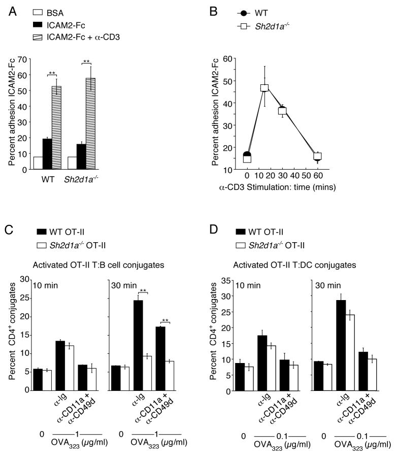
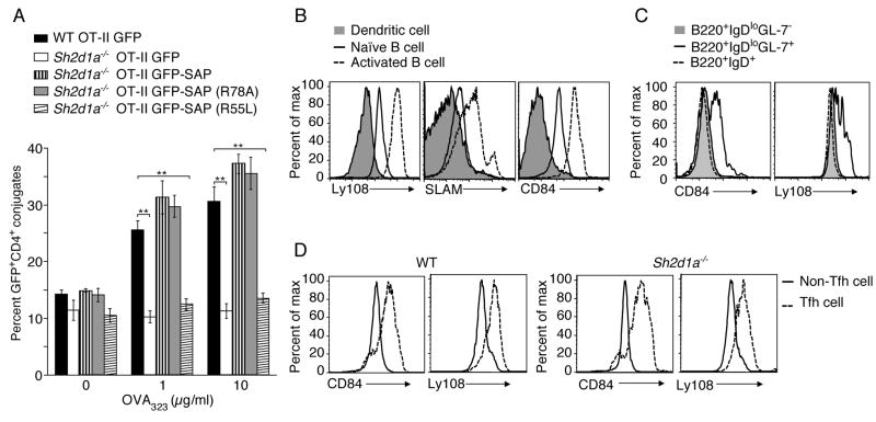
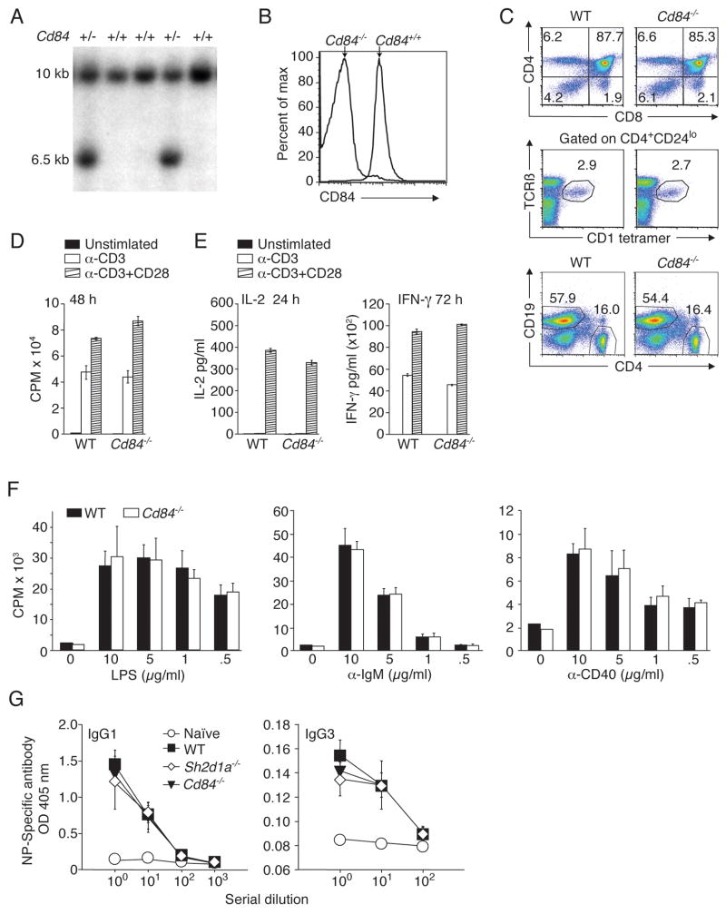
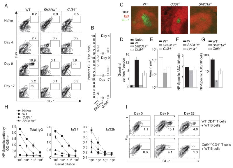
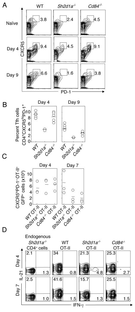
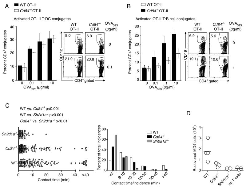
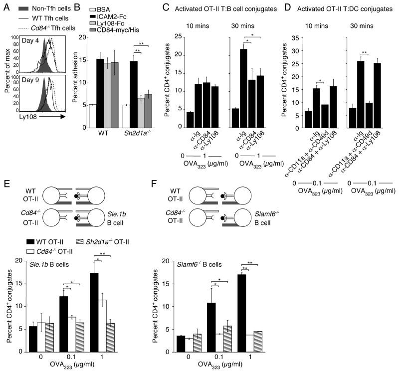
CD4(+) T cells deficient in signaling lymphocyte activation molecule (SLAM)-associated protein (SAP) exhibit a selective impairment in adhesion to antigen-presenting B cells but not dendritic cells (DCs), resulting in defective germinal center formation. However, the nature of this selective adhesion defect remained unclear. We found that whereas T cell:DC interactions were primarily integrin dependent, T cell:B cell interactions had both an early integrin-dependent phase and a sustained phase that also required SAP. We further found that the SLAM family member CD84 was required for prolonged T cell:B cell contact, optimal T follicular helper function, and germinal center formation in vivo. Moreover, both CD84 and another SLAM member, Ly108, mediated T cell adhesion and participated in stable T cell:B cell interactions in vitro. Our results reveal insight into the dynamic regulation of T cell:B cell interactions and identify SLAM family members as critical components of sustained T cell:B cell adhesion required for productive humoral immunity.


Figures

Figure 1
Figure 1. Integrin-mediated adhesion is critical for initial but not prolonged T:B cell conjugation
(A–B) Adhesion of pre-activated WT and Sh2d1a−/− CD4+ T cells to recombinant ICAM2-Fc following α-CD3 stimulation (A) 10 min. and (B) 15–60 min. Results presented as the mean percent adhesion (A) n=3 ± SEM. (B) n=6 ± SEM. (C–D) Conjugation efficiency after pre-incubation with blocking integrin antibodies then incubation for 10–30 min with OVA323 pulsed B cells (C), mean ± SEM frequency of CD4+CD19+ conjugates in total CD4+ events (n=3) or (D) DCs, mean ± SEM frequency of CD4+CD11c+ conjugates in total CD4+ events (n=3). **p<0.005.
Figure 2
Figure 2. Importance of SAP in T:B adhesion is consistent with SLAM family member expression on activated B cells
(A) OT-II T:B cell conjugation assays were conducted using CD4+ T cells transiently transfected with DNA constructs expressing either GFP, GFP-SAP, GFP-SAP(R78A), or GFP-SAP(R55L), n=6, mean ± SEM frequency of CD4+CD19+ conjugates in total GFP+CD4+ events, **p<0.005 (See Figure S2 for western blot and representative FACS plot). (B) CD11c+ splenic DCs, splenic B cells and LPS-activated B cells were assessed for SLAM, Ly108, and CD84 expression (n=3). (C–D) Day 9 post-NP-OVA in alum immunization, (C) B cells or (D) Tfh (CD4+CD44+CXCR5hiPD-1hi) and non-Tfh (CD4+CD44+CXCR5loPD-1lo) cells were evaluated for Ly108 and CD84 expression (n=2, 2–5 mice/genotype).
Figure 3
Figure 3. Targeted disruption of mouse Cd84 gene does not affect T and B cell development or in vitro stimulation
(A) Screening for homologous recombination by Southern blot of NcoI digested DNA. WT allele:10kb. Disrupted allele:6.5kb. Targeting vector and locus are shown in Figure S3A. (B) Surface CD84 expression on WT and Cd84−/− splenic B cells. (C) Top panel: thymocytes stained with α-CD4 and α-CD8. Middle panel: thymocytes stained with CD1d-αGalCer tetramers, α-CD4, α-CD24, and α–TCRβ to evaluate NKT cells. Bottom panel: splenocytes stained with α-CD19 and α-CD4 (n=3, 3 mice/genotype). (D) WT and Cd84−/− T cells were stimulated with α-CD3+/− α-CD28 and evaluated for proliferation and (E) IL-2 and IFN-γ production (n=3). (F) WT and Cd84−/− B cells were stimulated for 48 h with LPS, α-IgM, or α-CD40 and evaluated for proliferation (n=4). (G) WT, Sh2d1a−/−, and Cd84−/− mice were immunized with the type II T-independent antigen, NP-Ficoll, and assessed for NP-specific antibody production, day 21 (n=2, 5 mice/genotype).
Figure 4
Figure 4. Impaired humoral response in the absence of CD84
WT, Sh2d1a−/−, and Cd84−/− mice were immunized with T-dependent antigen NP-OVA in alum or Ribi. (A) GC B cell development evaluated by gating on B220+IgDlo cells: GC B cells are FashiGL-7+. (B) Percentage of B cells with GC markers at day 4, 9 and 17 post-immunization. (C) Splenic GC were detected via staining with IgD and GL-7 day 9. (D) GC number/section, day 9. (E) GC size, day 9. * Size of the rare (2) GC identified in Sh2d1a−/− sections. (F) NP-specific IgG ASCs in the spleen, day 9. (G) Long-lived NP-specific plasma cell responses were measured in bone marrow, day 30. (H) NP-specific [NP-(30)], day 30 (See Figure S4A for high affinity NP-specific [NP-(3)] responses). Results are shown from mice immunized with NP-OVA in alum (A, B and H, n=2, 3–5 mice/genotype/time point) or NP-OVA in Ribi (C, D, E, F and G n=3, 6–8 mice/genotype/time point). Similar results were obtained with either adjuvant. (I) Cd84−/− or WT sorted naïve CD4+CD62L+CD44lo T cells and WT B cells were co-transferred into Rag2−/− hosts. Day 25 following reconstitution, Rag2−/− mice were immunized with SRBCs and evaluated for GC development (See Figure S4B for antibody titres), n=2, 4 mice/genotype/time point.
Figure 5
Figure 5. Evaluation of Tfh cells
(A–B) Flow cytometric analysis of cells expressing Tfh markers from WT, Sh2d1a−/−, and Cd84−/− mice at day 4 and 9 post-immunization with NP-OVA in alum. (A) Representative FACS plots (gated on CD4+ cells). (B) Percent Tfh cells, n=2, 2–5 mice/genotype/time point. (C–D) WT, Sh2d1a−/−, or Cd84−/− OT-II GFP+ T cells were transferred into Sh2d1a−/− hosts subsequently immunized with NP-OVA in alum (n=2, 3 mice/genotype/time point). (C) Number of WT, Sh2d1a−/−, or Cd84−/− OT-II GFP+ Tfh (CD4+CXCR5hiPD-1hi) cells day 4 and 7 post-immunization. (D) IL-21 production from WT, Sh2d1a−/−, or Cd84−/− OT-II GFP+ cells. (See Figure S5 for Tfh IL-21 production)
Figure 6
Figure 6. Cd84−/− OT-II T cells are defective in adhesion to cognate B cells
(A-B) Conjugation efficiency of pre-activated WT and Cd84−/− OT-II T cells with OVA323 pulsed splenic (A) CD11c+ DCs, mean ± SEM frequency of CD4+CD11c+ conjugates in total CD4+ events (n=4, representative FACS plots, right panel), (B) LPS-activated B cells, mean ± SEM frequency of CD4+CD19+ conjugates in total CD4+ events (n=4, representative FACS plots, right panel) (*p<0.05, **p<0.005). (C) In vivo contact durations between T cells of indicated genotypes and MD4 B cells as measured by intravital microscopy between 60–72 h after immunization with HEL-OVA in alum. Individual contact durations (left) and their distribution (right) are shown. A total of 125, 120, and 49 contacts pooled from 3 experiments for WT, Cd84−/−, and Sh2d1a−/− T cells, respectively. Mean contact times: WT 14.5+/−1.2 min; Cd84−/− 8.5+/−2.1 min; Sh2d1a−/− 3.5+/−1.6 min. The p values were calculated by nonparametric one-way ANOVA. (D) Recovery of MD4 B cells from draining lymph nodes 96 h post-HEL-OVA immunization with or without exogenous OT-II T cells of indicated genotypes. Each symbol represents 1 of 4 mice/group. Data from 1 of 3 experiments with similar results are shown.
Figure 7
Figure 7. CD84 and Ly108 are adhesive receptors and contribute to long-lived stable T:B cell conjugates
(A) Ly108 expresssion on Tfh (CD4+CXCR5hiPD-1hi) cells from WT and Cd84−/− mice post-NP-OVA in alum immunization. (B) Evaluation of pre-activated WT and Sh2d1a−/− T cell adhesion to recombinant ICAM2-Fc, Ly108-Fc, and CD84-myc.HIS assessed for 30 min (n=4). (C-D) Conjugation efficiency of blasted WT OT-II T cells, pre-incubated with α-CD84 or α-Ly108, with OVA323-pulsed LPS-activated B cells (C), mean ± SEM frequency of CD4+CD19+ conjugates in total CD4+ events (n=4) or DCs (D), mean ± SEM frequency of CD4+CD11c+ conjugates in total CD4+ events (n=3). (E–F) Conjugation frequencies of pre-activated WT, Sh2d1a−/−, and Cd84−/− OT-II T cells with OVA323-pulsed LPS-activated B cells for 30 min from (E) Sle.1b congenic or (F) Slamf6−/− mice, mean ± SEM frequency of CD4+CD19+ conjugates in total CD4+ events, n=3 (See Figure S6 for representative FACS plot). (*p<0.05, **p<0.005).

References

    1. Al-Alwan MM, Rowden G, Lee TDG, West KA. The dendritic cell cytoskeleton is critical for the formation of the immunological synapse. J Immunol. 2001;166:1452–1456. - PubMed
    1. Allen CDC, Okada T, Cyster JG. Germinal-center organization and cellular dynamics. Immunity. 2007;27:190–202. - PMC - PubMed
    1. Barber DF, Long EO. Coexpression of CD58 or CD48 with intracellular adhesion molecule 1 on target cells enhances adhesion of resting NK cells. J Immunol. 2003;170:294–299. - PubMed
    1. Benvenuti F, Hugues S, Walmsley M, Ruf S, Fetler L, Popoff M, Tybulewicz VLJ, Amigorena S. Requirements of Rac1 and Rac2 expression by mature dendritic cells for T cell priming. Science. 2004;305:1150–1153. - PubMed
    1. Bryceson YT, March ME, Barber DF, Ljunggren HG, Long EO. Cytolytic granule polarization and degranulation controlled by different receptors in resting NK cells. J Exp Med. 2005;202:1101–1012. - PMC - PubMed

Publication types

MeSH terms

Substances