. 2010 Feb 17;17(2):198-212.
doi: 10.1016/j.ccr.2009.12.040.

A higher-order complex containing AF4 and ENL family proteins with P-TEFb facilitates oncogenic and physiologic MLL-dependent transcription

Affiliations

A higher-order complex containing AF4 and ENL family proteins with P-TEFb facilitates oncogenic and physiologic MLL-dependent transcription

Akihiko Yokoyama et al. Cancer Cell. .

Abstract

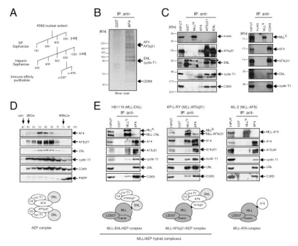
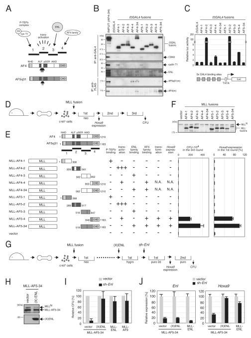
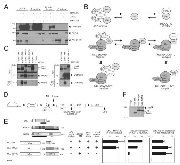
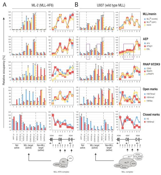
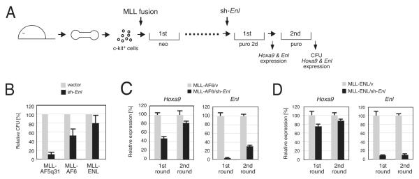
AF4 and ENL family proteins are frequently fused with MLL, and they comprise a higher order complex (designated AEP) containing the P-TEFb transcription elongation factor. Here, we show that AEP is normally recruited to MLL-target chromatin to facilitate transcription. In contrast, MLL oncoproteins fused with AEP components constitutively form MLL/AEP hybrid complexes to cause sustained target gene expression, which leads to transformation of hematopoietic progenitors. Furthermore, MLL-AF6, an MLL fusion with a cytoplasmic protein, does not form such hybrid complexes, but nevertheless constitutively recruits AEP to target chromatin via unknown alternative mechanisms. Thus, AEP recruitment is an integral part of both physiological and pathological MLL-dependent transcriptional pathways. Bypass of its normal recruitment mechanisms is the strategy most frequently used by MLL oncoproteins.


Figures

Figure 1
Figure 1
Heterologous associations of wild type and oncogenic AF4 and ENL family proteins (A) The scheme employed for purification of the AF4 complex. (B) A silver-stained image shows the proteins immuno-purified using anti-AF4 antibody, and subsequently identified by mass spectrometry as indicated by arrows on the right. Anti-GST antibody served as a negative control. (C) K562 nuclear extracts were analyzed by IP western blotting. IP was performed with the antibodies indicated on the top and the precipitates were immunoblotted with the antibodies indicated on the right. Anti-GST and anti-FLAG antibodies served as negative controls. Asterisks indicate signals from IgG used for IP. (D) Selected fractions from gel filtration analysis of K562 nuclear extracts were analyzed by western blotting for AF4-associated factors (PARP served as a negative control). Molecular weight standards are shown on the top. A cartoon of a putative AEP complex is depicted. C9, CDK9; cyc T1, cyclin T1. (E) IP western blot analysis was performed as in (C) on human leukemia cell lines that harbor MLL chromosomal translocations and express MLL chimeric oncoproteins (indicated at tops). Cartoons of putative MLL fusion complexes are depicted below. See also Figure S1.
Figure 2
Figure 2
Co-localization of MLL fusion proteins and AEP components on chromatin (A) Relative expression of various genes (indicated on the right) in seven human cell lines was analyzed by quantitative RT-PCR. Expression levels were normalized to GAPDH and depicted relative to the highest value among the seven cell lines arbitrarily set as 100. Error bars represent standard deviations of triplicate PCRs. (B) Genomic localizations of various proteins in HB1119 cells were determined by ChIP assay. Cross-linked chromatin was immunoprecipitated with antibodies specific for the indicated proteins and analyzed by quantitative PCR using primer/probe sets that target promoter-adjacent regions or other genomic regions indicated at the bottom. Occupancies are displayed relative to the highest value in the group arbitrarily set as 100. Error bars represent standard deviations of triplicate PCRs. Genes expressed more than 20% of the highest levels in panel A are defined as active genes. (C) A comparable analysis as (B) was performed for MV4-11 cells, which harbor a t(4;11) translocation and express MLL-AF4 proteins. The purple rectangle highlights a locus on which di-methyl H3K79 marks were absent, but the MLL-AF4/AEP complex was present. See also Figure S2.
Figure 3
Figure 3
Formation of an AEP-like complex is required for MLL-AF5q31-dependent myeloid transformation (A) The structures of AF4 and AF5q31 are schematically illustrated. Subregions (1-4) of AF4 and AF5q31 are indicated with associated functions. Upward arrows indicate the sites of fusion with MLL in human leukemia oncoproteins (Jansen et al. 2005). A9ID, AF9 interaction domain (Srinivasan et al. 2004). (B) The four subregions fused to GAL4 DNA binding domain were expressed in 293T cells (upper four panels) or co-expressed with myc-tagged AF4 or AF5q31 [AF4(m) or AF5q31(m)] (lower two panels) and analyzed by IP western blotting. IP antibodies are indicated on the left and proteins detected by western blotting are indicated on the right. (f)GAL4 fusions and myc-tagged AF4 family proteins were visualized with anti-FLAG and anti-myc antibodies, respectively. (C) Transactivation activity of respective GAL4 fusions was analyzed using the reporter gene shown below. Error bars represent standard deviations from triplicate analyses. (D) The experimental scheme of myeloid progenitor transformation assays to evaluate the oncogenic potentials of various MLL mutants shows the time points at which CFU (colony forming unit) activity or Hoxa9 expression was examined. (E) The structures of various MLL-AF4/AF5q31 mutants and their associated functions are summarized schematically. Hoxa9 levels were normalized to Gapdh and displayed relative to MLL-AF5-34-transduced cells arbitrarily set at 100 %. Error bars represent standard deviations of three independent analyses (left) or triplicate PCRs (right). N.A., not applicable due to unstable expression of MLL fusion proteins. (F) Protein levels of respective MLL mutants in virus packaging cells were examined by western blotting with anti-MLLN antibody. MLL-AF4-4 and MLL-AF4-34 proteins were not stably expressed. (G) The experimental scheme to evaluate the effect of Enl knockdown on MLL transformation is shown schematically. (X)ENL, Xpress-tagged human ENL. (H) Transduced myeloid progenitors were analyzed by western blotting with anti-MLLN (top) and anti-Xpress (bottom) antibodies to detect exogenous MLL-AF5q31 and human (X)ENL, respectively (I) The clonogenic potentials of MLL-AF5-34-transformed cells transduced with or without (X)ENL are shown at the second plating after sh-RNA transduction (vector or sh-Enl). MLL-ENL- or MLL-AF9-transformed cells were also subjected to sh-RNA transduction for comparison. CFUs are expressed relative to the vector control arbitrarily set as 100. Error bars represent standard deviations of three independent analyses. (J) Cells from first round colonies following sh-RNA transduction (vector or sh-Enl) were analyzed by RT-PCR for expression of endogenous Enl or Hoxa9. Expression levels were normalized to Gapdh and displayed relative to the vector/vector control cells arbitrarily set at 100. Error bars represent standard deviations of triplicate PCRs. See also Figure S3.
Figure 4
Figure 4
MLL-ENL and MLL-AF9 transform myeloid progenitors via the AHD, which is responsible for association with AF4 family proteins and DOT1L (A) The structures of ENL and AF9 are schematically illustrated with associated functions (Zeisig et al. 2005). Aligned amino acid sequences for the minimum transformation domain are also shown with the positions of deletion or substitution mutations and AHD. Upward arrows indicate the sites of fusion with MLL in human leukemia oncoproteins (Jansen et al., 2005). (B) Domain mapping of ENL family proteins for association with AF5q31 was performed with FLAG-tagged GAL4 fusion constructs of ENL (372-559 aa) and AF9 (478-568 aa). IP was performed with anti-GAL4 antibody and the precipitates were immunoblotted with anti-FLAG antibody for (f)GAL4 fusions or anti-AF5q31 antibody for endogenous AF5q31. (C) Transactivation activity of indicated GAL4 constructs was analyzed by luciferase assay as in Figure 3C. (D) The same set of GAL4 fusion proteins used in (B) and HA-tagged DOT1L [DOT1L(H)] were co-expressed in 293T cells and analyzed by IP western blotting. IP was performed with anti-FLAG antibody and the precipitates were immunoblotted with anti-HA antibody. (E) The experimental scheme is shown for myeloid progenitor transformation assays to evaluate the oncogenic potentials of MLL mutants. (F) The structures of MLL-ENL and MLL-AF9 mutants and their associated functions are summarized with schematic representations. Hoxa9 expression levels were normalized to Gapdh and displayed relative to the MLL-ENL-transduced cells arbitrarily set at 100 %. Error bars represent standard deviations of three independent analyses (left) or triplicate PCRs (right). N.A., not applicable due to unstable expression of MLL fusion proteins. The asterisk indicates that association of ENL Δ548-559 mutant with DOT1L was detected but reduced substantially compared to wt ENL. (G) Protein levels of respective MLL mutants in virus packaging cells were examined by western blotting with anti-MLLN antibody. MLL-ENL Δ523-547 was not stably expressed.
Figure 5
Figure 5
Associations of ENL family proteins with AF4 family proteins or DOT1L are mutually exclusive (A) AF5q31(m), (X)ENL, and DOT1L(H) were co-expressed in 293T cells and analyzed by IP western blotting. IP was performed with antibodies indicated on the top and the precipitates were immunoblotted with anti-myc, anti-Xpress or anti-HA antibody. (B) Putative conformations of various ENL complexes are shown schematically. ENL forms two distinct complexes: AEP and ENL/DOT1L. Similarly, MLL-ENL participates in two mutually exclusive associations to form the MLL-ENL/AEP and MLL-ENL/DOT1L complexes that are approximate to the MLL-AF5q31/AEP and MLL-DOT1L complexes, respectively. (C) FLAG-tagged MLL fusion proteins [(f) MLL fusions] were co-expressed with AF4(m) or DOT1L(H) in 293T cells and analyzed by IP western blotting. IP was performed with anti-FLAG antibody and the precipitates were immunoblotted with anti-MLLN, anti-myc, or anti-HA antibody. (D) The experimental scheme is shown for myeloid progenitor transformation assays to evaluate the oncogenic potentials of MLL mutants. (E) The structures of MLL-fusion proteins and their associated functions are summarized. Expression of MLL fusion genes or Hoxa9 was examined by RT-PCR in first round colonies. Expression levels were normalized to Gapdh levels and displayed relative to the transcript levels in MLL-ENL transduced cells arbitrarily set at 100. Error bars represent standard deviations of three independent analyses (left) or triplicate PCRs (middle and right). HMT, histone methyltransferase catalytic domain; EBD, ENL binding domain (Okada et al. 2005; Mueller et al. 2007). (F) Protein levels of MLL fusions in virus packaging cells were analyzed by western blotting with anti-MLLN antibody. See also Figure S4.
Figure 6
Figure 6
Indirect recruitment of AEP to MLL-AF6- or wt MLL-occupied loci (A & B) Genomic localizations of indicated proteins in ML-2 (A) and U937 (B) cells were determined by ChIP assay as in Figure 2B. The purple rectangle highlights regions where AEP is absent while the MLL complexes are present. ChIP data using anti-MLLN (rpN1) and anti-menin antibodies are partially adapted from a previous report (Yokoyama and Cleary, 2008). See also Figure S5.
Figure 7
Figure 7
ENL is required for MLL-AF6-dependent transactivation and transformaion (A) The experimental scheme to evaluate the effect of Enl knockdown on MLL transformation is shown. (B) Clonogenic potentials are shown for myeloid cells transformed by MLL oncogenes (indicated below) at the second plating after sh-RNA transduction (vector or sh-Enl). CFU numbers are displayed relative to the vector control arbitrarily set as 100. Error bars represent standard deviations of three independent analyses. (C) MLL-AF6-transformed cells from first and second round colonies following sh-RNA transduction (vector or sh-Enl) were analyzed by RT-PCR for expression of endogenous Enl or Hoxa9. Expression levels were normalized to GAPDH levels and displayed relative to the transcript levels in vector control cells arbitrarily set as 100. Error bars represent standard deviations of triplicate PCRs. (D) The same analysis as (C) was performed on MLL-ENL-transformed cells. Note that data in (B) and (D) are partially redundant with Figures 3I and 3J.
Figure 8
Figure 8
ENL functions downstream of physiologic MLL-dependent transcriptional pathways (A) Expression levels of Enl and Hoxc8 in wt or Men1-/- MEFs were determined by RT-PCR (normalized to β-actin levels and displayed relative to the vector control arbitrarily set as 100). Error bars represent standard deviations of triplicate PCRs. (B) Expression of Enl and Hoxc8 with or without Enl knockdown/rescue was determined by RT-PCR. Expression levels were normalized to β-actin levels and expressed relative to the vector/vector control arbitrarily set as 100. Error bars represent standard deviations of triplicate PCRs. Protein levels of the exogenously expressed (X)ENL (right panels) were assessed by western blotting with an anti-Xpress antibody (actin immunoblot served as a loading control). nsp, non-specific band. (C) ChIP assay was performed on wt or Men1-/- MEFs using anti-di-methyl H3K79 and histone H3 antibodies for the Hoxc8 promoter-adjacent region and results displayed as relative ratio (%) to the input DNA. Error bars represent standard deviations of triplicate PCRs. (D) The effects of ENL-knockdown are shown for two different sh-RNAs in U937 cells. Expression of various genes was analyzed by RT-PCR 4 d after transduction/puromycin selection. Expression values were normalized to GAPDH levels and displayed relative to the vector control arbitrarily set as 100. Error bars represent standard deviations of triplicate PCRs. (E) A three-step model of MLL-dependent transcription.

Similar articles

Cited by

References

    1. Ayton PM, Chen EH, Cleary ML. Binding to nonmethylated CpG DNA is essential for target recognition, transactivation, and myeloid transformation by an MLL oncoprotein. Mol Cell Biol. 2004;24:10470–10478. - PMC - PubMed
    1. Ayton PM, Cleary ML. Transformation of myeloid progenitors by MLL oncoproteins is dependent on Hoxa7 and Hoxa9. Genes Dev. 2003;17:2298–2307. - PMC - PubMed
    1. Bitoun E, Oliver PL, Davies KE. The mixed-lineage leukemia fusion partner AF4 stimulates RNA polymerase II transcriptional elongation and mediates coordinated chromatin remodeling. Hum Mol Genet. 2007;16:92–106. - PubMed
    1. Cheung N, Chan LC, Thompson A, Cleary ML, So CW. Protein arginine-methyltransferase-dependent oncogenesis. Nat Cell Biol. 2007;9:1208–1215. - PubMed
    1. Collins EC, Appert A, Ariza-McNaughton L, Pannell R, Yamada Y, Rabbitts TH. Mouse Af9 is a controller of embryo patterning, like Mll, whose human homologue fuses with Af9 after chromosomal translocation in leukemia. Mol Cell Biol. 2002;22:7313–7324. - PMC - PubMed

Publication types

MeSH terms

LinkOut - more resources