Review
. 2010 Apr;7(4):549-58.
doi: 10.1016/j.hrthm.2009.12.006. Epub 2009 Dec 11.

J wave syndromes

Affiliations
Review

J wave syndromes

Charles Antzelevitch et al. Heart Rhythm. 2010 Apr.

Abstract

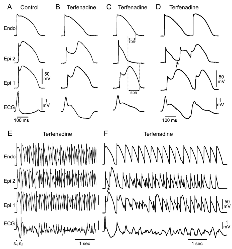
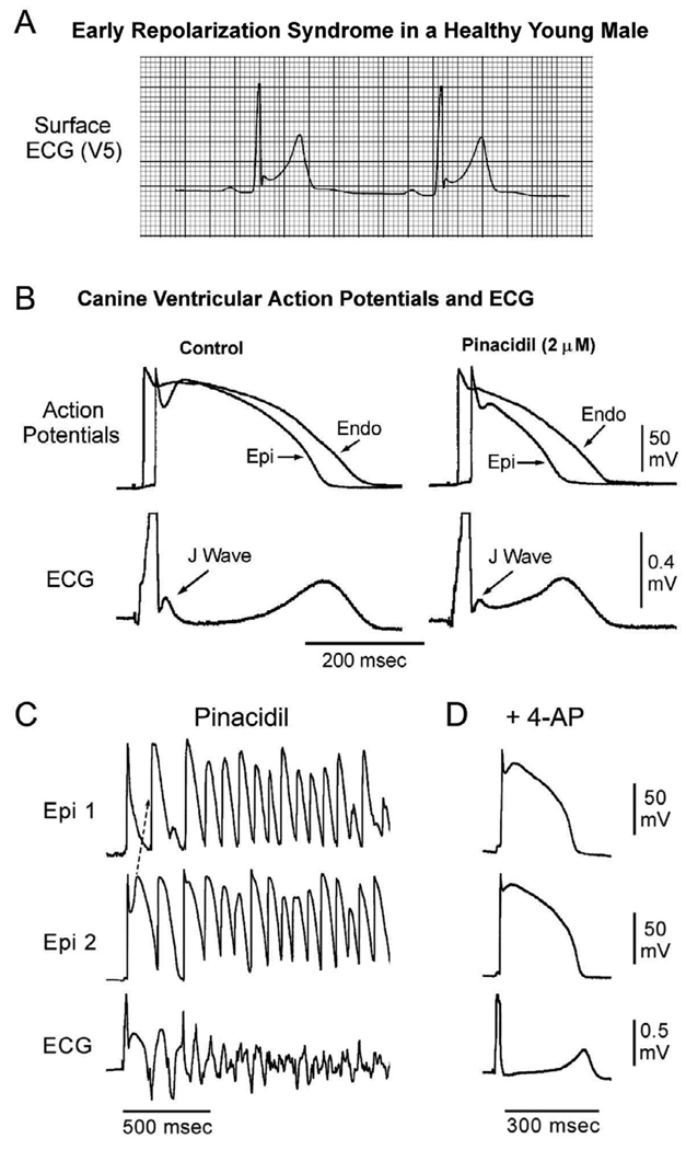
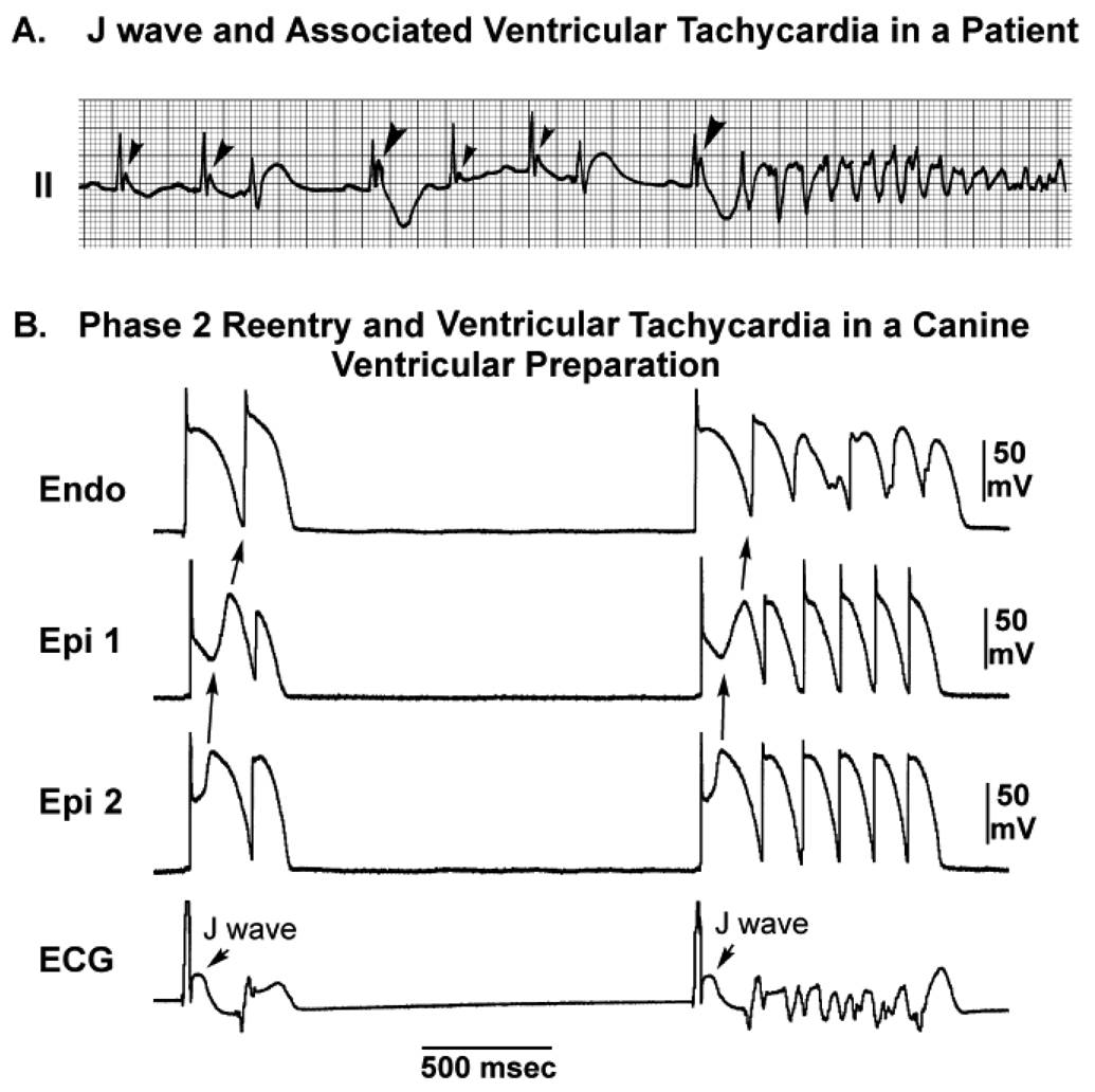
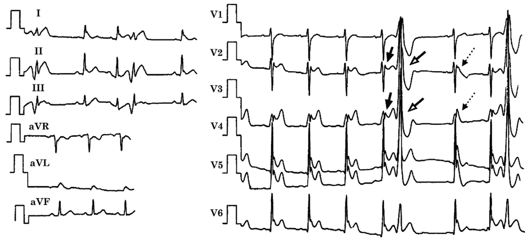
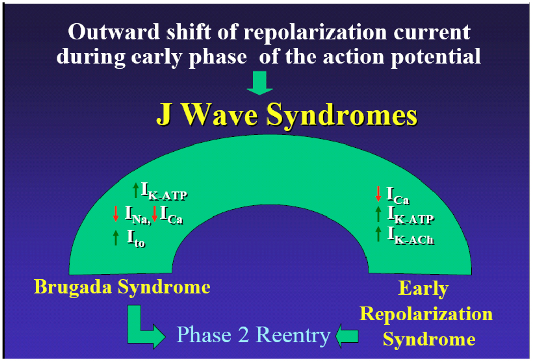
The J wave, also referred to as an Osborn wave, is a deflection immediately following the QRS complex of the surface ECG. When partially buried in the R wave, the J wave appears as J-point elevation or ST-segment elevation. Several lines of evidence have suggested that arrhythmias associated with an early repolarization pattern in the inferior or mid to lateral precordial leads, Brugada syndrome, or arrhythmias associated with hypothermia and the acute phase of ST-segment elevation myocardial infarction are mechanistically linked to abnormalities in the manifestation of the transient outward current (I(to))-mediated J wave. Although Brugada syndrome and early repolarization syndrome differ with respect to the magnitude and lead location of abnormal J-wave manifestation, they can be considered to represent a continuous spectrum of phenotypic expression that we propose be termed J-wave syndromes. This review summarizes our current state of knowledge concerning J-wave syndromes, bridging basic and clinical aspects. We propose to divide early repolarization syndrome into three subtypes: type 1, which displays an early repolarization pattern predominantly in the lateral precordial leads, is prevalent among healthy male athletes and is rarely seen in ventricular fibrillation survivors; type 2, which displays an early repolarization pattern predominantly in the inferior or inferolateral leads, is associated with a higher level of risk; and type 3, which displays an early repolarization pattern globally in the inferior, lateral, and right precordial leads, is associated with the highest level of risk for development of malignant arrhythmias and is often associated with ventricular fibrillation storms.


Conflict of interest statement

Conflicts of Interest: There are no conflicts of interest to disclose.

Figures

Figure 1
Figure 1
Effect of ventricular activation sequence on the J wave on the pseudo-ECG recorded for a coronary-perfused canine right ventricular wedge preparation. A. When the wedge preparation is stimulated from the endocardial (Endo) surface with epicardium (Epi) activated last, a J wave on the ECG is temporally aligned with Ito-mediated epicardial action potential notch. B. When the preparation is paced from the epicardial surface with endocardium activated last, the epicardial action potential notch is coincident with the QRS, and a J wave is no longer observed (reprinted from Yan and Antzelevitch with permission). C: and D: Hypothermia-induced J wave. Each panel shows transmembrane action potentials from the epicardial and endocardial regions of an arterially perfused canine left ventricular wedge and a transmural ECG simultaneously recorded. C: Because of the smaller notch in the LV epicardium, a distinct J wave is not seen under baseline conditions. The small action potential notch in epicardium but not in endocardium is associated with an elevated J-point at the R-ST junction (arrow) at 36°C. D: A decrease in the temperature of the perfusate to 29°C results in an increase in the amplitude and width of the action potential notch in epicardium but not endocardium, leading to the development of a transmural voltage gradient that manifests as a prominent J wave on the ECG (arrow). (Modified from Yan and Antzelevitch, with permission)
Figure 2
Figure 2
Cellular basis for electrocardiographic and arrhythmic manifestation of BrS. Each panel shows transmembrane action potentials from one endocardial (top) and two epicardial sites together with a transmural ECG recorded from a canine coronary-perfused right ventricular wedge preparation. A: Control (Basic cycle length (BCL) 400 msec). B: Combined sodium and calcium channel block with terfenadine (5 µM) accentuates the epicardial action potential notch creating a transmural voltage gradient that manifests as a ST segment elevation or exaggerated J wave in the ECG. C: Continued exposure to terfenadine results in all-or-none repolarization at the end of phase 1 at some epicardial sites but not others, creating a local epicardial dispersion of repolarization (EDR) as well as a transmural dispersion of repolarization (TDR). D: Phase 2 reentry occurs when the epicardial action potential dome propagates from a site where it is maintained to regions where it has been lost giving rise to a closely coupled extrasystole. E: Extrastimulus (S1–S2 = 250 msec) applied to epicardium triggers a polymorphic VT. F: Phase 2 reentrant extrasystole triggers a brief episode of polymorphic VT. (Modified from reference Fish and Antzelevitch, with permission)
Figure 3
Figure 3
Cellular basis for the early repolarization syndrome. A: Surface ECG (lead V5) recorded from a 17-year-old healthy African American male. Note the presence of a small J wave and marked ST segment elevation. B: Simultaneous recording of transmembrane action potentials from epicardial (Epi) and endocardial (Endo) regions and a transmural ECG in an isolated arterially perfused canine left ventricular wedge. A J wave in the transmural ECG is manifest due to the presence of an action potential notch in epicardium but not endocardium. Pinacidil (2 µM), an ATP-sensitive potassium channel opener, causes depression of the action potential dome in epicardium, resulting in ST segment elevation in the ECG resembling the early repolarization syndrome. Reprinted from Yan et al. with permission. C: IK-ATP activation in the canine right ventricular wedge preparation using 2.5 uM pinacidil produces heterogeneous loss of the AP dome in epicardium, resulting in ST segment elevation, phase 2 reentry and VT/VF (BrS phenotype). D: The Ito blocker, 4-aminopyridine (4-AP), restored the epicardial action potential (AP) dome, reduced both transmural and epicardial dispersion of repolarization, normalized the ST segment and prevented phase 2 reentry and VT/VF in the continued presence of pinacidil. (Modified from Di Diego et al. , with permission).
Figure 4
Figure 4
J wave-mediated arrhythmogenesis. A: Development of VF in a patient with prominent J waves in lead II (reprinted from Aizawa et al. , with permission). B: VF initiated by phase 2 reentry in a canine right ventricular wedge in the presence of 2.5 µmol/L of pinacidil. APs were simultaneously recorded from two epicardial sites (Epi1 and Epi2) and one Endo site. Loss of the AP dome in Epi1 but not in Epi2 led to phase 2 reentry capable of initiating VF (modified from Yan and Antzelevitch, with permission).
Figure 5
Figure 5
ECG obtained from a 34-year-old Chinese man who survived cardiac arrest displaying characteristics of BrS , ERS and IVF. Prominent J waves and ST segment elevation are seen in almost all leads. The echocardiogram and cardiac catheterization, were normal. Prominent J waves and ST segment elevation were observed in almost all ECG leads, including an ER pattern in leads of I, II, aVL, aVF and V4 to V6 and a saddleback ST segment elevation suggestive of BrS in V2 to V3 (thick arrows). R-on-T extrasystoles, likely due to phase 2 reentry (open arrows) are seen and the post-extrasystolic beat displays a coved ST segment elevation characteristic of BrS (thin arrows). Reprinted from Qi et al., with permission.
Figure 6
Figure 6
Twelve lead ECGs from an ERS3 patient initially diagnosed with IVF. A: ECGs obtained on Dec 19, 1998 B: Recorded Aug 18, 2003 4 hours before the VF storm. C: ECG taken 10 minutes before VFT episode showing prominent J waves across the precordial and inferior leads (arrows) as well as prominent ST revealed more prominent J waves across the precordial and inferior leads (arrows) as well as an ST segment elevation in the right precordial leads giving rise to an R on T extrasystole. D: One of several episodes of non-sustained polymorphic VT. Arrhythmias were always initiated by extrasystoles with a short-long-short sequence (Modified from Nam et al., with permission)
Figure 7
Figure 7
J wave Syndromes. Schematic depicts our working hypothesis that an outward shift in repolarizing current due to a decrease in sodium or calcium channel currents or an increase in Ito, IK-ATP or IK-ACh, or other outward currents can give rise to accentuated J waves associated with the BrS, Early Repolarization Syndrome and some forms of IVF. The particular phenotype depends on what part of the heart is principally affected and which ion channels are involved.

Similar articles

Cited by

References

    1. Osborn JJ. Experimental hypothermia: respiratory and blood pH changes in relation to cardiac function. Am J Physiol. 1953;175:389–398. - PubMed
    1. Yan GX, Antzelevitch C. Cellular basis for the electrocardiographic J wave. Circulation. 1996;93:372–379. - PubMed
    1. Gussak I, Antzelevitch C. Early repolarization syndrome: clinical characteristics and possible cellular and ionic mechanisms. J Electrocardiol. 2000;33:299–309. - PubMed
    1. Yan GX, Yao QH, Wang DQ, Cui CC. Electrocardiographic J wave and J wave syndromes. Chin J Cardiac Arrhyth. 2004;8:360–365.
    1. Shu J, Zhu T, Yang L, Cui C, Yan GX. ST-segment elevation in the early repolarization syndrome, idiopathic ventricular fibrillation, and the Brugada syndrome: cellular and clinical linkage. J Electrocardiol. 2005;38:26–32. - PubMed

Publication types

MeSH terms