. 2010 Apr 15;496(2):84-92.
doi: 10.1016/j.abb.2010.02.001. Epub 2010 Feb 11.

Size-dependent neurotoxicity of beta-amyloid oligomers

Affiliations

Size-dependent neurotoxicity of beta-amyloid oligomers

Paulius Cizas et al. Arch Biochem Biophys. .

Abstract

The link between the size of soluble amyloid beta (Abeta) oligomers and their toxicity to rat cerebellar granule cells (CGC) was investigated. Variation in conditions during in vitro oligomerization of Abeta(1-42) resulted in peptide assemblies with different particle size as measured by atomic force microscopy and confirmed by dynamic light scattering and fluorescence correlation spectroscopy. Small oligomers of Abeta(1-42) with a mean particle z-height of 1-2 nm exhibited propensity to bind to phospholipid vesicles and they were the most toxic species that induced rapid neuronal necrosis at submicromolar concentrations whereas the bigger aggregates (z-height above 4-5 nm) did not bind vesicles and did not cause detectable neuronal death. A similar neurotoxic pattern was also observed in primary cultures of cortex neurons whereas Abeta(1-42) oligomers, monomers and fibrils were non-toxic to glial cells in CGC cultures or macrophage J774 cells. However, both oligomeric forms of Abeta(1-42) induced reduction of neuronal cell densities in the CGC cultures.


Figures

Figure 1
Figure 1. Effect of differently prepared Aβ1-42 assemblies on neuronal cell viability in CGC cultures
A – Effect of Aβ1-42 oligomers (protocol I), fibrils (protocol III) and monomers on neuronal cell viability; B – Concentration dependence of Aβ1-42 oligomers (protocol I) to induce necrotic (PI-positive) and apoptotic (condensed/fragmented nuclei) cell death of neurons. C – Effect of Aβ1-42 oligomers (protocol I) on LDH release from cells into medium. LDH activity in the control group was equated to 100 %. * - statistically significant effect of Aβ if compared to control. Means ± standard errors of 5-7 experiments on separate CGC cultures are presented.
Figure 2
Figure 2. AFM results
Exemplary images of Aβ1-42 oligomers prepared by the different protocols: A - protocol I, B - protocol II, C - protocol III. The lateral size of the images is 4×4 μm. The horizontal bars in A and B shows the location at which hight profiles of the images were analyzed. D and E are the corresponding cross-sections of the particles obtained via protocols I and II.
Figure 3
Figure 3. Size distribution of differently prepared Aβ1-42 oligomers
A - DLS data: light gray: protocol I aggregates, dark gray: protocol II aggregates. The larger size oligomer preparation contained also a small contribution of sizes above 200 nm (not shown). B – FCS results for protocol I and protocol II oligomers with the same coding as in panel (A).
Figure 4
Figure 4. Toxicity of Aβ1-42 oligomers depends on aggregate size as determined with the AFM in terms of z-height
1-42 oligomers were prepared by protocol I and II. CGCs were treated with 1 μM Aβ1-42 oligomers for 24 h. Each point represents the effect of separate preparation of oligomers on viability of separate CGC culture (mean values are presented for each experiment).
Figure 5
Figure 5. Effect of Aβ1-42 peptides on neuronal cell viability in 10-12 DIV CGC cultures
Mixed neuronal-glial CGCs cultures were prepared from 7-8 day old Wistar rats as described in Material and Methods. Cells were grown in vitro for 10–14 days, then treated with 1 μM Aβ1-42 peptide for 24 h. The viability of neurons in the culture was measured by propidium iodide and Hoechst 33342 staining. * - statistically significant effect of Aβ peptide, if compared to control. Means ± standard errors of 4 separate experiments are presented.
Figure 6
Figure 6. Effect of Aβ1-42 on neuronal viability depends on the molecular mass of the peptide assemblies
1–42 oligomer preparations were fractionated through different Microcon filters as described in Materials and Methods. * - statistically significant effect of Aβ if compared to control. Means ± standard errors of 3-6 separate experiments are presented.
Figure 7
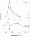
Figure 7. Aβ1-42 aggregate size causes FTIR spectral differences in the Amide I spectral region
A- Small oligomers (protocol I), B - larger oligomers (protocol II), and C difference spectrum. Transmission spectra were obtained from dried samples deposited on CaF2 substrates from 60 μM solution.
Figure 8
Figure 8. Different size Aβ1-42 oligomers bind differently to lipid membranes
FCS data (dots) fitted (continuous line) with single component 3D diffusion model for fluorescent labeled Aβ1-42 diffusing freely in bulk (red) and with two component 3D diffusion model for Aβ1-42 diffusing in the presence of non-fluorescent lipid vesicles (black). Residuals of the fits are shown at the top of each panel. All data are normalized for comparison purposes. A- small Aβ1-42 oligomers (protocol I), B- big Aβ1-42 oligomers (protocol II).
Figure 9
Figure 9. Aβ1-42 oligomers decrease the number of neurons in CGC cultures
After treatment of CGCs with 1 μM of Aβ1-42, the total number of viable, necrotic and apoptotic neurons was quantified in 5-7 randomly chosen microscopic fields in each well (two wells per treatment). Total number of cells counted per treatment varied between 1600-2500. * - statistically significant effect of Aβ1-42 if compared to control. Means ± standard errors of 5-7 separate experiments are presented.
Figure 10
Figure 10. Effect of Aβ1-42 peptides on neuronal cell viability in pure neuronal CGC cultures
CGCs cultures were prepared from 7-8 day old Wistar rats as described in Material and Methods. Cytosine arabinoside (10 μM) was added within 48 h of plating to prevent glial cell proliferation. Cells were grown in vitro for 10–14 days before exposure to Aβ1-42. The cultures contained 0.5% microglial cells and 2.1% astrocytes. Cultures were treated with 1 μM of Aβ1-42 peptide for 24 h. The viability of neurons in the culture was measured by propidium iodide and Hoechst 33342 staining. * - statistically significant effect of Aβ peptide, if compared to control. Means ± standard errors of 4 separate experiments are presented.

References

    1. Hardy J, Selkoe DJ. Science. 2002;297:353–356. - PubMed
    1. Chen YR, Glabe CG. Journal of Biological Chemistry. 2006;281:24414–24422. - PubMed
    1. McLean CA, Cherny RA, Fraser FW, Fuller SJ, Smith MJ, Beyreuther K, Bush AI, Masters CL. Annals of Neurology. 1999;46:860–866. - PubMed
    1. Cleary JP. Nat.Neurosci. 2005;8:79–84. - PubMed
    1. Lesne S, Koh MT, Kotilinek L, Kayed R, Glabe CG, Yang A, Gallagher M, Ashe KH. Nature. 2006;440:352–357. - PubMed

Publication types

LinkOut - more resources