Pharmacokinetics and bioactivity of glial cell line-derived factor (GDNF) and neurturin (NTN) infused into the rat brain
- PMID: 20153340
- PMCID: PMC2849906
- DOI: 10.1016/j.neuropharm.2010.02.002
Pharmacokinetics and bioactivity of glial cell line-derived factor (GDNF) and neurturin (NTN) infused into the rat brain
Abstract
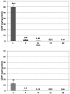
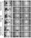
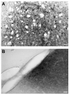
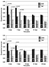
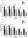
Convection-enhanced delivery (CED) of GDNF and NTN was employed to determine the tissue clearance of these factors from the rat striatum and the response of the dopaminergic system to a single infusion. Two doses of GDNF (15 and 3 microg) and NTN (10 microg and 2 microg) were infused into the rat striatum. Animals were euthanized 3, 7, 14, 21, and 28 days post-infusion. Brains were processed for ELISA, HPLC, and immunohistochemistry (IHC). Both doses of the infused GDNF resulted in a sharp increase in striatal GDNF levels followed by a rapid decrease between day 3 and 7. Interestingly, IHC revealed GDNF in the septum and the base of the brain 14 days after GDNF administration. Dopamine (DA) turnover was significantly increased in a dose-dependent manner for more than 7 days after a single GDNF infusion. NTN persisted in the brain for at least two weeks longer than GDNF. It also had more persistent effects on DA turnover, probably due to its precipitation in the brain at neutral pH after infusion. Our data suggest that daily or continuous dosing may not be necessary for delivering growth factors into the CNS.
(c) 2010 Elsevier Ltd. All rights reserved.
Figures


Similar articles
-
Neurturin protects against 6-hydroxydopamine-induced reductions in evoked dopamine overflow in rat striatum.Neurochem Int. 2010 Nov;57(5):540-6. doi: 10.1016/j.neuint.2010.06.019. Epub 2010 Jul 6. Neurochem Int. 2010. PMID: 20615442 Free PMC article.
-
Neurturin effects on nigrostriatal dopamine release and content: comparison with GDNF.Neurochem Res. 2010 May;35(5):727-34. doi: 10.1007/s11064-010-0128-0. Epub 2010 Jan 30. Neurochem Res. 2010. PMID: 20119638 Free PMC article.
-
GDNF revisited: A novel mammalian cell-derived variant form of GDNF increases dopamine turnover and improves brain biodistribution.Neuropharmacology. 2019 Mar 15;147:28-36. doi: 10.1016/j.neuropharm.2018.05.014. Epub 2018 May 29. Neuropharmacology. 2019. PMID: 29857941 Review.
-
Differential in vivo effects of neurturin and glial cell-line-derived neurotrophic factor.Exp Neurol. 1999 Nov;160(1):235-43. doi: 10.1006/exnr.1999.7175. Exp Neurol. 1999. PMID: 10630208
-
Other neurotrophic factors: glial cell line-derived neurotrophic factor (GDNF).Microsc Res Tech. 1999 May 15-Jun 1;45(4-5):292-302. doi: 10.1002/(SICI)1097-0029(19990515/01)45:4/5<292::AID-JEMT13>3.0.CO;2-8. Microsc Res Tech. 1999. PMID: 10383122 Review.
Cited by
-
Evaluation of an AAV2-based rapamycin-regulated glial cell line-derived neurotrophic factor (GDNF) expression vector system.PLoS One. 2011;6(11):e27728. doi: 10.1371/journal.pone.0027728. Epub 2011 Nov 21. PLoS One. 2011. PMID: 22132130 Free PMC article.
-
Mesencephalic Astrocyte-Derived Neurotrophic Factor (MANF) Elevates Stimulus-Evoked Release of Dopamine in Freely-Moving Rats.Mol Neurobiol. 2018 Aug;55(8):6755-6768. doi: 10.1007/s12035-018-0872-8. Epub 2018 Jan 18. Mol Neurobiol. 2018. PMID: 29349573 Free PMC article.
-
Neurturin overexpression in dopaminergic neurons induces presynaptic and postsynaptic structural changes in rats with chronic 6-hydroxydopamine lesion.PLoS One. 2017 Nov 27;12(11):e0188239. doi: 10.1371/journal.pone.0188239. eCollection 2017. PLoS One. 2017. PMID: 29176874 Free PMC article.
-
Trial of magnetic resonance-guided putaminal gene therapy for advanced Parkinson's disease.Mov Disord. 2019 Jul;34(7):1073-1078. doi: 10.1002/mds.27724. Epub 2019 May 30. Mov Disord. 2019. PMID: 31145831 Free PMC article.
-
Neurturin protects against 6-hydroxydopamine-induced reductions in evoked dopamine overflow in rat striatum.Neurochem Int. 2010 Nov;57(5):540-6. doi: 10.1016/j.neuint.2010.06.019. Epub 2010 Jul 6. Neurochem Int. 2010. PMID: 20615442 Free PMC article.
References
-
- Airaksinen MS, Saarma M. The GDNF family: signalling, biological functions and therapeutic value. Nat Rev Neurosci. 2002;3:383–394. - PubMed
-
- Airaksinen MS, Titievsky A, Saarma M. GDNF family neurotrophic factor signaling: four masters, one servant? Mol Cell Neurosci. 1999;13:313–325. - PubMed
-
- Choi-Lundberg DL, Lin Q, Chang YN, Chiang YL, Hay CM, Mohajeri H, Davidson BL, Bohn MC. Dopaminergic neurons protected from degeneration by GDNF gene therapy. Science. 1997;275:838–841. - PubMed
-
- Curtis MA, Kam M, Nannmark U, Anderson MF, Axell MZ, Wikkelso C, Holtas S, van Roon-Mom WM, Bjork-Eriksson T, Nordborg C, Frisen J, Dragunow M, Faull RL, Eriksson PS. Human neuroblasts migrate to the olfactory bulb via a lateral ventricular extension. Science. 2007;315:1243–1249. - PubMed
Publication types
MeSH terms
Substances
Grants and funding
LinkOut - more resources
Full Text Sources
Other Literature Sources
Research Materials

