

Host-microbe interactions in the developing zebrafish
- PMID: 20153622
- PMCID: PMC3030977
- DOI: 10.1016/j.coi.2010.01.006
Host-microbe interactions in the developing zebrafish
Abstract
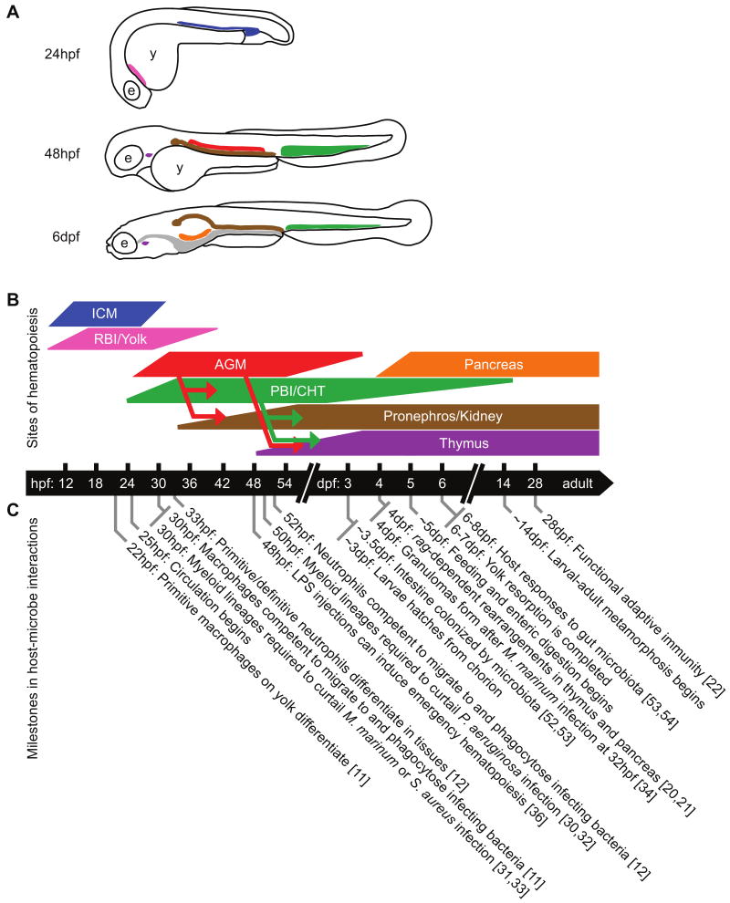
The amenability of the zebrafish to in vivo imaging and genetic analysis has fueled expanded use of this vertebrate model to investigate the molecular and cellular foundations of host-microbe relationships. Study of microbial encounters in zebrafish hosts has concentrated on developing embryonic and larval stages, when the advantages of the zebrafish model are maximized. A comprehensive understanding of these host-microbe interactions requires appreciation of the developmental context into which a microbe is introduced, as well as the effects of that microbial challenge on host ontogeny. In this review, we discuss how in vivo imaging and genetic analysis in zebrafish has advanced our knowledge of host-microbe interactions in the context of a developing vertebrate host. We focus on recent insights into immune cell ontogeny and function, commensal microbial relationships in the intestine, and microbial pathogenesis in zebrafish hosts.
Copyright 2010 Elsevier Ltd. All rights reserved.
Figures
References
-
- Grunwald D, Eisen J. Headwaters of the zebrafish -- emergence of a new model vertebrate. Nat Rev Genet. 2002;3:717–724. - PubMed
-
- Lieschke GJ, Currie PD. Animal models of human disease: zebrafish swim into view. Nat Rev Genet. 2007;8:353–367. - PubMed
-
- Meeker N, Trede N. Immunology and zebrafish: spawning new models of human disease. Dev Comp Immunol. 2008;32:745–757. - PubMed
-
- Sullivan C, Kim CH. Zebrafish as a model for infectious disease and immune function. Fish Shellfish Immunol. 2008;25:341–350. - PubMed
-
- Hall CJ, Flores MV, C KE, Crosier PS. Live imaging early immune cell ontogeny and function in zebrafish Danio rerio. Journal of Fish Biology. 2008:1833–1871.
Publication types
MeSH terms
Grants and funding
LinkOut - more resources
Full Text Sources
Other Literature Sources
