

Inhibition of human arginase I by substrate and product analogues
- PMID: 20153713
- PMCID: PMC2850953
- DOI: 10.1016/j.abb.2010.02.004
Inhibition of human arginase I by substrate and product analogues
Abstract
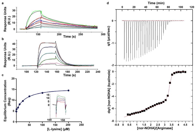
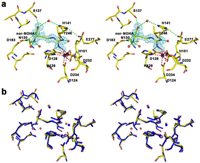
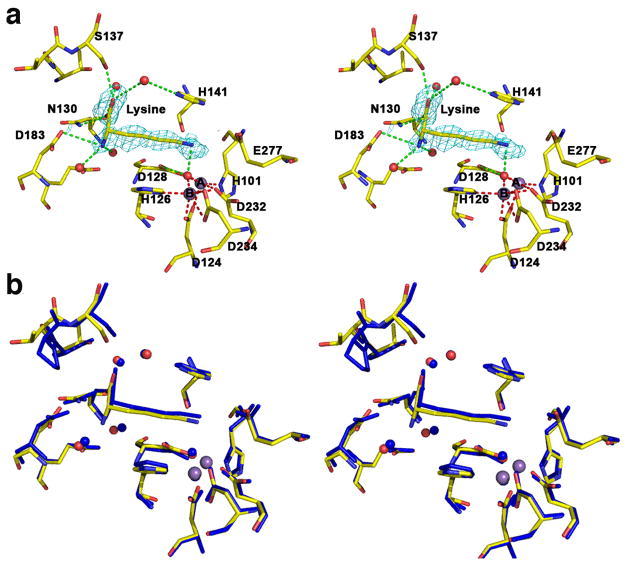
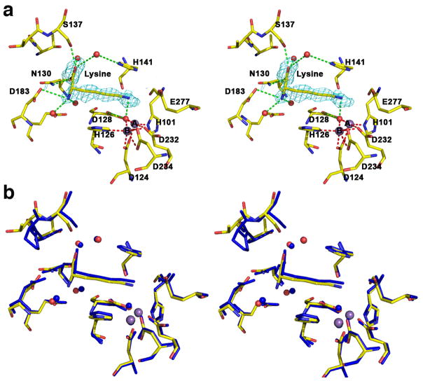
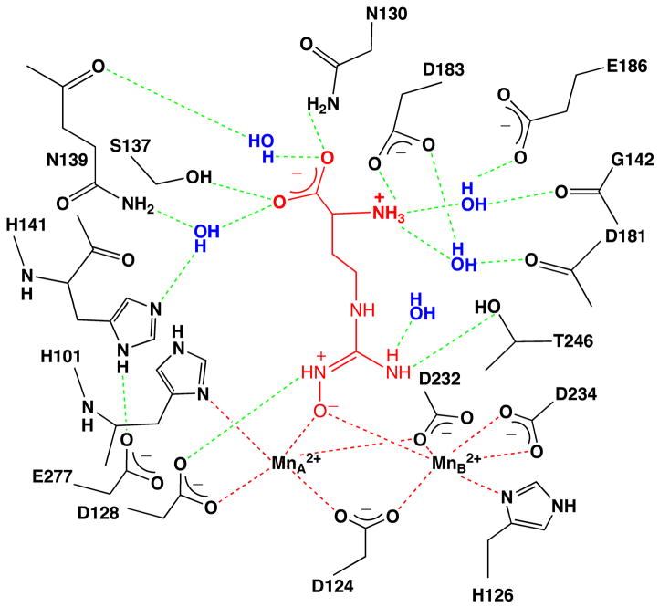
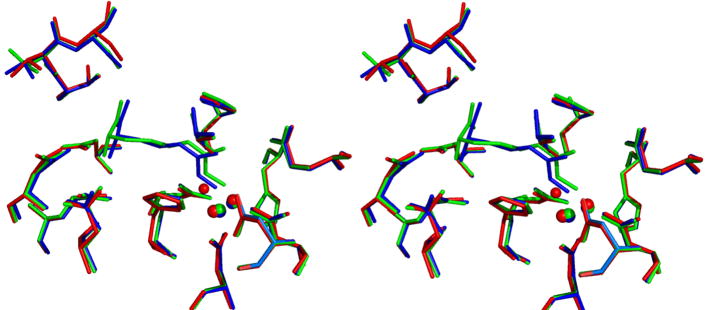
Human arginase I is a binuclear manganese metalloenzyme that catalyzes the hydrolysis of L-arginine to generate L-ornithine and urea. We demonstrate that N-hydroxy-L-arginine (NOHA) binds to this enzyme with K(d)=3.6 microM, and nor-N-hydroxy-L-arginine (nor-NOHA) binds with K(d)=517 nM (surface plasmon resonance) or K(d) approximately 50 nM (isothermal titration calorimetry). Crystals of human arginase I complexed with NOHA and nor-NOHA afford 2.04 and 1.55 A resolution structures, respectively, which are significantly improved in comparison with previously-determined structures of the corresponding complexes with rat arginase I. Higher resolution structures clarify the binding interactions of the inhibitors. Finally, the crystal structure of the complex with L-lysine (K(d)=13 microM) is reported at 1.90 A resolution. This structure confirms the importance of hydrogen bond interactions with inhibitor alpha-carboxylate and alpha-amino groups as key specificity determinants of amino acid recognition in the arginase active site.
2010 Elsevier Inc. All rights reserved.
Figures
Similar articles
-
Mechanistic and metabolic inferences from the binding of substrate analogues and products to arginase.Biochemistry. 2001 Mar 6;40(9):2689-701. doi: 10.1021/bi002318+. Biochemistry. 2001. PMID: 11258880
-
L-arginine binding to liver arginase requires proton transfer to gateway residue His141 and coordination of the guanidinium group to the dimanganese(II,II) center.Biochemistry. 1998 Jun 9;37(23):8539-50. doi: 10.1021/bi972874c. Biochemistry. 1998. PMID: 9622506
-
Inhibitor coordination interactions in the binuclear manganese cluster of arginase.Biochemistry. 2004 Jul 20;43(28):8987-99. doi: 10.1021/bi0491705. Biochemistry. 2004. PMID: 15248756
-
Arginase Inhibitors: A Rational Approach Over One Century.Med Res Rev. 2017 May;37(3):475-513. doi: 10.1002/med.21419. Epub 2016 Nov 15. Med Res Rev. 2017. PMID: 27862081 Review.
-
Structure and function of arginases.J Nutr. 2004 Oct;134(10 Suppl):2760S-2764S; discussion 2765S-2767S. doi: 10.1093/jn/134.10.2760S. J Nutr. 2004. PMID: 15465781 Review.
Cited by
-
Arginase: shedding light on the mechanisms and opportunities in cardiovascular diseases.Cell Death Discov. 2022 Oct 8;8(1):413. doi: 10.1038/s41420-022-01200-4. Cell Death Discov. 2022. PMID: 36209203 Free PMC article. Review.
-
Macrophage Infiltration and Alternative Activation during Wound Healing Promote MEK1-Induced Skin Carcinogenesis.Cancer Res. 2016 Feb 15;76(4):805-817. doi: 10.1158/0008-5472.CAN-14-3676. Epub 2016 Jan 11. Cancer Res. 2016. PMID: 26754935 Free PMC article.
-
Arginase as a Potential Biomarker of Disease Progression: A Molecular Imaging Perspective.Int J Mol Sci. 2020 Jul 25;21(15):5291. doi: 10.3390/ijms21155291. Int J Mol Sci. 2020. PMID: 32722521 Free PMC article. Review.
-
Arginase of Helicobacter Gastric Pathogens Uses a Unique Set of Non-catalytic Residues for Catalysis.Biophys J. 2017 Mar 28;112(6):1120-1134. doi: 10.1016/j.bpj.2017.02.009. Biophys J. 2017. PMID: 28355540 Free PMC article.
-
Pathophysiology of Arginases in Cancer and Efforts in Their Pharmacological Inhibition.Int J Mol Sci. 2024 Sep 10;25(18):9782. doi: 10.3390/ijms25189782. Int J Mol Sci. 2024. PMID: 39337272 Free PMC article. Review.
References
Publication types
MeSH terms
Substances
Associated data
- Actions
- Actions
- Actions
Grants and funding
LinkOut - more resources
Full Text Sources
