

Matrix metalloproteinase-3 in articular cartilage is upregulated by joint immobilization and suppressed by passive joint motion
- PMID: 20153826
- PMCID: PMC2902573
- DOI: 10.1016/j.matbio.2010.02.004
Matrix metalloproteinase-3 in articular cartilage is upregulated by joint immobilization and suppressed by passive joint motion
Abstract
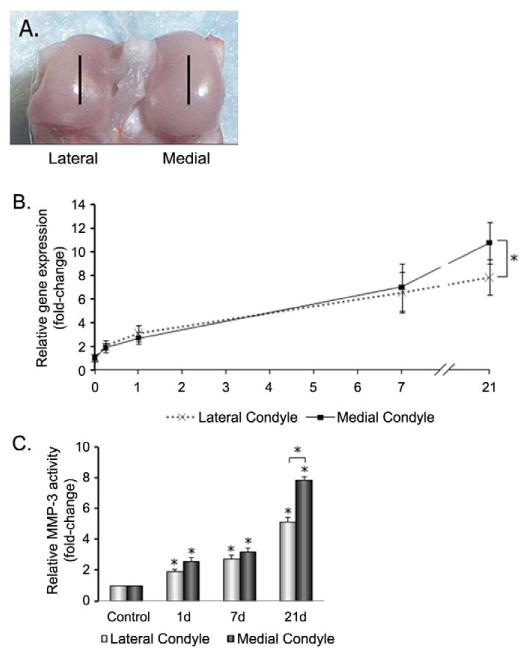
Both underloading and overloading of joints can lead to articular cartilage degradation, a process mediated in part by matrix metalloproteinases (MMPs). Here we examine the effects of reduced loading of rat hindlimbs on articular cartilage expression of MMP-3, which not only digests matrix components but also activates other proteolytic enzymes. We show that hindlimb immobilization resulted in elevated MMP-3 mRNA expression at 6h that was sustained throughout the 21day immobilization period. MMP-3 upregulation was higher in the medial condyle than the lateral, and was greatest in the superficial cartilage zone, followed by middle and deep zones. These areas also showed decreases in safranin O staining, consistent with reduced cartilage proteoglycan content, as early as 7days after immobilization. One hour of daily moderate mechanical loading, applied as passive joint motion, reduced the MMP-3 and ADAMTS-5 increases that resulted from immobilization, and also prevented changes in safranin O staining. Intra-articular injections of an MMP-3 inhibitor, N-isobutyl-N-(4-methoxyphenylsulfonyl)-glycylhydroxamic acid (NNGH), dampened the catabolic effects of a 7day immobilization period, indicating a likely requirement for MMP-3 in the regulation of proteoglycan levels through ADAMTS-5. These results suggest that biomechanical forces have the potential to combat cartilage destruction and can be critical in developing effective therapeutic strategies.
Copyright (c) 2010 International Society of Matrix Biology. Published by Elsevier B.V. All rights reserved.
Figures


Similar articles
-
Mechanotransduction and cartilage integrity.Ann N Y Acad Sci. 2011 Dec;1240:32-7. doi: 10.1111/j.1749-6632.2011.06301.x. Ann N Y Acad Sci. 2011. PMID: 22172037 Free PMC article. Review.
-
Increased expression of metalloproteinase-8 and -13 on articular cartilage in a rat immobilized knee model.Tohoku J Exp Med. 2009 Apr;217(4):271-8. doi: 10.1620/tjem.217.271. Tohoku J Exp Med. 2009. PMID: 19346731
-
Matrix metalloproteinase-3 inhibitor retards treadmill running-induced cartilage degradation in rats.Arthritis Res Ther. 2011;13(6):R192. doi: 10.1186/ar3521. Epub 2011 Nov 24. Arthritis Res Ther. 2011. PMID: 22114772 Free PMC article.
-
Potential role of hyaluronic acid on bone in osteoarthritis: matrix metalloproteinases, aggrecanases, and RANKL expression are partially prevented by hyaluronic acid in interleukin 1-stimulated osteoblasts.J Rheumatol. 2014 May;41(5):945-54. doi: 10.3899/jrheum.130378. Epub 2014 Apr 15. J Rheumatol. 2014. PMID: 24737908
-
[Destruction of the articular cartilage in osteoarthritis].Clin Calcium. 2013 Dec;23(12):1705-13. Clin Calcium. 2013. PMID: 24292524 Review. Japanese.
Cited by
-
Effects of Electrical Stimulation on Articular Cartilage Regeneration with a Focus on Piezoelectric Biomaterials for Articular Cartilage Tissue Repair and Engineering.Int J Mol Sci. 2023 Jan 17;24(3):1836. doi: 10.3390/ijms24031836. Int J Mol Sci. 2023. PMID: 36768157 Free PMC article. Review.
-
Impact of Abnormal Mechanical Stress on Chondrocyte Death in Osteoarthritis.Med Sci Monit. 2025 Jun 10;31:e948290. doi: 10.12659/MSM.948290. Med Sci Monit. 2025. PMID: 40493524 Free PMC article. Review.
-
Mechanotransduction and cartilage integrity.Ann N Y Acad Sci. 2011 Dec;1240:32-7. doi: 10.1111/j.1749-6632.2011.06301.x. Ann N Y Acad Sci. 2011. PMID: 22172037 Free PMC article. Review.
-
Optimizing artificial meniscus by mechanical stimulation of the chondrocyte-laden acellular meniscus using ad hoc bioreactor.Stem Cell Res Ther. 2022 Jul 30;13(1):382. doi: 10.1186/s13287-022-03058-w. Stem Cell Res Ther. 2022. PMID: 35908010 Free PMC article.
-
Mechanical intervention for maintenance of cartilage and bone.Clin Med Insights Arthritis Musculoskelet Disord. 2011;4:65-70. doi: 10.4137/CMAMD.S6982. Epub 2011 Jun 29. Clin Med Insights Arthritis Musculoskelet Disord. 2011. PMID: 21792344 Free PMC article.
References
-
- Aigner T, Zien A, Gehrsitz A, Gebhard PM, McKenna L. Anabolic and catabolic gene expression pattern analysis in normal versus osteoarthritic cartilage using complementary DNA-array technology. Arthritis Rheum. 2001;44:2777–2789. - PubMed
-
- Andriacchi TP, Mundermann A, Smith RL, Alexander EJ, Dyrby CO, Koo S. A framework for the in vivo pathomechanics of osteoarthritis at the knee. Ann Biomed Eng. 2004;32:447–457. - PubMed
-
- Arokoski JP, Jurvelin JS, Vaatainen U, Helminen HJ. Normal and pathological adaptations of articular cartilage to joint loading. Scand J Med Sci Sports. 2000;10:186–198. - PubMed
-
- Blom AB, van Lent PL, Libregts S, Holthuysen AE, van der Kraan PM, van Rooijen N, van den Berg WB. Crucial role of macrophages in matrix metalloproteinase-mediated cartilage destruction during experimental osteoarthritis: involvement of matrix metalloproteinase 3. Arthritis Rheum. 2007;56:147–157. - PubMed
Publication types
MeSH terms
Substances
Grants and funding
LinkOut - more resources
Full Text Sources
Miscellaneous
