. 2010 Apr 16;285(16):12011-27.
doi: 10.1074/jbc.M109.097790. Epub 2010 Feb 12.

High temperature requirement A3 (HtrA3) promotes etoposide- and cisplatin-induced cytotoxicity in lung cancer cell lines

Affiliations

High temperature requirement A3 (HtrA3) promotes etoposide- and cisplatin-induced cytotoxicity in lung cancer cell lines

Daniah Beleford et al. J Biol Chem. .

Abstract

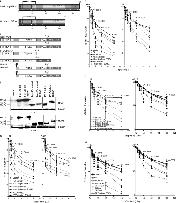
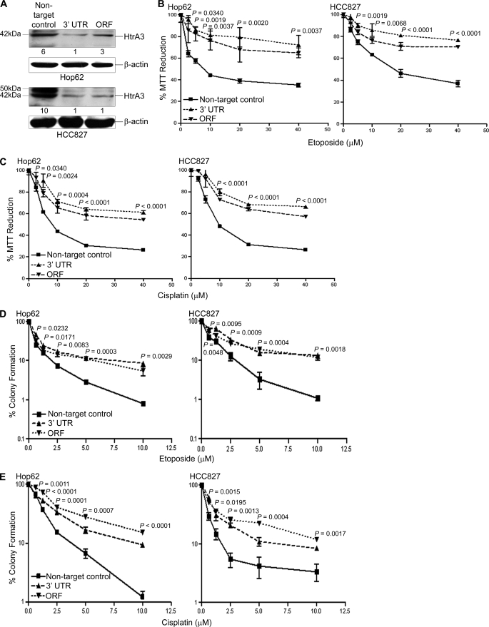
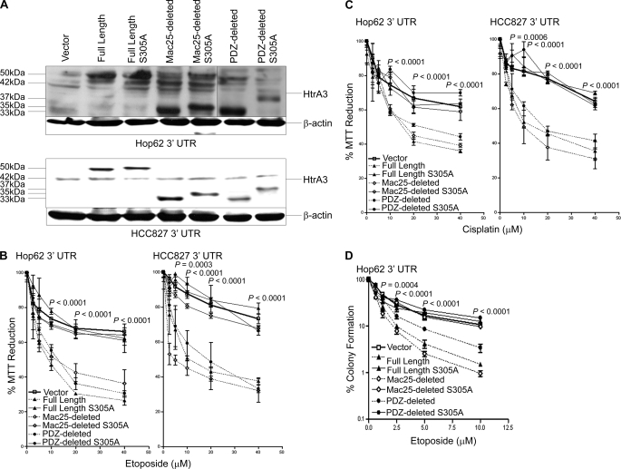
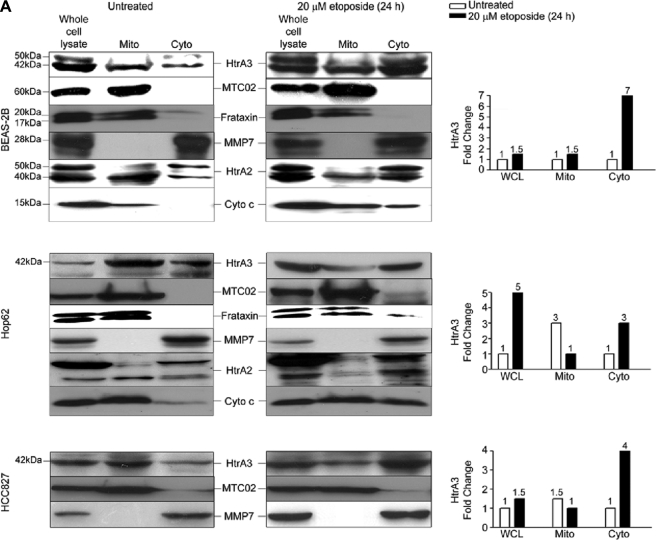
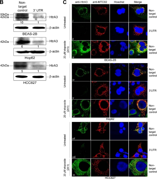
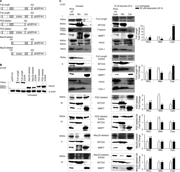
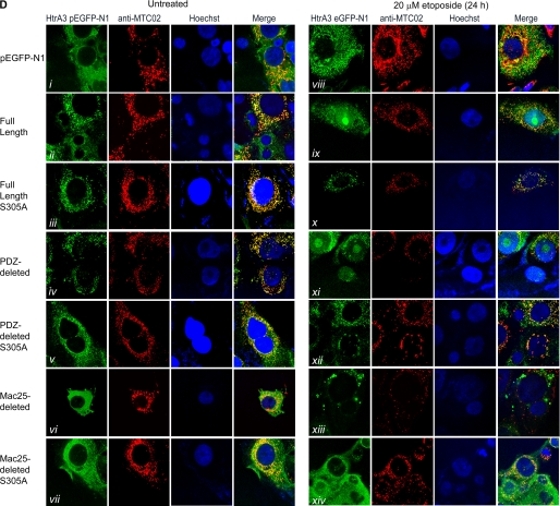
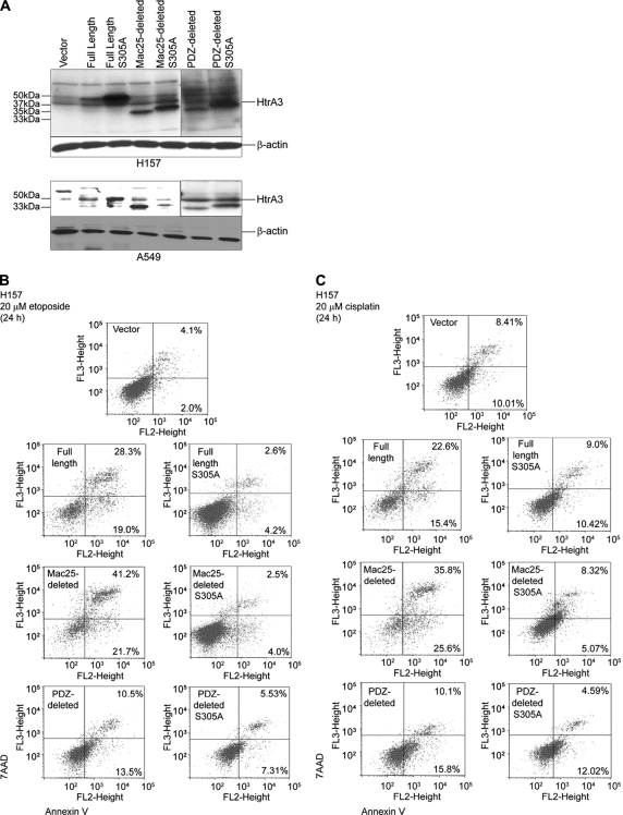
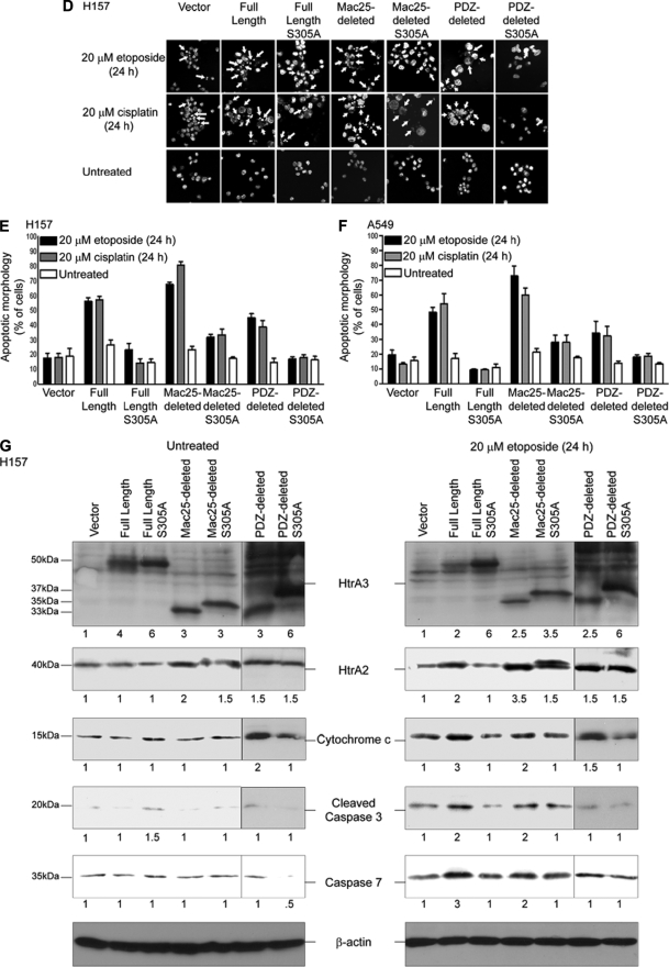
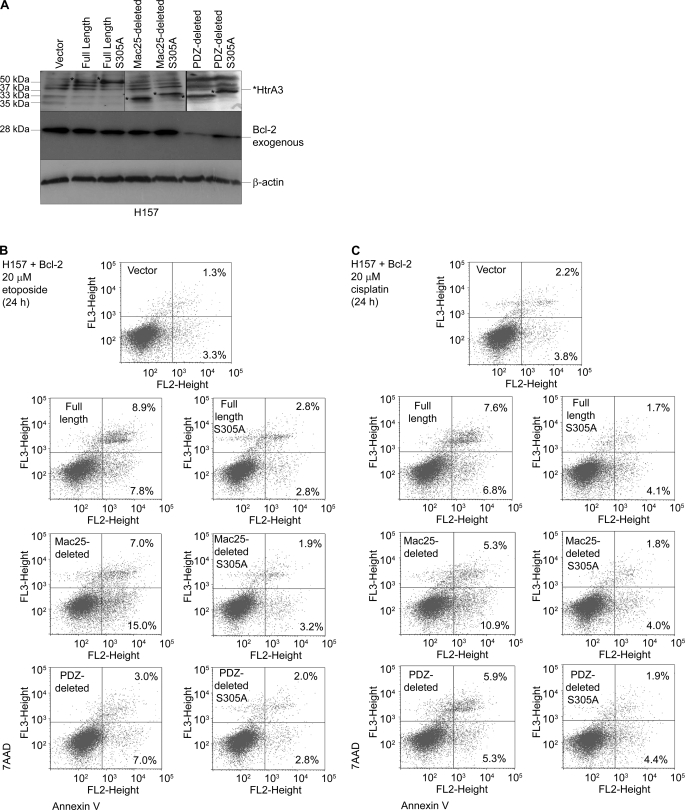
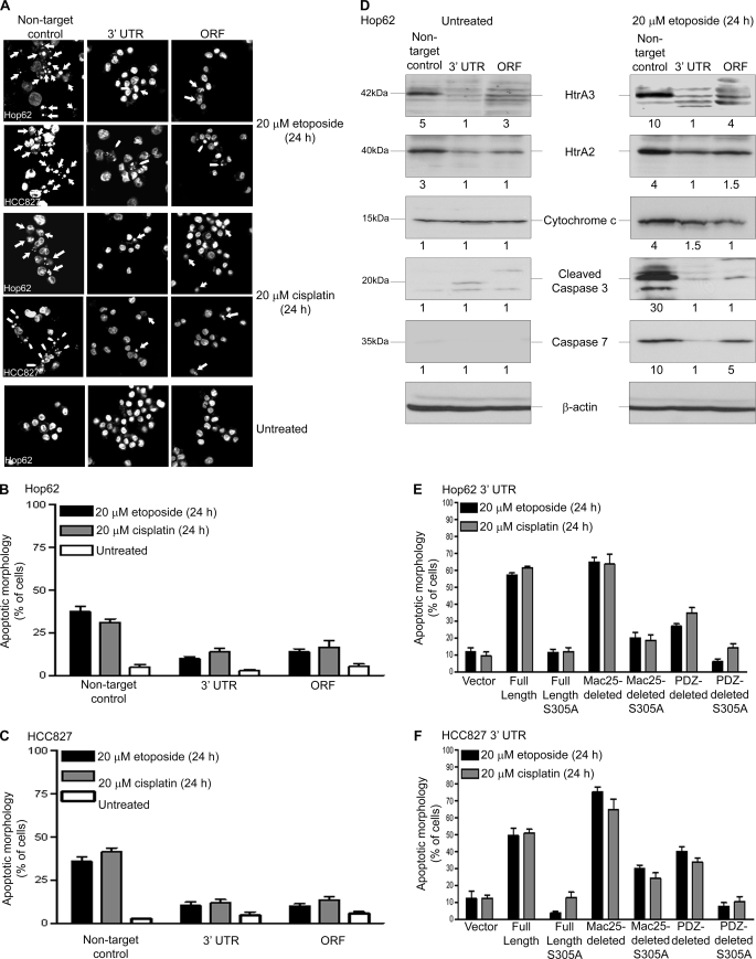
Lung cancer is the leading cause of cancer-related deaths worldwide. Here we show for the first time that HtrA3 is a mitochondrial stress-response factor that promotes cytotoxicity to etoposide and cisplatin in lung cancer cell lines. Exogenous expression of wild type HtrA3 domain variants significantly attenuated cell survival with etoposide and cisplatin treatment in lung cancer cell lines H157 and A549 compared with expression of protease inactive mutants (S305A) or vector control. Conversely, HtrA3 suppression promoted cell survival with etoposide and cisplatin treatment in lung cancer cell lines Hop62 and HCC827. Survival was attenuated by re-expression of wild type HtrA3 variants during treatment but not by protease inactive mutants or vector control. HtrA3 also co-fractionated and co-localized with mitochondrial markers with both endogenous and exogenous expression in normal lung and lung cancer cell lines but was translocated from mitochondria following etoposide treatment. Moreover, HtrA3 translocation from mitochondria correlated with an increase in cell death that was attenuated by either HtrA3 suppression or Bcl-2 overexpression. Taken together, these results suggest that HtrA3 may be a previously uncharacterized mitochondrial cell death effector whose serine protease function may be crucial to modulating etoposide- and cisplatin-induced cytotoxicity in lung cancer cell lines.


Figures

FIGURE 1.
FIGURE 1.
WT HtrA3 variants modulate cytotoxicity to etoposide and cisplatin with exogenous expression in lung cancer cell lines. A, domain structural schematics of the long and short HtrA3 splice variants. The long variant is 453 amino acids in length and has an N-terminal signal peptide (SP, amino acids 1–17), an insulin/insulin-like growth factor-binding domain (amino acids 29–94), a Kazal-type serine protease-inhibitor domain (amino acids 89–126), a trypsin protease domain (amino acids 176–341), and one C-terminal PDZ domain (amino acids 384–440). The insulin/insulin-like growth factor-binding domain (IBD) and Kazal domains are homologous to Mac25. The short isoform is 357 amino acids in length and is identical to the long isoform with two exceptions: the short isoform lacks the PDZ domain, and the last seven C-terminal residues in the short form (APSLAVH) differ from the corresponding residues in the long isoform (DWKKRFI). B, domain structural schematics of WT and protease inactive full length, PDZ-deleted, and Mac25-deleted HtrA3 variants that are C-terminally myc- and polyhistidine-tagged. The WT PDZ-deleted variant was not tagged. Protease inactive HtrA3 variants contain a serine to alanine point mutation at residue 305 (S305A) within the conserved trypsin catalytic triad. C, immunoblots showing expression of HtrA3 variants, vector control, and β-actin loading controls in HtrA3-deficient lung cancer cell lines H157 and A549. PDZ-deleted variants were detected using an HtrA3 polyclonal antibody. D and E, MTT survival assays showing statistically significant attenuation of MTT reduction (p < 0.05) with exogenous expression of WT HtrA3 variants during etoposide (D) or cisplatin (E) treatment. The data were expressed as the means ± S.E. and represented at least two independent trials performed in quadruplicate. The p values were calculated using unpaired two-tailed Student's t test for two groups and ANOVA. F and G, clonogenic survival assays showing decreased clonogenic survival with exogenous expression of WT HtrA3 variants following etoposide (F) or cisplatin (G) treatment. The data are expressed as the means ± S.E. and represent independent trials performed in triplicate. The p values were calculated using unpaired two-tailed Student's t test for two groups and ANOVA.
FIGURE 2.
FIGURE 2.
Down-regulation of endogenous HtrA3 expression attenuates etoposide- and cisplatin-induced cytotoxicity. A, immunoblots showing endogenous HtrA3 expression in pooled clonal lung cancer cell lines stably transduced with a nontargeted control vector and HtrA3 down-regulation in pooled clonal lines stably transduced with shRNA targeting either the open reading frame (ORF) or 3′-UTR. For each cell line, densitometric analysis of fold changes in HtrA3 intensity normalized by β-actin loading controls is given below each band. B and C, MTT survival assays showing statistically significant decrease in MTT reduction with endogenous HtrA3 expression with either etoposide (B) or cisplatin (C) treatment compared with decrease in MTT reduction with HtrA3 down-regulation (p < 0.05). The data are expressed as the means ± S.E. and represent at least two independent trials performed in quadruplicate. The p values were calculated using unpaired two-tailed Student's t test for two groups and ANOVA. D and E, clonogenic survival assays showing significantly greater clonogenic survival with HtrA3 down-regulation following etoposide (D) or cisplatin (E) treatment compared with survival with normal endogenous HtrA3 expression (p < 0.05). The data are expressed as the means ± S.E. and represent independent trials performed in triplicate. The p values were calculated using unpaired two-tailed Student's t test for two groups and ANOVA.
FIGURE 3.
FIGURE 3.
HtrA3 re-expression promotes etoposide and cisplatin cytotoxicity in lung cancer cell lines. A, immunoblots showing exogenous expression of WT and protease inactive full length, Mac25-deleted, and PDZ-deleted HtrA3 variants, vector control, and β-actin loading controls in Hop62 and HCC827 clonal cell lines stably down-regulated for HtrA3 expression through shRNA-mediated targeting of the 3′-UTR. Endogenous HtrA3 is expressed at 42 kDa. B and C, MTT survival assays showing statistically significant attenuation of MTT reduction with re-expression of WT HtrA3 variants during etoposide (B) or cisplatin (C) treatment compared with expression of protease inactive variants or vector control (p < 0.05). The data are expressed as the means ± S.E. and represent at least two independent trials performed in quadruplicate. p values were calculated using unpaired two-tailed Student's t test for two groups and ANOVA. D, clonogenic survival assay showing significant decrease in clonogenic survival in Hop62 with exogenous expression of WT HtrA3 variants following etoposide treatment compared with expression of protease inactive mutants or vector control. The data are expressed as the means ± S.E. and represent independent trials performed in triplicate. The p values were calculated using unpaired two-tailed Student's t test for two groups and ANOVA.
FIGURE 4.
FIGURE 4.
Endogenous HtrA3 is localized to mitochondria and is translocated from mitochondria by etoposide-induced cytotoxic stress. A, immunoblots showing endogenous expression of HtrA3, mitochondrial marker MTC02, human frataxin, MMP7, HtrA2, and cytochrome c in whole cell lysates, mitochondria-enriched fractions (Mito), and postmitochondrial cytoplasmic fractions (Cyto) before and after etoposide treatment in the human bronchial cell line BEAS-2B and in lung cancer cell lines Hop62 and HCC827. Both the mature (40 kDa) and immature (50 kDa) HtrA2 forms are shown. Densitometric analysis shows fold changes in HtrA3 intensity for each cell line. B, immunoblots showing endogenous HtrA3 expression, stable HtrA3 down-regulation by 3′ UTR-targeted shRNA, and β-actin loading controls in pooled stable clonal cell lines. A polyclonal antibody was used for detection of endogenous HtrA3. Densitometric analysis of fold changes in HtrA3 intensity normalized by β-actin loading controls is given for each cell line. C, immunocytochemistry using an HtrA3 polyclonal antibody showing HtrA3 and MTC02 co-localization before and after etoposide-mediated apoptotic stimulation. Hoechst stain binds nuclear material. Original magnification, ×100.
FIGURE 4.
FIGURE 4.
Endogenous HtrA3 is localized to mitochondria and is translocated from mitochondria by etoposide-induced cytotoxic stress. A, immunoblots showing endogenous expression of HtrA3, mitochondrial marker MTC02, human frataxin, MMP7, HtrA2, and cytochrome c in whole cell lysates, mitochondria-enriched fractions (Mito), and postmitochondrial cytoplasmic fractions (Cyto) before and after etoposide treatment in the human bronchial cell line BEAS-2B and in lung cancer cell lines Hop62 and HCC827. Both the mature (40 kDa) and immature (50 kDa) HtrA2 forms are shown. Densitometric analysis shows fold changes in HtrA3 intensity for each cell line. B, immunoblots showing endogenous HtrA3 expression, stable HtrA3 down-regulation by 3′ UTR-targeted shRNA, and β-actin loading controls in pooled stable clonal cell lines. A polyclonal antibody was used for detection of endogenous HtrA3. Densitometric analysis of fold changes in HtrA3 intensity normalized by β-actin loading controls is given for each cell line. C, immunocytochemistry using an HtrA3 polyclonal antibody showing HtrA3 and MTC02 co-localization before and after etoposide-mediated apoptotic stimulation. Hoechst stain binds nuclear material. Original magnification, ×100.
FIGURE 5.
FIGURE 5.
HtrA3 localization is regulated by domain composition and protease function with etoposide-induced cytotoxic stress. A, domain structural schematics of GFP-tagged WT and protease inactive HtrA3 variants. Inactive variants contain a single serine to alanine point mutation at amino acid residue 305 (S305A) within the conserved trypsin catalytic triad. B, immunoblots showing expression of GFP-tagged HtrA3 variants in lung cancer cell line H157. PDZ-deleted variants were detected using an HtrA3 polyclonal antibody. β-actin shows equal loading. C, panels I–VI, immunoblots showing expression of HtrA3 variants, mitochondrial marker MTC02 and MMP7 in whole cell lysates (WCL) and in mitochondria-enriched (Mito) and postmitochondrial cytoplasmic fractions (Cyto) before and after etoposide treatment. Endogenous expression of human frataxin, HtrA2, and cytochrome c are given in panels I and II. Both the mature (40 kDa) and immature (50 kDa) HtrA2 forms are shown. Densitometric analysis shows fold changes in HtrA3 intensity for each HtrA3 variant. D, immunocytochemistry showing subcellular localization of MTC02 and GFP-tagged HtrA3 variants before and after etoposide treatment. Hoechst stain binds nuclear material. Original magnification, ×100.
FIGURE 5.
FIGURE 5.
HtrA3 localization is regulated by domain composition and protease function with etoposide-induced cytotoxic stress. A, domain structural schematics of GFP-tagged WT and protease inactive HtrA3 variants. Inactive variants contain a single serine to alanine point mutation at amino acid residue 305 (S305A) within the conserved trypsin catalytic triad. B, immunoblots showing expression of GFP-tagged HtrA3 variants in lung cancer cell line H157. PDZ-deleted variants were detected using an HtrA3 polyclonal antibody. β-actin shows equal loading. C, panels I–VI, immunoblots showing expression of HtrA3 variants, mitochondrial marker MTC02 and MMP7 in whole cell lysates (WCL) and in mitochondria-enriched (Mito) and postmitochondrial cytoplasmic fractions (Cyto) before and after etoposide treatment. Endogenous expression of human frataxin, HtrA2, and cytochrome c are given in panels I and II. Both the mature (40 kDa) and immature (50 kDa) HtrA2 forms are shown. Densitometric analysis shows fold changes in HtrA3 intensity for each HtrA3 variant. D, immunocytochemistry showing subcellular localization of MTC02 and GFP-tagged HtrA3 variants before and after etoposide treatment. Hoechst stain binds nuclear material. Original magnification, ×100.
FIGURE 6.
FIGURE 6.
HtrA3 promotes cell death in response to etoposide- and cisplatin-induced cytotoxic stress. A, immunoblots showing expression of exogenous WT and inactive HtrA3 variants in HtrA3-deficient lung cancer cell lines H157 and A549. PDZ-deleted variants were detected using an HtrA3 polyclonal antibody. β-actin shows equal loading. B and C, analysis of apoptotic activity with exogenous overexpression of WT and protease inactive HtrA3 variants with etoposide (B) or cisplatin (C) treatment in lung cancer cell line H157. Apoptosis activity was assessed by annexin V labeling followed by flow microfluorimetry. The percentage of cells in early (bottom right quarters) and late (top right quarters) stages of apoptosis is given. The results show greater cell death with overexpression of WT variants than with expression of protease mutants or vector control. D, Hoechst nuclear staining contrasting the normal nuclear morphology of untreated cells with nuclear condensation and fragmentation characteristic of apoptosis with transient expression of WT HtrA3 variants during either etoposide or cisplatin treatment. Original magnification, ×100. E and F, quantitative analyses of Hoechst staining showing more apoptotic nuclei with exogenous expression of WT HtrA3 variants in lung cancer cell lines H157 (E) and A549 (F) with either etoposide or cisplatin treatment than with exogenous expression of protease inactive mutants or vector control. The error bars represent ± S.D. of three independent trials performed at least in triplicate. G, immunoblots showing up-regulation of apoptosis effectors with exogenous expression of WT HtrA3 variants during etoposide treatment in H157. An HtrA3 polyclonal antibody was used to detect PDZ-deleted variants. Densitometric analysis showing fold changes in intensity normalized by β-actin loading controls is given below each blot.
FIGURE 6.
FIGURE 6.
HtrA3 promotes cell death in response to etoposide- and cisplatin-induced cytotoxic stress. A, immunoblots showing expression of exogenous WT and inactive HtrA3 variants in HtrA3-deficient lung cancer cell lines H157 and A549. PDZ-deleted variants were detected using an HtrA3 polyclonal antibody. β-actin shows equal loading. B and C, analysis of apoptotic activity with exogenous overexpression of WT and protease inactive HtrA3 variants with etoposide (B) or cisplatin (C) treatment in lung cancer cell line H157. Apoptosis activity was assessed by annexin V labeling followed by flow microfluorimetry. The percentage of cells in early (bottom right quarters) and late (top right quarters) stages of apoptosis is given. The results show greater cell death with overexpression of WT variants than with expression of protease mutants or vector control. D, Hoechst nuclear staining contrasting the normal nuclear morphology of untreated cells with nuclear condensation and fragmentation characteristic of apoptosis with transient expression of WT HtrA3 variants during either etoposide or cisplatin treatment. Original magnification, ×100. E and F, quantitative analyses of Hoechst staining showing more apoptotic nuclei with exogenous expression of WT HtrA3 variants in lung cancer cell lines H157 (E) and A549 (F) with either etoposide or cisplatin treatment than with exogenous expression of protease inactive mutants or vector control. The error bars represent ± S.D. of three independent trials performed at least in triplicate. G, immunoblots showing up-regulation of apoptosis effectors with exogenous expression of WT HtrA3 variants during etoposide treatment in H157. An HtrA3 polyclonal antibody was used to detect PDZ-deleted variants. Densitometric analysis showing fold changes in intensity normalized by β-actin loading controls is given below each blot.
FIGURE 7.
FIGURE 7.
Exogenous Bcl-2 expression attenuates HtrA3-induced cell death in response to etoposide and cisplatin. A, immunoblots showing expression of exogenous WT and inactive HtrA3 variants and exogenous Bcl-2 in HtrA3-deficient lung cancer cell line H157. PDZ-deleted variants were detected using an HtrA3 polyclonal antibody. β-Actin shows equal loading. B and C, analysis of apoptosis activity with exogenous overexpression of WT and protease inactive HtrA3 variants and Bcl-2 with etoposide (B) or cisplatin (C) treatment in lung cancer cell line H157. Apoptosis activity was assessed by annexin V labeling followed by flow microfluorimetry. The percentage of cells in early (bottom right quarters) and late (top right quarters) stages of apoptosis is given.
FIGURE 8.
FIGURE 8.
HtrA3 down-regulation attenuates the apoptotic response to etoposide- and cisplatin-induced cytotoxicity in lung cancer cell lines. A, Hoechst nuclear staining contrasting the normal nuclear morphology of untreated Hop62 and HCC827 cells and of pooled stable clones in which HtrA3 expression was down-regulated with nuclear condensation and fragmentation characteristic of apoptosis with endogenous HtrA3 expression during either etoposide or cisplatin treatment. Original magnification, ×100. B and C, quantitative analysis of Hoechst staining showing fewer apoptotic nuclei with HtrA3 down-regulation in Hop62 (B) and HCC827 (C) with either etoposide or cisplatin treatment than with normal endogenous HtrA3 expression. The error bars represent ± S.D. of three independent trials performed at least in triplicate. D, immunoblots showing decrease in expression of apoptosis effectors with HtrA3 down-regulation during etoposide treatment in Hop62. Densitometric analysis showing fold changes in intensity normalized by β-actin loading controls is given below each blot. E and F, quantitative analyses of Hoechst staining showing a greater number of apoptotic nuclei with transient re-expression of catalytically active HtrA3 variants in a pooled Hop62 (E) and HCC827 (F) clonal lines stably down-regulated for HtrA3 expression with either etoposide or cisplatin treatment. The error bars represent ± S.D. of three independent trials performed at least in triplicate. ORF, open reading frame.

Similar articles

Cited by

References

    1. Jemal A., Siegel R., Ward E., Hao Y., Xu J., Murray T., Thun M. J. (2008) CA-Cancer J. Clin. 58, 71–96 - PubMed
    1. Minna J. D., Schiller J. H. (2008) Harrison's Principles of Internal Medicine, 17th Ed., McGraw-Hill Book Co., New York
    1. Ciombor K. K., Rocha Lima C. M. (2006) Curr. Treat. Options Oncol. 7, 59–68 - PubMed
    1. Ferraldeschi R., Baka S., Jyoti B., Faivre-Finn C., Thatcher N., Lorigan P. (2007) Drugs 67, 2135–2152 - PubMed
    1. Fischer B., Arcaro A. (2008) Rev. Recent Clin. Trials 3, 40–61 - PubMed

Publication types

MeSH terms