. 2010 Apr 9;285(15):11498-507.
doi: 10.1074/jbc.M109.090209. Epub 2010 Feb 12.

Celastrol, a triterpene, enhances TRAIL-induced apoptosis through the down-regulation of cell survival proteins and up-regulation of death receptors

Affiliations

Celastrol, a triterpene, enhances TRAIL-induced apoptosis through the down-regulation of cell survival proteins and up-regulation of death receptors

Bokyung Sung et al. J Biol Chem. .

Retraction in

Abstract

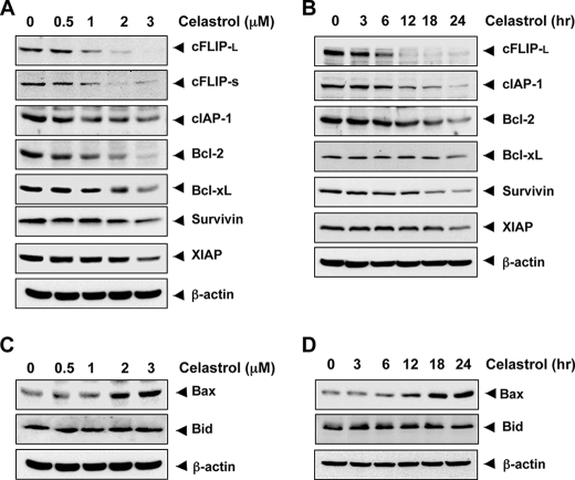
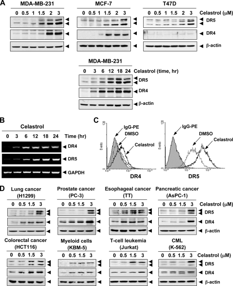
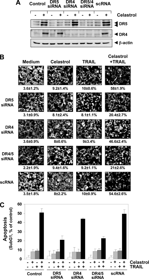
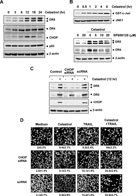
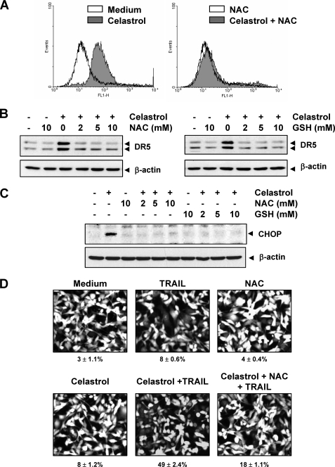
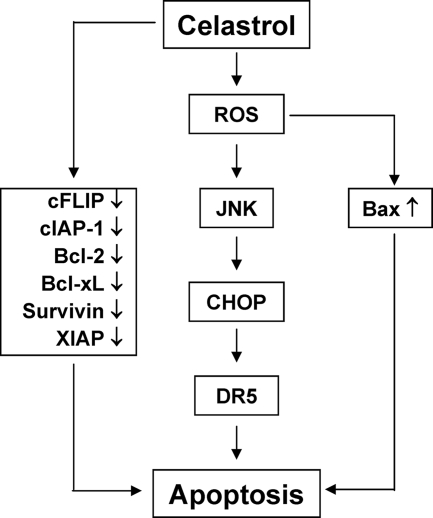
Whether celastrol, a triterpene from traditional Chinese medicine, can modulate the anticancer effects of TRAIL, the cytokine that is currently in clinical trial, was investigated. As indicated by assays that measure plasma membrane integrity, phosphatidylserine exposure, mitochondrial activity, and activation of caspase-8, caspase-9, and caspase-3, celastrol potentiated the TRAIL-induced apoptosis in human breast cancer cells, and converted TRAIL-resistant cells to TRAIL-sensitive cells. When examined for its mechanism, we found that the triterpene down-regulated the expression of cell survival proteins including cFLIP, IAP-1, Bcl-2, Bcl-xL, survivin, and XIAP and up-regulated Bax expression. In addition, we found that celastrol induced the cell surface expression of both the TRAIL receptors DR4 and DR5. This increase in receptors was noted in a wide variety of cancer cells including breast, lung, colorectal, prostate, esophageal, and pancreatic cancer cells, and myeloid and leukemia cells. Gene silencing of the death receptor abolished the effect of celastrol on TRAIL-induced apoptosis. Induction of the death receptor by the triterpenoid was found to be p53-independent but required the induction of CAAT/enhancer-binding protein homologous protein (CHOP), inasmuch as gene silencing of CHOP abolished the induction of DR5 expression by celastrol and associated enhancement of TRAIL-induced apoptosis. We found that celastrol also induced reactive oxygen species (ROS) generation, and ROS sequestration inhibited celastrol-induced expression of CHOP and DR5, and consequent sensitization to TRAIL. Overall, our results demonstrate that celastrol can potentiate the apoptotic effects of TRAIL through down-regulation of cell survival proteins and up-regulation of death receptors via the ROS-mediated up-regulation of CHOP pathway.


Figures

FIGURE 1.
FIGURE 1.
Celastrol sensitizes breast cancer cell to TRAIL. A, left, cells (3000 cells/well) were incubated various concentrations of TRAIL. After 24 h, cell viability was determined by the MTT assay. Right, cells were pretreated with celastrol for 6 h, washed with PBS to remove celastrol, and then were exposed to the indicated concentrations of soluble TRAIL for 24 h. The cell viability was determined by the MTT assay. Points, mean percentage relative to control-treated cells (n = 5); bars, standard deviation. B, MDA-MB-231 and T74D cells were treated with 2 μmol/liter celastrol for 6 h and washed with PBS to remove celastrol. Then cells were treated with the indicated concentration of TRAIL for 24 h. Apoptosis was determined by the Live/Dead Assay. C, cells were exposed to 2 μmol/liter celastrol for 6 h, and then the celastrol was removed. Then cells were treated with TRAIL (10 ng/ml) for 24 h. Cells were stained with PI, and the sub-G1 fraction was analyzed using flow cytometry. D, cells were pretreated with celastrol for 6 h and washed out. Then cells were treated with TRAIL for 24 h. Whole cell extracts were prepared and analyzed by Western blotting using antibodies against caspase-3, caspase-8, caspase-9, and PARP.
FIGURE 2.
FIGURE 2.
Celastrol modulates antiapoptotic protein expression. A and B, MDA-MB-231 cells were treated with the indicated dose of celastrol for 24 h. For determining time-dependent modulation of antiapoptotic proteins by celastrol, cells were treated with 3 μmol/liter celastrol for indicated time intervals. Whole cell extracts were prepared and analyzed by Western blotting using relevant antibodies against antiapoptotic proteins. C and D, cells were treated with the indicated dose of cleastrol at the indicated times; whole cell extracts were prepared, and Western blotting performed using antibodies against pro-apoptotic proteins. The same blots were stripped and reprobed with β-actin antibody to verify equal protein loading.
FIGURE 3.
FIGURE 3.
Celastrol up-regulates DR5 and DR4 expression. A, human breast cancer cells (5 × 10⁵ cells/well) were treated with indicated doses of celastrol for 24 h (upper panel). Whole cell extracts were then prepared and analyzed for DR4 and DR5 expression by Western blotting. For time-dependent assessment, cells (5 × 10⁵ cells/well) were treated 3 μmol/liter of celastrol for indicated times and analyzed for DR4 and DR5 expression (lower panel). B, celastrol induces DR5 gene mRNA expression. Cells (1 × 10⁶/ml) were treated with 3 μmol/liter of celastrol for indicated times, and total RNA was extracted and examined for expression of DR4 and DR5 by RT-PCR. GAPDH was used as an internal control to show equal RNA loading. C, celastrol increases cell surface expression of DR4 and DR5. Cell surface expression of DR4 and DR5 was measured by flow cytometry on MDA-MB-231 cells following celastrol treatment for 24 h using anti-DR4 and anti-DR5 antibodies conjugated with phycoerythrin. The filled gray peaks represent cells stained with a matched control PE-conjugated IgG isotype antibody. The open peaks are cells stained with PE-conjugated antibody against an individual DR. D, celastrol up-regulates DR5 and DR4 in various types of cancer cells. Cells were treated with indicated concentrations of celastrol for 24 h, and whole cell extracts were analyzed by Western blotting using antibodies against DR5 and DR4.The same blots were stripped and reprobed with β-actin antibody to verify equal protein loading.
FIGURE 4.
FIGURE 4.
Blockage of DR induction reverses the ability of celastrol to augment TRAIL-induced apoptosis. MDA-MB-231 cells were cultured in 6-well plates and the next day transfected with DR5 siRNA, DR4 siRNA, and control siRNA alone or combined. Twenty-four hours after the transfection, cells were re-seeded in 6-well plates (A and C) or chamber slides (B) and treated with 3 μmol/liter celastrol (A). After 24 h, the cells were subjected to preparation of whole cell lysates and Western blotting analysis. B and C, cells were exposed to 2 μmol/liter celastrol for 6 h, washed with PBS to remove celastrol, and then treated with 10 ng/ml TRAIL. Cell death was determined by Live/Dead assay (B) and flow cytometry to measure sub-G1 (C).
FIGURE 5.
FIGURE 5.
Up-regulation of DR5 by celastrol requires CHOP. A, MDA-MB-231 cells (5 × 10⁵) were incubated with 3 μmol/liter celastrol for the indicated times, and whole cell lysates were subjected to Western blotting analysis using relevant antibodies. B, upper panel, cells were incubated with 3 μmol/liter celastrol for indicated times, and whole cell extracts were immunoprecipitated with anti-JNK1 antibody and subjected to kinase assay as described under “Experimental Procedures.” The same protein extracts were subjected to Western blotting analysis using anti-JNK1 antibody. Lower panel, cells were pretreated with JNK inhibitor (SP600125) for 1 h and then exposed to 3 μmol/liter celastrol for 24 h. Whole cell extracts were prepared and analyzed for the expression of DR4 and DR5 using relevant antibodies. C, MDA-MB-231 cells (3 × 10⁶/well) were transfected with either CHOP siRNA or control siRNA. Twenty-four hours after the transfection, cells were re-seeded in 6-well plates or chamber slides. Cells were treated with 3 μmol/liter celastrol for 24 h, and whole cell lysates were analyzed by Western blotting (C). The same blots were stripped and reprobed with β-actin antibody to verify equal protein loading. D, cells were exposed to 2 μmol/liter celastrol for 6 h, washed with PBS to remove celastrol, and then treated with 10 ng/ml TRAIL. Cell death was determined by Live/Dead assay.
FIGURE 6.
FIGURE 6.
Celstrol-induced ROS is involved in CHOP induction leading to DR5 up-regulation. A, MDA-MB-231 cells were treated with 3 μmol/liter celastrol with or without 10 mmol/liter NAC. Twelve hours later, intracellular ROS levels were measured by flow cytometry using CM-H2DCFDA, as described under “Experimental Procedures.” B, cells (5 × 10⁵ cells) were pretreated with various concentrations of NAC or GSH for 1 h and then treated with 3 μmol/liter celastrol for 24 h. Whole cell extracts were prepared and analyzed by Western blotting using DR5 antibody. C, cells were treated with either NAC or GSH for 1 h and exposed to 3 μmol/liter celastrol for 12 h, and then whole cell extracts were subjected to Western blotting for CHOP. The same blot was stripped and reprobed with β-actin antibody to verify equal protein loading. D, MDA-MB-231 cells were pretreated with NAC for 1 h and then treated with celastrol for 6 h. Next cells were washed with PBS to remove celastrol and treated with TRAIL for 24 h. Cell death was determined by the Live/Dead Assay.
FIGURE 7.
FIGURE 7.
A flowsheet describing the mechanism by which celastrol potentiates the effect of TRAIL on apoptosis.

Similar articles

Cited by

References

    1. Wiley S. R., Schooley K., Smolak P. J., Din W. S., Huang C. P., Nicholl J. K., Sutherland G. R., Smith T. D., Rauch C., Smith C. A. (1995) Immunity 3, 673–682 - PubMed
    1. Pitti R. M., Marsters S. A., Ruppert S., Donahue C. J., Moore A., Ashkenazi A. (1996) J. Biol. Chem. 271, 12687–12690 - PubMed
    1. Bhardwaj A., Aggarwal B. B. (2003) J. Clin. Immunol. 23, 317–332 - PubMed
    1. Rowinsky E. K. (2005) J. Clin. Oncol. 23, 9394–9407 - PubMed
    1. Wagner K. W., Punnoose E. A., Januario T., Lawrence D. A., Pitti R. M., Lancaster K., Lee D., von Goetz M., Yee S. F., Totpal K., Huw L., Katta V., Cavet G., Hymowitz S. G., Amler L., Ashkenazi A. (2007) Nat. Med. 13, 1070–1077 - PubMed

Publication types