. 2010 Apr 9;285(15):11433-44.
doi: 10.1074/jbc.M109.077065. Epub 2010 Feb 12.

Distinct role of Kruppel-like factor 11 in the regulation of prostaglandin E2 biosynthesis

Affiliations

Distinct role of Kruppel-like factor 11 in the regulation of prostaglandin E2 biosynthesis

Navtej S Buttar et al. J Biol Chem. .

Abstract

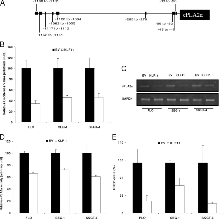
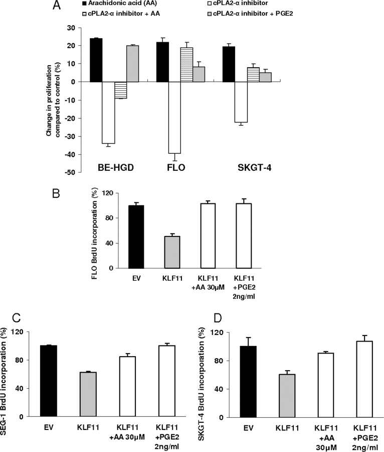
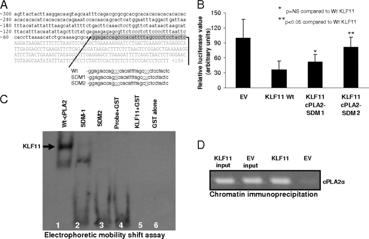
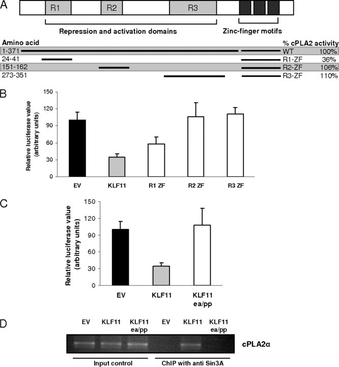
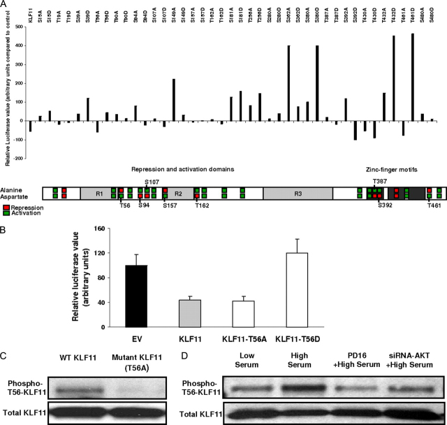
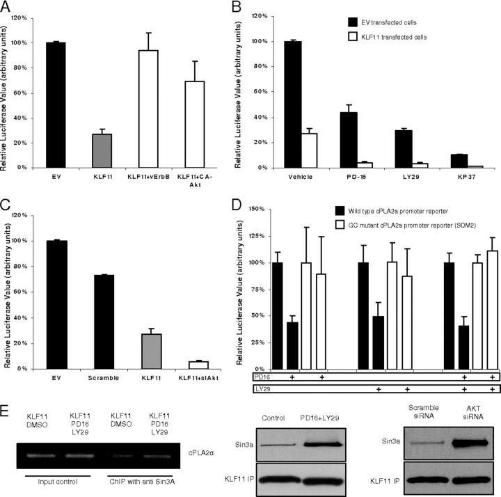
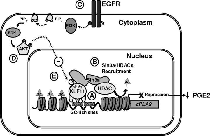
Kruppel-like factor (KLF) proteins are emerging as key regulators of lipid metabolism, diabetes, and the biosynthesis of immunological cytokines. However, their role in the synthesis of prostaglandins, widely known biochemical mediators that act in a myriad of cell biological processes remain poorly understood. Consequently, in this study a comprehensive investigation at the cellular, biochemical, and molecular levels reveal that KLF11 inhibits prostaglandin E(2) synthesis via transcriptional silencing of the promoter of its biosynthetic enzyme, cytosolic phospholipase A2alpha. Mechanistically, KLF11 accomplishes this function by binding to the promoter via specific GC-rich sites and recruiting the Sin3-histone deacetylase chromatin remodeling complex. Further functional characterization reveals that this function of KLF11 can be reversed by epidermal growth factor receptor-AKT-mediated post-translational modification of threonine 56, a residue within its Sin3-binding domain. This is the first evidence supporting a relevant role for any KLF protein in doing both: transcriptionally inhibiting prostaglandin biosynthesis and its reversibility by an epidermal growth factor receptor-AKT signaling-mediated posttranslational mechanisms.


Figures

FIGURE 1
FIGURE 1
KLF11 represses the rate-limiting enzyme cPLA2α and down-regulates PGE2 synthesis.A, the promoter region of cPLA2α contains several GC-rich, putative KLF11 binding sites. B, esophageal adenocarcinoma (FLO, SEG-1, and SKGT-4) cell lines were co-transfected with a cPLA2α promoter luciferase reporter construct (−1200 to +150 relative to transcription start site) along with full-length KLF11 or the EV construct for 48 h. Luciferase levels normalized to lysate protein concentrations show that compared with EV, co-transfection with KLF11 decreased cPLA2α promoter activity in FLO by 66 ± 3.3%, SEG-1 by 54.4 ± 5.8%, and SKGT-4 by 45 ± 9% (p < 0.05). As a negative control, the cyclin B1 promoter was used where KLF11 failed to decrease the promoter activity (data shown in supplemental Fig. S1). C and D, compared with empty vector, adenoviral infection of cells for 48 h with KLF11 decreased cPLA2α expression in all three cell lines and significantly reduced cPLA2α activity in FLO cells by 34% (474 ± 13 versus 319 ± 2.4 arbitrary units (AU), p < 0.05), in SEG-1 cells by 28% (572 ± 34 versus 407 ± 3 AU, p < 0.05), and in SKGT-4 by 39% (509 ± 12.8 versus 309 ± 1.6 AU, p < 0.05). E, compared with empty vector, adenoviral infection for 48 h with KLF11 also significantly reduced PGE2 production in FLO cells by 78% (86.7 ± 22 versus 19.4 ± 8.9 pg, p < 0.05), in SEG-1 cells by 46% (19.7 ± 9 versus 10.7 ± 3.1 pg, p < 0.05), and in SKGT-4 by 81% (100.4 ± 35 versus 19 ± 2.7 pg, p < 0.05). GAPDH, glyceraldehyde-3-phosphate dehydrogenase.
FIGURE 2
FIGURE 2
KLF11-mediated down-regulation of cPLA2α-PGE2 inhibit cell proliferation.A, to test the cell biological significance of the cPLA2α-PGE2 pathway, FLO cells were treated for 48 h with 30 μm arachidonic acid, a catalytic product of cPLA2α, and increased proliferation by 22 ± 2.6%, whereas AACOCF3, a cPLA2α inhibitor, decreased proliferation by 39.5 ± 0.4% compared with control (BrdUrd incorporation, p < 0.05). The effect of AACOCF3α on FLO cell proliferation was reversed by the catalytic product of cPLA2α namely arachidonic acid and PGE2. Similar results were noted using other esophageal cancer cells (SKGT-4 and BE-HGD). These findings support that the effect of cPLA2α on cell proliferation is mediated by the release of arachidonic acid and production of PGE2. B–D, compared with EV, adenoviral infection of FLO (multiplicity of infection 30), SEG-1 (multiplicity of infection 100), and SKGT-4 (multiplicity of infection 100) cells for 48 h with KLF11 significantly (p < 0.05) reduced BrdUrd incorporation in cells that were treated with vehicle (49.5 ± 4.7, 38.5 ± 1.6, and 40 ± 5.9%, respectively for FLO, SEG-1 cells, and SKGT-4, p < 0.05), however, this growth inhibitory effect of KLF11 was abrogated in the cells that were treated with 30 μm arachidonic acid (AA, a catalytic product of cPLA2α and substrate of PGE2) or 2 ng/ml of PGE2 suggesting that the growth inhibitory effect of KLF11 is mediated via down-regulation of cPLA2α-PGE2 pathway.
FIGURE 3
FIGURE 3
KLF11-mediated regulation of cPLA2α requires defined promoter site recognition and KLF11 binds to cPLA2α promoter in vivo.A, outlining of site-directed mutagenesis in GC-rich areas of the cPLA2α promoter reporter construct. B, compared with control, KLF11 was able to repress the cPLA2α-WT promoter but failed to repress the cPLA2α promoter that had cc to tt mutations in the distal GC-rich site (cPLA2α-WT (36.29 ± 17.7%) versus cPLA2α-SDM2 (80.8 ± 20.9%), p < 0.05). The mutations in the more proximal GC-rich site (SDM1) only partially relived the KLF11-dependent repression (52 ± 14.7%), which was not significantly different compared with cPLA2α-WT. C, electrophoretic mobility shift assay shows that binding of the KLF11-GST recombinant protein and digoxigenin-labeled fragment of the cPLA2α core promoter sequence was partially disrupted with SDM-1 mutations (lane 2) and completely disrupted by SDM-2 mutations (lane 3) in the GC-rich sequence of the cPLA2α promoter. Lane 1 represents the fragments containing the wild-type cPLA2α core promoter sequence (Wt-cPLA2α) and lanes 4–6 are negative controls. D, ChIP assay using FLO cell lysates shows that the cis-regulatory cPLA2α promoter sequence is enriched in immunoprecipitated samples from cells infected with KLF11-carrying adenovirus and absent in EV control-infected cells demonstrating that KLF11 can bind to the promoter of cPLA2α endogenously.
FIGURE 4
FIGURE 4
R1 repressor domain of KLF11 is critical in the cPLA2α promoter repression via binding and function of the Sin3a-HDAC chromatin remodeling complex.A, the top panel shows the outline of repressor and DNA binding domains of the KLF11 protein. The lower panel is a summary as described in the legend to Fig. 3B. B, FLO cells were co-transfected with the cPLA2α promoter reporter construct along with either empty vector or full-length KLF11 or KLF11 deletions containing distinct regulatory domains. Compared with empty vector control, both full-length and R1-ZF KLF11 significantly repressed cPLA2α promoter activity (65.6 ± 12 and 42.4 ± 9.8%, p < 0.05) but R2-ZF and R3-ZF failed to repress the cPLA2α promoter activity. C, in FLO cells, compared with control, wild-type KLF11 repressed the cPLA2α promoter activity by 65.6 ± 12% (p < 0.05), the ΔE29P/ΔA30P-KLF11 (the mutant to disrupt Sin3a-HDAC binding) completely abolished cPLA2α repression by KLF11. D, ChIP assay using FLO cell lysates shows that the cis-regulatory cPLA2α promoter sequence is enriched in anti-Sin3a antibody (SC-994) immunoprecipitated samples from cells infected with adenovirus carrying wild-type KLF11 (fifth lane from the left) but absent in adenovirus carrying the ΔE29P/ΔA30P-KLF11 mutation (sixth lane from the left) demonstrating that the KLF11-mediated recruitment of Sin3a to the cPLA2α promoter can be abrogated by the ΔE29P/ΔA30P mutation in KLF11. The input controls are in the first three lanes on the left and similar results were noted in SEG-1 cells (data not shown). Together, the data support that the R1 domain of KLF11 is critical in repression of the cPLA2α promoter and this repression is Sin3a-HDAC-dependent.
FIGURE 5
FIGURE 5
Post-translational modification of threonine at position 56 in KLF11, a target of phosphorylation by AKT, is crucial in KLF11-mediated repression of the cPLA2α promoter.A, Chinese hamster ovary cells were co-transfected with the cPLA2α promoter reporter construct along with either EV or KLF11 constructs from a library of mutant KLF11 proteins where serines and threonines were replaced with either alanines or aspartic acids as indicated. The eight phosphomimetic and non-phosphorylatable KLF11 mutants with opposing effects on cPLA2α promoter activity are displayed along with a KLF11 protein domain outline. B, FLO cells co-transfected with the cPLA2α promoter reporter construct along with either empty vector or a phosphomimetic (T56D) or non-phosphorylatable (T56A) KLF11 mutant in the R1 domain (in close proximity of its Sin3a binding site) shows that at 48 h, compared with control, wild-type KLF11 repressed the cPLA2α promoter activity to 43 ± 6% but the phosphomimetic T56D-KLF11 mutant resulted in a complete release of cPLA2α promoter repression by KLF11 (120 ± 23%). The repression of cPLA2α persisted with the T56A-KLF11 mutant (42 ± 7%). C, lysates from Chinese hamster ovary cells transfected with wild-type KLF11 or T56A mutant KLF11 after immunoprecipitation of His-tagged KLF11 followed by Western blot with phospho-Thr-56-KLF11 antibody shows the specificity of this antibody as it does not bind to non-phosphorylatable T56A-KLF11. D, KLF11-transfected FLO cells were treated with either scrambled siRNA + vehicle or siRNA against AKT (AKT-1, -2, and -3) transfection or PD168393 (EGFR blocker) to inhibit AKT. 24 h later cells were either maintained in 5% FBS (low serum) or given a 90-min pulse of high serum medium (10% FBS to activate EGFR-AKT pathway). After immunoprecipitating His-tagged KLF11 protein, resolving by 10% SDS-PAGE, and immunoblotting with anti-phospho-Thr-56-KLF11 and total KLF11, as a loading control, we found that the high serum pulse that activates AKT (data shown in supplemental Fig. S6, 2 and 3) results in phosphorylation of Thr-56 in KLF11 and that the siRNA against AKT, as well as PD168393 to inhibit AKT, markedly reduced the phosphorylation of this site.
FIGURE 6
FIGURE 6
EGFR-AKT signaling is involved in KLF11-mediated cPLA2α promoter repression.A, FLO cells were co-transfected with the cPLA2α promoter reporter construct along with either EV or KLF11, with or without vErbB (constitutively active EGFR), or CA-AKT. KLF11 overexpression reduced the cPLA2α promoter activity by 63 ± 4.3% (p < 0.05), however, this repression was released in the presence of vErbB and CA-AKT (6 ± 0.9 and 31 ± 7% repression, respectively, p < 0.05 compared with KLF11). B, FLO cells co-transfected with cPLA2α along with either EV or KLF11 were treated with either vehicle or the blockers of EGFR-AKT pathway (10 μm PD168393, 100 μm LY294002, or 1 μm KP372–1). 48 h later, KLF11 decreased the cPLA2α promoter activity in the presence of vehicle by 3.7-fold (100 ± 1.3 versus 27 ± 4.3%), with PD168393 by 10.7-fold (43 ± 6.5 versus 4 ± 1%, p < 0.05 compared with KLF11 with vehicle), with LY294002 by 9.5-fold (29.7 ± 1.6 versus 3 ± 0.9%, p < 0.05 compared with KLF11 with vehicle), and with KP372-1 by 10-fold (10 ± 1 versus 1 ± 0.05%, p < 0.05 compared with KLF11 with vehicle). C, FLO cells were co-transfected with cPLA2α along with either EV or KLF11 and siRNA against AKT or scramble RNA. Cells were maintained in 10% FBS for 48 h. KLF11 decreased cPLA2α promoter activity by 12-fold (73 ± 1 versus 6 ± 1%) in the presence of AKT siRNA compared with a 3.7-fold reduction (100 ± 1.3 versus 27 ± 4.3%) with scramble siRNA (p < 0.05). D, FLO cells transfected with either cPLA2α-WT promoter or cPLA2α-SDM2 (mutation in GC-rich sequence to which KLF11 binds). Cells were treated with vehicle, 10 μm PD168393, or 100 μm LY294002 or both PD168393 and LY294002 for 24 h in 10% FBS. PD168393 decreased the cPLA2α-WT promoter activity by 66% (100 ± 10 versus 44 ± 6%, p < 0.05) but had no significant effect on the cPLA2α-SDM2 promoter activity (100 ± 33 versus 89 ± 35%). A similar pattern was also noted with LY294002 alone or with both PD168393 and LY294002. E, KLF11-transfected FLO cells were treated with either vehicle or 10 μm PD168393 plus 100 μm LY294002 for 24 h in the presence of 10% FBS. Chromatin immunoprecipitation with anti-Sin3a antibody showed a slight increase in cPLA2α promoter enrichment in the blocker-treated group. To compliment this, FLO cells were either co-transfected with KLF11 and AKT siRNA (or scramble RNA control) or treated with 10 μm PD168393 plus 100 μm LY294002 (or vehicle control). Western blots after immunoprecipitation of His-tagged KLF11 followed by probing with the anti-Sin3a antibody shows that compared with control there was increased KLF11-Sin3a complexing in AKT blockers as well as AKT-siRNA-treated cells compared with the control. DMSO, dimethyl sulfoxide.
FIGURE 7
FIGURE 7
Mechanistic model of KLF11-mediated tumor suppression and its antagonism by an oncogenic pathway. KLF11 binds to the GC-rich consensus sequences in the promoter region of cPLA2α, the key rate-limiting enzyme of the oncogenic PGE2 cascade. A, KLF11 represses the cPLA2α promoter by recruiting the chromatin remodeling complex, Sin3a-HDAC (B). As a consequence, KLF11 behaves as a tumor suppressor in Barrett's epithelial cells, at least in part, by repression of the cPLA2α-PGE2 pathway. EGFR-AKT signaling, which is up-regulated in a subset of patients during carcinogenesis in Barrett's esophagus (C) phosphorylates threonine at position 56 in the R1 domain of KLF11 in the immediate vicinity of its Sin3a interacting domain (D), and reverses the KLF11-dependent repression of the cPLA2α promoter (E). Together, this model outlines mechanistic links, namely KLF11-SIN3a/HDAC-cPLA2α-PGE2 and EGFR/AKT-KLF11 (Thr-56 phosphorylation)-SIN3a/HDAC-cPLA2α-PGE2.

References

    1. Murakami M., Kudo I. Prog. Lipid Res. 2004;43:3–35. - PubMed
    1. Murakami M., Shimbara S., Kambe T., Kuwata H., Winstead M.V., Tischfield J.A., Kudo I. J. Biol. Chem. 1998;273:14411–14423. - PubMed
    1. Leslie C.C. J. Biol. Chem. 1997;272:16709–16712. - PubMed
    1. Funk C.D. Science. 2001;294:1871–1875. - PubMed
    1. Bonventre J.V., Huang Z., Taheri M.R., O'Leary E., Li E., Moskowitz M.A., Sapirstein A. Nature. 1997;390:622–625. - PubMed

Publication types

MeSH terms