. 2010 Apr 2;285(14):10252-64.
doi: 10.1074/jbc.M109.083857. Epub 2010 Feb 12.

Endogenous proteolytic cleavage of disease-associated prion protein to produce C2 fragments is strongly cell- and tissue-dependent

Affiliations

Endogenous proteolytic cleavage of disease-associated prion protein to produce C2 fragments is strongly cell- and tissue-dependent

Michel Dron et al. J Biol Chem. .

Abstract

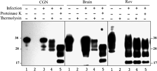
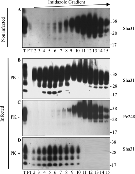
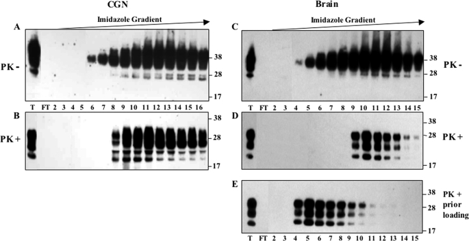
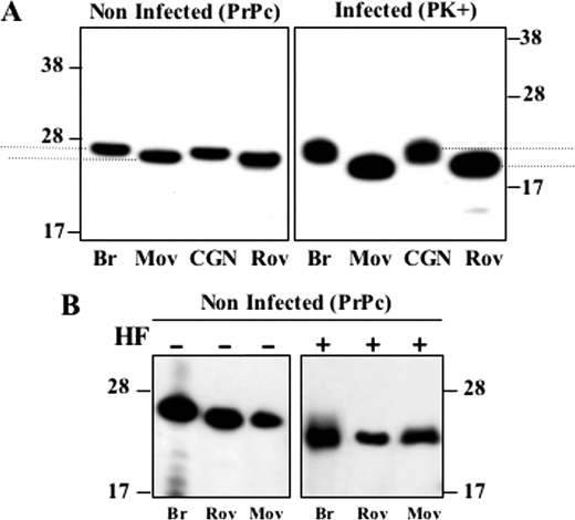
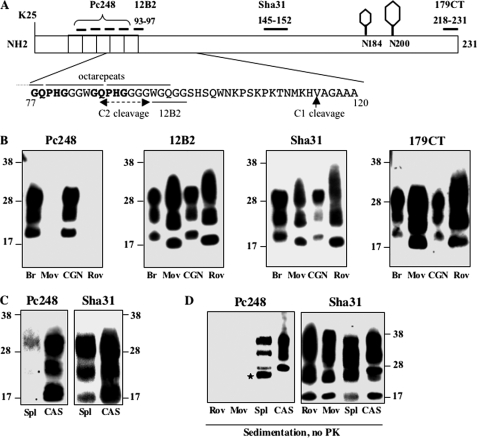
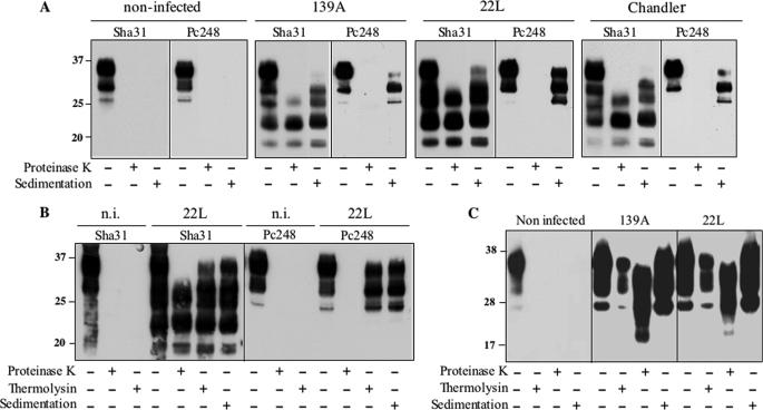
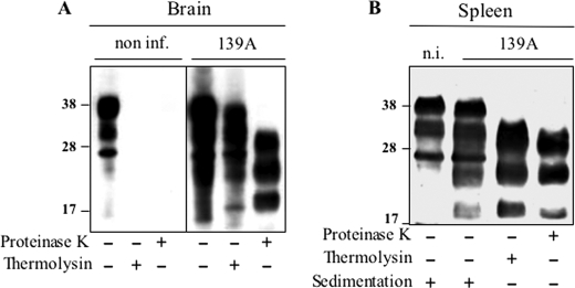
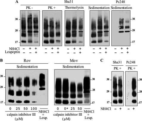
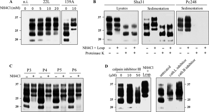
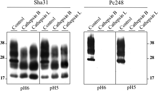
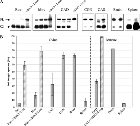
The abnormally folded form of the prion protein (PrP(Sc)) accumulating in nervous and lymphoid tissues of prion-infected individuals can be naturally cleaved to generate a N-terminal-truncated fragment called C2. Information about the identity of the cellular proteases involved in this process and its possible role in prion biology has remained limited and controversial. We investigated PrP(Sc) N-terminal trimming in different cell lines and primary cultured nerve cells, and in the brain and spleen tissue from transgenic mice infected by ovine and mouse prions. We found the following: (i) the full-length to C2 ratio varies considerably depending on the infected cell or tissue. Thus, in primary neurons and brain tissue, PrP(Sc) accumulated predominantly as untrimmed species, whereas efficient trimming occurred in Rov and MovS cells, and in spleen tissue. (ii) Although C2 is generally considered to be the counterpart of the PrP(Sc) proteinase K-resistant core, the N termini of the fragments cleaved in vivo and in vitro can actually differ, as evidenced by a different reactivity toward the Pc248 anti-octarepeat antibody. (iii) In lysosome-impaired cells, the ratio of full-length versus C2 species dramatically increased, yet efficient prion propagation could occur. Moreover, cathepsin but not calpain inhibitors markedly inhibited C2 formation, and in vitro cleavage by cathepsins B and L produced PrP(Sc) fragments lacking the Pc248 epitope, strongly arguing for the primary involvement of acidic hydrolases of the endolysosomal compartment. These findings have implications on the molecular analysis of PrP(Sc) and cell pathogenesis of prion infection.


Figures

FIGURE 1.
FIGURE 1.
PrPSc is truncated in Rov and MovS cells but remains in a full-length form in tg338 CGN and the brain. A, cultures of Rov and MovS cell lines, and primary neurons (CGN) and astrocytes (CAS) derived from tg338 mice were infected (lanes 2, 4, and 6) or not (lanes 1, 3, and 5) by the ovine prion 127S strain. Crude lysates (lanes 1 and 2), PK-treated (lanes 3 and 4), or sedimented materials (lanes 5–7) were analyzed by immunoblotting. Lane 7 in the CGN panel corresponds to PK digestion of the sedimented material shown in lane 6. PrPSc sedimented as the truncated species from Rov and MovS cells and as FL species from CGN cells. Both truncated and FL PrPSc were detected in CAS cells. B, analysis of PrPSc in the brain and spleen homogenates from 127S-infected tg338 mouse (lanes 2 and 4) before (lanes 1 and 2) and after PK treatment (lanes 3 and 4). Mostly FL PrPSc is detected in the brain, whereas spleen PrPSc mainly contains the truncated species (lanes 2). All immunoblots were revealed with Sha31 anti-PrP mAb. Equivalent protein amounts were loaded per lane for each sample, except for the sedimented spleen lysate for which 10-fold more material was needed to detect PrPSc. The molecular profiles shown have been observed in multiple experiments on multiple blots. The positions of molecular mass markers (in kDa) are shown on both sides of the figure for all gels. The arrows designate the PrP 26-kDa aglycosylated species.
FIGURE 2.
FIGURE 2.
CGN accumulate thermolysin-resistant, full-length PrPSc. Whole lysates from uninfected (lanes 1 and 2 of each panel) and 127S-infected (lanes 3–5) tg338 CGN, brain, and Rov cultures were digested with thermolysin (≥4 experiments) using conditions in which PrPC was completely proteolyzed (lanes 2 and 4). Immunoblots using the Sha31 mAb show that thermolysin-resistant PrPSc from CGN cells and the brain migrates with the mobility of FL species (lanes 4), not with PrPres generated by the PK treatment (lanes 5). In contrast, the profiles of thermolysin- and PK-resistant PrPSc from Rov cells are similar (compare lanes 4 and 5 in Rov panel). The positions of molecular mass markers are indicated.
FIGURE 3.
FIGURE 3.
Truncated PrPSc from MovS cells binds to the IMAC-Cu2+ column. Lysates of MovS cells (lane T) were loaded onto an IMAC-Cu2+ column. After recovery of unbound proteins (FT, flow-through), bound proteins were eluted by application of a 3–200 mm linear imidazole gradient and fractions (numbers 2–15) were analyzed by immunoblotting using either Sha31 mAb that recognizes the PrP core (A, B, and D) or anti-octarepeat Pc248 mAb (C). PrP elution profiles obtained with non-infected (A) and infected cells before (B and C) or after PK digestion of the eluted fractions (D) are shown. Pc248 mAb detects non-truncated PrP in infected cells (C), whereas Sha31 detects both truncated and FL molecules (B). PK digestion (D) proves that the population of truncated PrP corresponds to the bulk of PrPSc produced by MovS cells.
FIGURE 4.
FIGURE 4.
PrPSc from CGN and the brain binds to the IMAC-Cu2+ column and elutes as full-length PrP. Total lysates (lane T) of 127S-infected CGN (A) or the tg338 brain (C) were applied to the IMAC-Cu2+-charged column. The flow-through fraction (FT) was collected and bound proteins were eluted by application of a 3–200 mm linear imidazole gradient and analyzed by immunoblotting for the presence of PrP molecules before (A and C) and after PK treatment (B and D). In E, the lysate of the infected brain was first digested with PK prior to loading onto the column and the eluted fractions were analyzed. Immunoblots were revealed with Sha31 mAb. The binding and elution of FL PrPC and PrPSc produced in CGN and in the brain are similar (compare A to B and C to D). PK-digested brain PrPSc (E) is also retained on a copper column but elutes at lower imidazole concentrations than FL PrPSc (compare D to E). The positions of molecular mass markers are shown on the right of each immunoblot.
FIGURE 5.
FIGURE 5.
Higher electrophoretic mobility of PrPC and PrPres from MovS and Rov cells compared with CGN or brain tissue. A, the comparative mobility of PK-resistant PrPSc (right panel) and PrPC (left panel) from tg338 mouse brain, and MovS, CGN, and Rov lysates is shown on immunoblots revealed with ICSM4, an antibody specific for the non-glycosylated form of PrP. To compare the mobility differences accurately, in these experiments the runs were performed so that the migration distances of unglycosylated species PrPC and PrPSc were the same. B, treatment with hydrofluoric acid (HF) of non-infected brain tissue, Rov, and MovS cells generates a PrP species with identical mobility (immunodetection with ICSM4). The electrophoretic mobility differences shown were observed in three (B) or more (A) independent experiments.
FIGURE 6.
FIGURE 6.
N-terminal trimming of ovine PrPSc produced in Rov, MovS, CAS cells and in the spleen occurs downstream of the main PK cleavage sites. A, schematic diagram of ovine PrPC indicating the two glycosylation sites (N184 and N200, sheep numbering) and the location of epitopes used for mapping. The deduced region of cleavage to produce C2 (dashed line) is within the Pc248 epitope or just upstream of the 12B2 epitope (underlined), as indicated on the highlighted amino acid sequence. The Pc248 epitope is indicated in bold. B, PK-digested PrPSc from infected materials was tested for reactivity to the four antibodies indicated on the scheme. Only the anti-octarepeat mAb Pc248 discriminates PrPSc produced in CGN and the brain from PrPSc accumulated in Rov and MovS cells. C, PK-digested PrPSc from the spleen (Spl) of infected tg338 mice and from infected CAS was analyzed by immunoblotting with both Pc248 and Sha31 mAbs. D, sedimented, and non-PK-treated materials from infected Rov, MovS, spleen, and CAS were immunoblotted with Pc248 and Sha31 mAbs. The positions of molecular mass markers are indicated. The star on panel D points to a nonspecific band detected with Pc248 in the spleen-sedimented sample.
FIGURE 7.
FIGURE 7.
Mouse PrPSc is truncated in CAD cells but remains full-length in CGN. A, CAD cells were infected with mouse-adapted scrapie prion (139A, 22L, or Chandler strain) or mock-infected and analyzed for their PrPC and PrPSc content. Immunoreactive PrP species were revealed in whole cell extracts, PK-digested and sedimented materials, as indicated, using either Sha31 or Pc248 mAbs. Note that both truncated and FL, Pc248-positive PrP species are recovered in sedimented material from infected samples, whatever the strain. B, whole cell lysate of 22L- or mock-infected CAD cells and thermolysin- or PK-digested samples were analyzed using Sha31 or Pc248 antibody. Note the Pc248-positive bands in thermolysin-resistant and sedimented materials, reflecting the presence of FL PrPSc. C, CGN from C57Bl/6 mice were mock-infected or infected with 139A or 22L scrapie strains. Cell lysates performed 24 days post-infection were centrifuged or digested either with thermolysin or PK as indicated, and analyzed by Western blot using Sha31 mAb. The molecular profiles have been observed in multiple experiments on multiple blots. n.i., non-infected.
FIGURE 8.
FIGURE 8.
Mouse PrPSc is efficiently truncated in the spleen but not the brain. Analysis of PrPSc in brain (A) and spleen (B) homogenates from non-infected or 139A-infected tga20 mice. Tissue lysates (from at least three animals) were centrifuged (spleen only) or digested either by thermolysin or PK as indicated, and analyzed by Western blot using Sha31 mAb. PrPSc detected in the spleen is essentially truncated prior to PK digestion (lane 3). Note the presence of sedimentable PrPC in both infected and non-infected tga20 mouse spleen, which was consistently observed with mouse PrPC in these conditions. n.i., non-infected.
FIGURE 9.
FIGURE 9.
N-terminal trimming of PrPSc is inhibited in lysosome-impaired Rov and MovS cells but is not calpain-dependent. A, infected Rov cells were left untreated or cultivated for 7 days in the presence of 10 mm NH4Cl or 10 mm NH4Cl + 100 μg/ml of leupeptin, as indicated. The PrP profiles of undigested, PK-digested, and thermolysin-digested samples are shown after Sha31 mAb immunoblotting. Lysates were also sedimented without protease treatment and the pellets were revealed with either Sha31 or Pc248 mAbs (right two panels). B, infected Rov and MovS cells were treated with various concentrations of calpain inhibitor III or NH4Cl plus leupeptin, as indicated (0*: vehicle only). Sedimented materials from cell lysates were analyzed by Western blotting with Sha31 mAb. C, infected Rov cells treated or not with NH4Cl were digested with PK before immunoblot analysis using Sha31 or Pc248 mAbs. The above experiments have been repeated three times with consistent results.
FIGURE 10.
FIGURE 10.
Lysosome inhibition blocks PrPSc N-terminal trimming but not prion propagation in CAD cells. A, non-infected, 22L- and 139A-infected cells were either left untreated or treated with NH4Cl as indicated, and cell lysates were centrifuged. The pellets were analyzed for their PrPSc content by immunoblotting using Sha31 mAb. B, 22L-infected CAD cells cultivated in the absence or presence of 10 mm NH4Cl plus 10 μg/ml of leupeptin for two consecutive passages. Whole cell lysates and sedimented materials (right two panels) were PK-digested or not before immunoblotting using Sha31 or Pc248 mAb. C, 22L-infected CAD cells were cultivated in the absence or presence of 10 mm NH4Cl for 4 days and cells were passaged twice a week in the absence or presence of the lysosomal inhibitor. Cell lysates were immunoblotted using Sha31 mAb. Molecular mass markers in kDa are shown. D, infected CAD cells were treated with various concentrations of calpain inhibitor III, NH4Cl plus leupeptin, cathepsin L inhibitor I (cath.L), or cathepsin B inhibitor I (cath.B), as indicated. Sedimented materials from cell lysates were analyzed by Western blotting with Sha31 mAb. These experiments have been repeated three times with consistent results. n.i., non-infected.
FIGURE 11.
FIGURE 11.
In vitro cleavage of full-length PrPSc by cathepsin B and L remove the Pc248 epitope. PrPSc from a lysate of infected Rov cells treated with NH4Cl and leupeptin was purified by sedimentation, resuspended in two different cathepsin buffers (pH 6 or 5), and digested or not (control lanes) with the indicated proteases as described under “Experimental Procedures.” The same samples were revealed with either Sha31 or Pc248 mAbs.
FIGURE 12.
FIGURE 12.
Proportion of untrimmed PrPSc in infected cells and tissues. A, Western blot analysis (Sha31 mAb) of PNGase-treated samples: cell lines (Rov, Mov CAD), primary cultures of neurons (CGN) or astrocytes (CAS), and brain and spleen tissues. Each of the three left panels show three samples left untreated and one sample treated with lysosomal inhibitors (10 mm NH4Cl plus 10 μg/ml leupeptin); the four right panels each show two different samples. Sedimented material from lysates of cell lines and primary cultures were resuspended and treated with PNGase (similar results were obtained using samples that were digested with thermolysin before PNGase; not shown). Lysates from tg338 brain and spleen tissues were treated with thermolysin and further centrifuged in the case of spleen material to concentrate PrPSc. Arrows indicate deglycosylated full-length (FL) and N-terminal-truncated (C2) PrPSc species. B, the histogram shows the percentage of full-length species over total PrPSc as determined by quantification of samples such as seen in A. Average values and standard deviation were determined from four to seven different experiments.

Similar articles

Cited by

References

    1. Prusiner S. B. (1998) Proc. Natl. Acad. Sci. U.S.A. 95, 13363–13383 - PMC - PubMed
    1. Westergard L., Christensen H. M., Harris D. A. (2007) Biochim. Biophys. Acta 1772, 629–644 - PMC - PubMed
    1. Aguzzi A., Sigurdson C., Heikenwaelder M. (2008) Annu. Rev. Pathol. 3, 11–40 - PubMed
    1. Gambetti P., Kong Q., Zou W., Parchi P., Chen S. G. (2003) Br. Med. Bull. 66, 213–239 - PubMed
    1. Wadsworth J. D., Collinge J. (2007) Biochim. Biophys. Acta 1772, 598–609 - PubMed

Publication types

MeSH terms