

Ubiquilin modifies TDP-43 toxicity in a Drosophila model of amyotrophic lateral sclerosis (ALS)
- PMID: 20154090
- PMCID: PMC2856981
- DOI: 10.1074/jbc.C109.078527
Ubiquilin modifies TDP-43 toxicity in a Drosophila model of amyotrophic lateral sclerosis (ALS)
Abstract
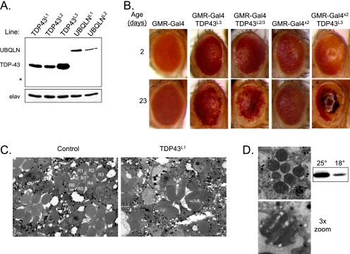
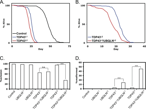
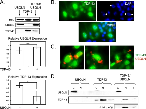
TDP-43 (43-kDa TAR DNA-binding protein) is a major constituent of ubiquitin-positive cytosolic aggregates present in neurons of patients with amyotrophic lateral sclerosis (ALS) and ubiquitin-positive fronto-temporal lobar degeneration (FTLD-U). Inherited mutations in TDP-43 have been linked to familial forms of ALS, indicating a key role for TDP-43 in disease pathogenesis. Here, we describe a Drosophila melanogaster model of TDP-43 proteinopathy. Expression of wild-type human TDP-43 protein in Drosophila motor neurons led to motor dysfunction and dramatic reduction of life span. Interestingly, coexpression of ubiquilin 1, a previously identified TDP-43-interacting protein with suspected functions in autophagy and proteasome targeting, reduced steady-state TDP-43 expression but enhanced the severity of TDP-43 phenotypes. Finally, ectopically expressed TDP-43 was largely localized to motor neuron nuclei, suggesting that expression of wild-type TDP-43 alone is detrimental even in the absence of cytosolic aggregation. Our findings demonstrate that TDP-43 exerts cell-autonomous neurotoxicity in Drosophila and further imply that dose-dependent alterations of TDP-43 nuclear function may underlie motor neuron death in ALS.
Figures
Similar articles
-
Motor neurons and glia exhibit specific individualized responses to TDP-43 expression in a Drosophila model of amyotrophic lateral sclerosis.Dis Model Mech. 2013 May;6(3):721-33. doi: 10.1242/dmm.010710. Epub 2013 Feb 1. Dis Model Mech. 2013. PMID: 23471911 Free PMC article.
-
Potentiation of amyotrophic lateral sclerosis (ALS)-associated TDP-43 aggregation by the proteasome-targeting factor, ubiquilin 1.J Biol Chem. 2009 Mar 20;284(12):8083-92. doi: 10.1074/jbc.M808064200. Epub 2008 Dec 26. J Biol Chem. 2009. PMID: 19112176 Free PMC article.
-
Motor neuron-specific disruption of proteasomes, but not autophagy, replicates amyotrophic lateral sclerosis.J Biol Chem. 2012 Dec 14;287(51):42984-94. doi: 10.1074/jbc.M112.417600. Epub 2012 Oct 24. J Biol Chem. 2012. PMID: 23095749 Free PMC article.
-
[FTLD/ALS as TDP-43 proteinopathies].Rinsho Shinkeigaku. 2010 Nov;50(11):1022-4. doi: 10.5692/clinicalneurol.50.1022. Rinsho Shinkeigaku. 2010. PMID: 21921552 Review. Japanese.
-
[Component of ubiquitin-positive inclusions in ALS].Brain Nerve. 2007 Oct;59(10):1171-7. Brain Nerve. 2007. PMID: 17969358 Review. Japanese.
Cited by
-
Mitochondrion-Dependent Cell Death in TDP-43 Proteinopathies.Biomedicines. 2021 Apr 2;9(4):376. doi: 10.3390/biomedicines9040376. Biomedicines. 2021. PMID: 33918437 Free PMC article. Review.
-
TDP-43 induces mitochondrial damage and activates the mitochondrial unfolded protein response.PLoS Genet. 2019 May 17;15(5):e1007947. doi: 10.1371/journal.pgen.1007947. eCollection 2019 May. PLoS Genet. 2019. PMID: 31100073 Free PMC article.
-
Disease animal models of TDP-43 proteinopathy and their pre-clinical applications.Int J Mol Sci. 2013 Oct 9;14(10):20079-111. doi: 10.3390/ijms141020079. Int J Mol Sci. 2013. PMID: 24113586 Free PMC article. Review.
-
Nuclear TAR DNA-binding protein 43: A new target for amyotrophic lateral sclerosis treatment.Neural Regen Res. 2013 Dec 15;8(35):3284-95. doi: 10.3969/j.issn.1673-5374.2013.35.003. Neural Regen Res. 2013. PMID: 25206650 Free PMC article.
-
TDP-43 and Cytoskeletal Proteins in ALS.Mol Neurobiol. 2018 Apr;55(4):3143-3151. doi: 10.1007/s12035-017-0543-1. Epub 2017 May 2. Mol Neurobiol. 2018. PMID: 28466273 Review.
References
-
- Mitsumoto H., Chad D. A., Pioro E. P. (1998) Amyotrophic Lateral Sclerosis, F.A. Davis, Philadelphia
-
- Andersen P. M., Sims K. B., Xin W. W., Kiely R., O'Neill G., Ravits J., Pioro E., Harati Y., Brower R. D., Levine J. S., Heinicke H. U., Seltzer W., Boss M., Brown R. H., Jr. (2003) Amyotroph. Lateral Scler. Other Motor Neuron Disord. 4, 62–73 - PubMed
-
- Gurney M. E. (1997) J. Neurol Sci. 152, Suppl. 1, S67–S73 - PubMed
-
- Benatar M. (2007) Neurobiol. Dis. 26, 1–13 - PubMed
Publication types
MeSH terms
Substances
Grants and funding
LinkOut - more resources
Full Text Sources
Other Literature Sources
Medical
Molecular Biology Databases
Miscellaneous
