. 2010 Jun;192(12):3011-23.
doi: 10.1128/JB.01571-09. Epub 2010 Feb 12.

Di-adenosine tetraphosphate (Ap4A) metabolism impacts biofilm formation by Pseudomonas fluorescens via modulation of c-di-GMP-dependent pathways

Affiliations

Di-adenosine tetraphosphate (Ap4A) metabolism impacts biofilm formation by Pseudomonas fluorescens via modulation of c-di-GMP-dependent pathways

Russell D Monds et al. J Bacteriol. 2010 Jun.

Abstract

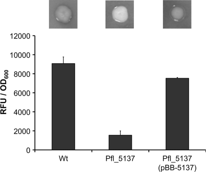
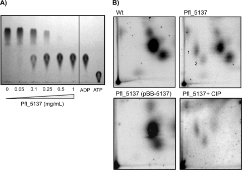
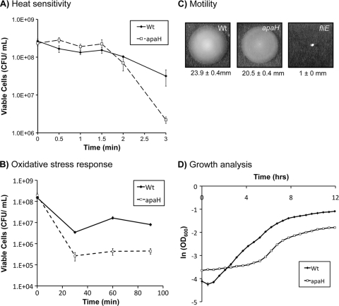
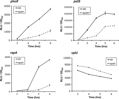
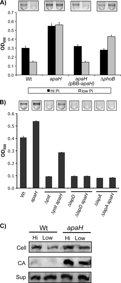
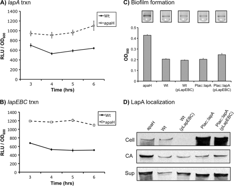
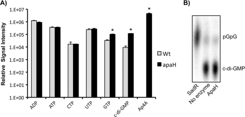
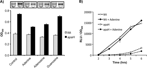
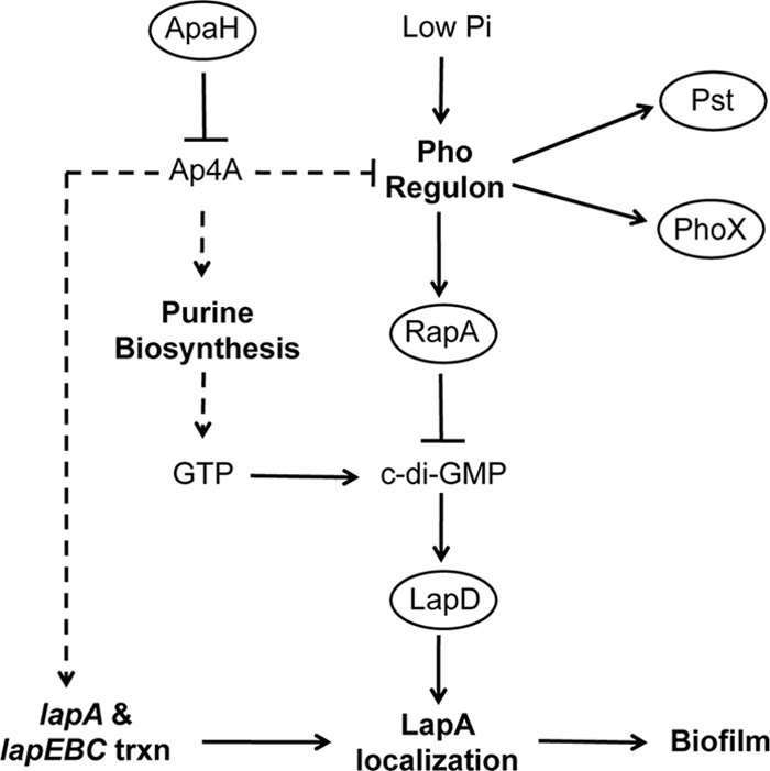
Dinucleoside tetraphosphates are common constituents of the cell and are thought to play diverse biological roles in organisms ranging from bacteria to humans. In this study we characterized two independent mechanisms by which di-adenosine tetraphosphate (Ap4A) metabolism impacts biofilm formation by Pseudomonas fluorescens. Null mutations in apaH, the gene encoding nucleoside tetraphosphate hydrolase, resulted in a marked increase in the cellular level of Ap4A. Concomitant with this increase, Pho regulon activation in low-inorganic-phosphate (P(i)) conditions was severely compromised. As a consequence, an apaH mutant was not sensitive to Pho regulon-dependent inhibition of biofilm formation. In addition, we characterized a Pho-independent role for Ap4A metabolism in regulation of biofilm formation. In P(i)-replete conditions Ap4A metabolism was found to impact expression and localization of LapA, the major adhesin regulating surface commitment by P. fluorescens. Increases in the level of c-di-GMP in the apaH mutant provided a likely explanation for increased localization of LapA to the outer membrane in response to elevated Ap4A concentrations. Increased levels of c-di-GMP in the apaH mutant were associated with increases in the level of GTP, suggesting that elevated levels of Ap4A may promote de novo purine biosynthesis. In support of this suggestion, supplementation with adenine could partially suppress the biofilm and c-di-GMP phenotypes of the apaH mutant. We hypothesize that changes in the substrate (GTP) concentration mediated by altered flux through nucleotide biosynthetic pathways may be a significant point of regulation for c-di-GMP biosynthesis and regulation of biofilm formation.


Figures

FIG. 1.
FIG. 1.
Alkaline phosphatase activity assays. For the top panel, strains were grown on a low-Pi (K10Tπ) medium containing the chromogenic phosphatase substrate 5-bromo-4-chloro-3-indolylphosphate (BCIP). The darkness of a colony indicates the relative phosphatase activity of the strain. For the bottom panel, phosphatase activity was quantified for each strain grown in low-Pi medium using fluorescent detection of substrate cleavage normalized to cell density and was expressed in relative fluorescence units (RFU)/OD600. The error bars indicate standard errors (n = 4). Wt, wild type.
FIG. 2.
FIG. 2.
The Pfl_5137 protein is a di-adenosine tetraphosphatase. (A) In vitro reactions to assess enzymatic cleavage of di-adenosine tetraphosphate (Ap4A) were performed with different concentrations of purified Pfl_5137 protein and commercially available Ap4A as the substrate. Reaction products were separated and visualized by 1D-TLC, and the standards included were ATP and ADP. Purified ApaH protein was shown to cleave Ap4A in a dose-dependent fashion. Cleavage was symmetrical, producing ADP as the sole cleavage product. (B) In vivo analysis of Ap4A concentrations. Cells were grown in Pi-limiting medium for 6 h before they were labeled with sodium dihydrogen [32P]orthophosphate, which was followed by acid extraction. Shown are autoradiographs of whole-cell acid extracts separated by 2D-TLC. The preparations analyzed were the wild-type strain (Wt), the Pfl_5137 mutant, the Pfl_5137 mutant complemented in trans with pBB-5137, and Pfl_5137 mutant extract treated with calf intestinal phosphatase (CIP) to cleave phosphate monoester bonds. The numbers indicate the reported locations of Ap4A (spot 1) and Ap4G (spot 2).
FIG. 3.
FIG. 3.
Conservation of known apaH mutant phenotypes in P. fluorescens. (A) Heat sensitivity. The wild-type (Wt) and apaH strains were challenged with a 50°C heat shock. Samples were recovered every 30 s, and plate counting was performed to ascertain the number of CFU. (B) Oxidative stress response. The wild type and the apaH mutant were challenged with 10 mM H2O2 for the times indicated, aliquots were recovered and neutralized, and plate counting was performed to ascertain the number of CFU. (C) Flagellum-mediated swimming. The wild type and the apaH mutant were inoculated onto 0.3% high-Pi (K10T-1) agar, and their abilities to swim away from the inoculation point were assessed. Swim diameters (mean ± standard error; n = 10) are indicated below the images. (D) Growth analysis. The optical density at 600 nm was measured for cultures of both the wild type and the apaH mutant during growth in high-Pi (K10T-1) medium. The natural log of OD600 was plotted against time. The growth rate, lag time, and yield were calculated as described in Materials and Methods.
FIG. 4.
FIG. 4.
apaH mutation inhibits Pho regulon expression. Transcriptional fusions coupling the promoters of phoX, pstS, rapA, and rplU to luciferase expression were constructed. Luciferase activity was recorded over time for each fusion in the wild-type (Wt) and apaH backgrounds during growth in low-Pi (K10Tπ) medium. The results are expressed in relative light units (RLU) normalized to the optical density of the culture at the time of analysis. The rate of the increase in light production is an indirect measure of transcriptional induction from a specific promoter. The phoX, rapA, and pstS genes are known members of the Pho regulon. The rplU transcriptional fusion was used as a Pho-independent control.
FIG. 5.
FIG. 5.
Analysis of biofilm formation by the apaH mutant. (A) Biofilm formation by the apaH mutant was assessed by comparison to biofilm formation by the wild type (Wt) for organisms grown in both high- and low-Pi conditions after incubation for 10 h. The biofilm phenotypes of the apaH strain were complemented by providing a wild-type copy of apaH on a plasmid (pBB-apaH). The phoB mutant was used as a positive control because it is unable to express the Pho regulon and forms biofilms regardless of the Pi concentration. The error bars indicate standard errors (n = 10). (B) The apaH allele was tested to determine its ability to rescue the biofilm defects of strains with null mutations in pst, lapD, and lapA. Strains were grown in high-Pi conditions for 6 h before attached bacteria were stained. The Δpst apaH strain showed partial rescue of biofilm formation compared to the pst single mutant. The ΔlapA apaH and ΔlapD apaH strains did not show increases in biofilm formation compared to the lapA and lapD single mutants, respectively. The error bars indicate standard errors (n = 10). (C) The apaH mutation results in accumulation of LapA at the cell surface: Western blot detection of LapA-HA for whole-cell (Cell), cell surface-associated (CA), and supernatant (Sup) fractions prepared from both the wild type and the apaH mutant grown in both high- and low-Pi-conditions.
FIG. 6.
FIG. 6.
Increased lapA and lapEBC transcription does not explain increased biofilm formation by the apaH mutant. (A and B) Effects of the apaH mutation on lapA transcription (trxn) (A) and lapEBC transcription (B). Luciferase fusions were constructed for the lapA and lapEBC promoters and integrated into the native chromosomal location to generate a merodiploid. Luciferase activity was measured over time for strains grown in high-Pi (K10T-1) medium. The error bars indicate standard errors (n = 3). RLU, relative light units; Wt, wild type. (C) Effect of expression of lapA and lapEBC from heterologous promoters on biofilm formation. Biofilms were incubated for 8 h before visualization by crystal violet staining. Levels of biofilm formation are shown. The error bars indicate standard errors (n = 10). (D) Effect of expression of lapA and lapEBC from heterologous promoters on LapA secretion and localization: Western blot detection of LapA-HA for whole-cell (Cell), cell surface-associated (CA), and supernatant (Sup) fractions prepared from both the wild type and the apaH mutant grown in high-Pi conditions.
FIG. 7.
FIG. 7.
Effect of the apaH mutation on nucleotide pools. (A) Analysis of nucleotide pools by mass spectrometry. Wild-type (Wt) and apaH mutant cultures were grown in triplicate for 5 to 6 h in high-Pi (K10T-1) medium before extraction of nucleotides and relative quantification by liquid chromatography-tandem mass spectrometry (LC-MS/MS). Extracts were analyzed to determine the levels of ADP, ATP, CTP, UTP, GTP, c-di-GMP, and Ap4A. The error bars indicate standard errors (n = 3). For statistical inference with Student's t tests, alpha (α) was set at 0.0071 after adjustment for multiple comparisons using a Bonferroni correction (α = 0.05/7). Statistically significant differences are indicated by an asterisk. Ap4A could not be detected in wild-type extracts. Using this assay, the limit of detection for Ap4A is ∼20 ng/ml. (B) Assay for c-di-GMP phosphodiesterase activity. Purified ApaH (5 mg/ml) was unable to cleave c-di-GMP. SadR was used as a positive control for PDE activity, which completely cleaved c-di-GMP to generate pGpG.
FIG. 8.
FIG. 8.
Purine metabolism and apaH mutant phenotypes. (A) Supplementation of biofilm formation assay mixtures with exogenous adenine, adenosine, and guanosine to a final concentration of 1 mM. Biofilm assay mixtures were incubated for 8 h before visualization by crystal violet staining. The levels of biofilm formation are shown. The error bars indicate standard errors (n = 10). (B) Effect of addition of adenine on Pho regulon activation. The wild type and the apaH mutant expressing the phoX luciferase promoter fusion were monitored for Pho activation during growth in low-Pi (K10Tπ) medium. The error bars indicate standard errors (n = 3). RLU, relative light units; Wt, wild type.
FIG. 9.
FIG. 9.
Summary of the current model for the role of ApaH in biofilm formation by P. fluorescens. Loss of the ApaH function and subsequent accumulation of Ap4A promote biofilm formation by two mechanisms. (i) Accumulation of Ap4A prevents efficient recycling of ADP, which in turn promotes de novo purine biosynthesis. This leads to increased levels of GTP and subsequent increases in the levels of c-di-GMP through the action of diguanylate cyclases. Higher levels of c-di-GMP result in increased biofilm formation by promoting localization of the adhesin LapA to the cell surface via LapD. Ap4A also promotes expression of LapA and its transporter, LapEBC, which in conjunction with increases in the level of c-di-GMP, contribute to increased biofilm formation. (ii) In low-Pi environments biofilm formation is inhibited through expression of RapA, a c-di-GMP phosphodiesterase that is a member of the Pho regulon. High levels of Ap4A inhibit activation of the Pho regulon and suppress the loss of biofilm formation. Ellipses indicate a protein, bold type indicates a biological process, and normal type indicates a small molecule. Solid arrows indicate that there is experimental evidence for direct interactions, whereas dashed lines indicate interactions that may be either direct or indirect.

Similar articles

Cited by

References

    1. Baker, J. C., and M. K. Jacobson. 1986. Alteration of adenyl dinucleotide metabolism by environmental stress. Proc. Natl. Acad. Sci. U. S. A. 83:2350-2352. - PMC - PubMed
    1. Bertani, G. 2004. Lysogeny at mid-twentieth century: P1, P2, and other experimental systems. J. Bacteriol. 186:595-600. - PMC - PubMed
    1. Blanchin-Roland, S., S. Blanquet, J. M. Schmitter, and G. Fayat. 1986. The gene for Escherichia coli diadenosine tetraphosphatase is located immediately clockwise to folA and forms an operon with ksgA. Mol. Gen. Genet. 205:515-522. - PubMed
    1. Bochner, B. R., and B. N. Ames. 1982. Complete analysis of cellular nucleotides by two-dimensional thin layer chromatography. J. Biol. Chem. 257:9759-9769. - PubMed
    1. Bochner, B. R., P. C. Lee, S. W. Wilson, C. W. Cutler, and B. N. Ames. 1984. AppppA and related adenylylated nucleotides are synthesized as a consequence of oxidation stress. Cell 37:225-232. - PubMed

Publication types

MeSH terms

LinkOut - more resources