. 2010 Apr;30(8):1898-909.
doi: 10.1128/MCB.01199-09. Epub 2010 Feb 12.

The chaperone-like protein HYPK acts together with NatA in cotranslational N-terminal acetylation and prevention of Huntingtin aggregation

Affiliations

The chaperone-like protein HYPK acts together with NatA in cotranslational N-terminal acetylation and prevention of Huntingtin aggregation

Thomas Arnesen et al. Mol Cell Biol. 2010 Apr.

Abstract

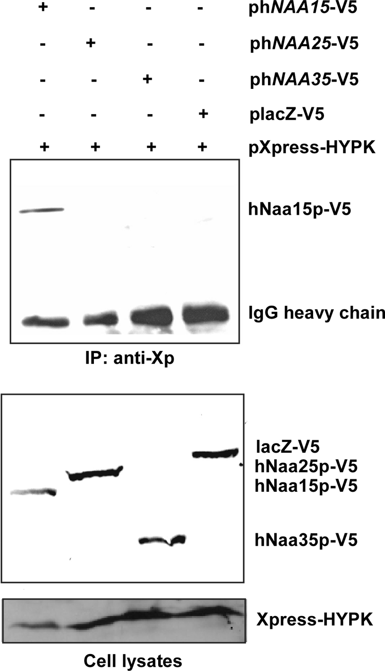
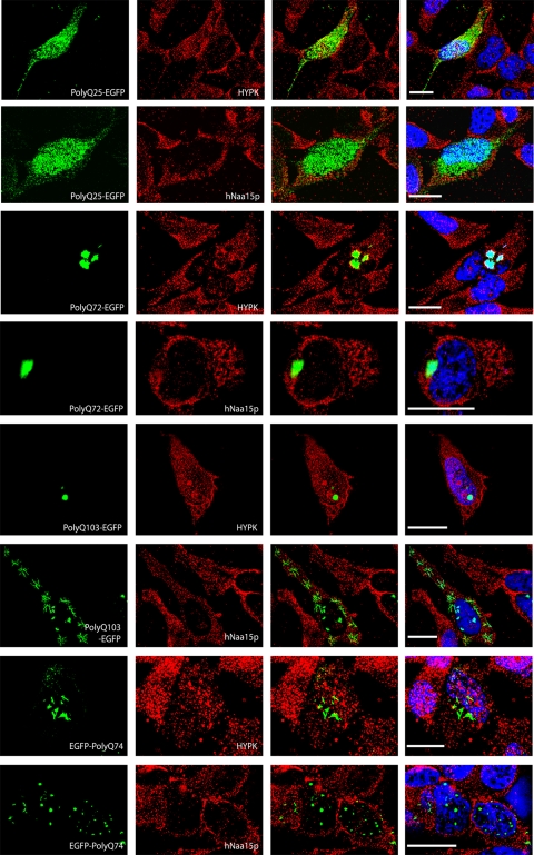
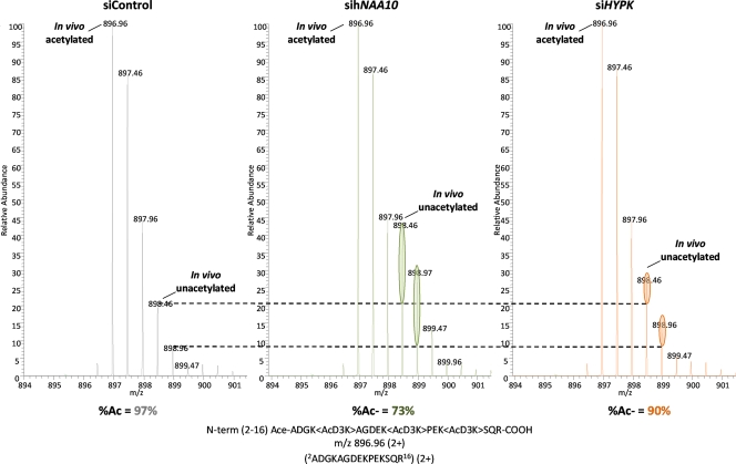
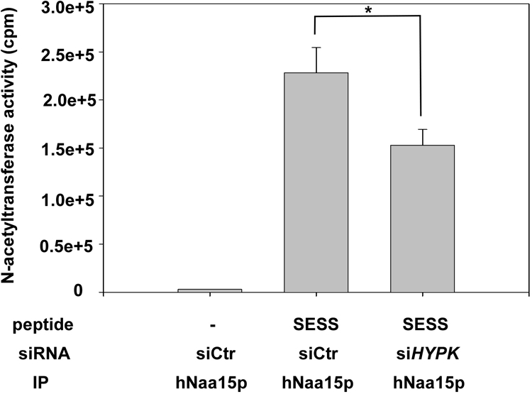
The human NatA protein N(alpha)-terminal-acetyltransferase complex is responsible for cotranslational N-terminal acetylation of proteins with Ser, Ala, Thr, Gly, and Val N termini. The NatA complex is composed of the catalytic subunit hNaa10p (hArd1) and the auxiliary subunit hNaa15p (hNat1/NATH). Using immunoprecipitation coupled with mass spectrometry, we identified endogenous HYPK, a Huntingtin (Htt)-interacting protein, as a novel stable interactor of NatA. HYPK has chaperone-like properties preventing Htt aggregation. HYPK, hNaa10p, and hNaa15p were associated with polysome fractions, indicating a function of HYPK associated with the NatA complex during protein translation. Knockdown of both hNAA10 and hNAA15 decreased HYPK protein levels, possibly indicating that NatA is required for the stability of HYPK. The biological importance of HYPK was evident from HYPK-knockdown HeLa cells displaying apoptosis and cell cycle arrest in the G(0)/G(1) phase. Knockdown of HYPK or hNAA10 resulted in increased aggregation of an Htt-enhanced green fluorescent protein (Htt-EGFP) fusion with expanded polyglutamine stretches, suggesting that both HYPK and NatA prevent Htt aggregation. Furthermore, we demonstrated that HYPK is required for N-terminal acetylation of the known in vivo NatA substrate protein PCNP. Taken together, the data indicate that the physical interaction between HYPK and NatA seems to be of functional importance both for Htt aggregation and for N-terminal acetylation.


Figures

FIG. 1.
FIG. 1.
The C terminus of hNaa15p interacts directly with HYPK. (A) Purified MBP-hNaa15p fusion proteins containing N-terminal MBP and a His tag fused to a segment of the hNaa15p protein, as indicated, were purified and bound to Ni-affinity beads. HYPK was separately fully purified and added to the beads containing different MBP-hNaa15p fusions. After incubation with rotation for 10 min and several washing steps, the MBP-hNaa15p beads were analyzed for HYPK binding by SDS-PAGE and Western blotting using anti-HYPK. A positive signal indicates that purified HYPK binds to the MBP-hNaa15p fusion protein. Equal amounts of the different MBP-hNaa15p fusions used were confirmed by SDS-PAGE and Coomassie staining. (B) Schematic model of the predicted domains of hNaa15p and HYPK and the potential regions involved in the hNaa15p-HYPK interaction. TPR, tetratricopeptide repeat.
FIG. 2.
FIG. 2.
Subcellular localization of HYPK. (A) (Left) HeLa cells were transfected with a plasmid encoding Xpress-HYPK, and at 48 h posttransfection, cells were fixed and anti-Xpress antibodies and Alexa 488-conjugated anti-mouse antibodies were used to visualize Xpress-HYPK. (Middle) DAPI (4′,6-diamidino-2-phenylindole) staining was used to visualize the nuclei of the cells. (Right) Overlay of DAPI and Xpress-HYPK signals. More than 200 transfected cells were inspected, and representative cells are shown. (B) Twenty-four hours after being seeded, HeLa cells were fixed and anti-HYPK and Alexa 488-conjugated anti-rabbit antibodies were used to visualize endogenous HYPK. Rabbit IgG was used as an unspecific negative control. DAPI staining is presented next to each sample to visualize nuclei and verify the presence of cells. Pictures are representative of three independent experiments in which at least 100 cells were taken into account.
FIG. 3.
FIG. 3.
HYPK cosediments with polysomal fractions in a salt-sensitive manner. Polysomal pellets from HeLa cells were resuspended in buffer containing increasing concentrations of KCl. Cell lysate and polysomal pellets after KCl treatment were analyzed by SDS-PAGE and Western blotting. The membrane was incubated with anti-HYPK, anti-hNaa15p, anti-L26 (ribosomal protein), and anti-CytC antibodies. Molecular mass markers (in kDa) are indicated on the left. Results shown are representative of three independent experiments.
FIG. 4.
FIG. 4.
HYPK-hNaa15p interaction is independent of active translation. HEK293 cells were cotransfected with plasmids expressing Xpress-HYPK and hNaa15p-V5. At 48 h posttransfection, cells were treated with 50 μg/ml cycloheximide (CHX) or solvent (control) for 30 min. The cells were then harvested and subjected to immunoprecipitation (IP), using anti-Xpress or negative-control antibodies. The presence of hNaa15-V5 in complex with Xpress-HYPK was analyzed by SDS-PAGE and Western blotting, using anti-V5. The inhibitory effect of CHX on protein translation was verified in parallel samples detecting the reduction of unstable proteins. The results shown are representative of three independent experiments.
FIG. 5.
FIG. 5.
HYPK interacts specifically with hNaa15p of hNatA, not with hNaa25p of hNatB and hNaa35p of hNatC. HEK293 cells were cotransfected with plasmids encoding Xpress-HYPK plus hNaa15p-V5, hNaa25p-V5, hNaa35p-V5, or lacZ-V5, as indicated. At 48 h posttransfection, cells were harvested and subjected to immunoprecipitation using anti-Xpress (anti-Xp) antibodies. The presence of hNaa15p-V5, hNaa25p-V5, and hNaa35p-V5 in complex with Xpress-HYPK was investigated by SDS-PAGE and Western blotting, using anti-V5. (Top) Western blot of immunoprecipitates. (Bottom) Western blot of cell lysates prior to immunoprecipitation. The results shown are representative of three independent experiments.
FIG. 6.
FIG. 6.
siRNA-mediated knockdown of HYPK, hNAA10, and hNAA15. (A) Five individual siRNAs potentially targeting HYPK expression (H1 to H5) were tested by siRNA transfection of HeLa cells and subsequent RT-PCR analysis at 48 h posttransfection, using primers specific for HYPK and β-actin. G, siGAPDH (negative control). (B) siRNA-mediated knockdown in HeLa cells of the specific genes HYPK, hNAA10, and hNAA15, as indicated. siLamin and siGAPDH were used as negative controls. siHYPK is an equal mixture of the four effective siHYPK siRNAs shown in panel A (H1, H2, H4, and H5; 50 nM total concentration). RT-PCR analysis was performed at 72 h posttransfection. (C) SDS-PAGE and Western blotting, using anti-HYPK, anti-hNaa10p, anti-hNaa15p, and antiactin, of HeLa samples treated identically to those described for panel B. Protein levels were quantified using a Fuji Film IR LAS 1000 documentation system and Image Gauge 3.45. Protein levels in siLamin- and siGAPDH-treated cells (the mean for these two negative controls) were set to 1.0, and protein levels in sihNAA10-, sihNAA15-, and siHYPK-treated cells were estimated relative to this and normalized to actin levels. HYPK appeared as one or two specific bands, and the sum of the two was used for quantification. All results are representative of at least three independent experiments.
FIG. 7.
FIG. 7.
HYPK knockdown induces cell death and G1/G0 cell cycle arrest. Flow cytometric cell cycle analysis of HYPK, hNAA10, and hNAA15 knockdown cells was performed at 72 h posttransfection. siLamin- and siGAPDH-treated samples were used as negative controls. (Top panels) No ZVAD treatment. (Bottom panels) ZVAD treatment to inhibit caspase activity and, thereby, induction of caspase-mediated cell death. Experiments were performed three times, and representative values are given.
FIG. 8.
FIG. 8.
HYPK and hNAA10 knockdown increases Huntingtin aggregation. HeLa cells were transfected with the indicated siRNAs, and at 24 h posttransfection, the medium was replaced and the cells were transfected with plasmids encoding the indicated Htt-EGFP fusion proteins. Twenty-four hours after plasmid transfection, the cells were fixed, cells displaying Htt-EGFP aggregates were counted, and the percentage of cells with aggregates among the transfected cells was calculated. At least 200 transfected cells were counted per sample, and values presented are means ± standard deviations (SD) for three to six independent experiments. P values for independent t tests for samples versus control are indicated with asterisks (P < 0.05).
FIG. 9.
FIG. 9.
HYPK and hNaa15p do not colocalize with Htt aggregates. HeLa cells expressing various EGFP-tagged polyQ versions (left column) were incubated with HYPK- or hNaa15p-specific antibodies, as indicated in the figure (second column from left). Alexa 594-conjugated anti-mouse antibodies were used to visualize HYPK and hNaa15p. The third column from left presents an overlay of polyQ-EGFP and HYPK or hNaa15p. DAPI staining was used to visualize DNA, as included in the overlay shown in the right column. Images were processed by deconvolution (Leica 4000 software). Bar, 25 μm.
FIG. 10.
FIG. 10.
HYPK knockdown reduces NatA-mediated N-terminal acetylation. HeLa cells were transfected with siRNAs targeting hNAA10, HYPK, or a control. At 24 h posttransfection, the medium was replaced and cells transfected with pPCNP-V5 plasmid. Forty-eight hours after plasmid transfection, the cells were harvested and lysed, and PCNP-V5 was immunoprecipitated and processed as described in Materials and Methods. ZVAD was added every 24 h to prevent induction of apoptosis. MS spectra of doubly charged peptide ions originating from the N terminus of PCNP (GenBank accession no. Q8WW12) are shown. The peptide was identified as 2-ADGKAGDEKPEKSQR-16. The extent of PCNP Nα acetylation, as determined by an MS isotope pattern calculator (http://prospector.ucsf.edu), was calculated as being 97%, 73%, and 90% in the control, sihNAA10, and siHYPK samples, respectively. The ellipsoids indicate the relative increases of the in vivo unacetylated N terminus of PCNP in the sihNAA10- and siHYPK-treated samples compared to that in the control sample. The data are representative of two independent experiments.
FIG. 11.
FIG. 11.
hNatA immunoprecipitated from HYPK knockdown cells displays reduced in vitro acetylation activity. HeLa cells (approximately 5 × 10⁶ per sample) were transfected with the indicated siRNAs, and at 24 h posttransfection, ZVAD was added to prevent induction of apoptosis. After 48 h, the cells were harvested and lysed, and the lysate was subjected to immunoprecipitation (IP) using an anti-hNaa15p specific antibody. The beads containing functional hNatA complexes were analyzed for Nα-acetyltransferase activity, using [14C]acetyl-CoA and a Ser-Asp-Ser-Ser (SESS)-starting 24-mer peptide known to be acetylated by NatA in vitro. The amount of acetyl incorporation was determined by isolation of the peptides followed by scintillation counting. Verification of knockdown and the presence of equal levels of hNaa15p in the immunoprecipitates were routinely confirmed. Experiments were performed three times, and values are means ± SD. P values for independent t tests for siHYPK samples versus control are indicated with an asterisk (P < 0.05).

Similar articles

Cited by

References

    1. Aiken, C. T., J. S. Steffan, C. M. Guerrero, H. Khashwji, T. Lukacsovich, D. Simmons, J. M. Purcell, K. Menhaji, Y. Z. Zhu, K. Green, F. Laferla, L. Huang, L. M. Thompson, and J. L. Marsh. 2009. Phosphorylation of threonine-3: implications for huntingtin aggregation and neurotoxicity. J. Biol. Chem. 284:29427-29436. - PMC - PubMed
    1. Ametzazurra, A., E. Larrea, M. P. Civeira, J. Prieto, and R. Aldabe. 2008. Implication of human N-alpha-acetyltransferase 5 in cellular proliferation and carcinogenesis. Oncogene 27:7296-7306. - PubMed
    1. Anderson, D. C., W. Li, D. G. Payan, and W. S. Noble. 2003. A new algorithm for the evaluation of shotgun peptide sequencing in proteomics: support vector machine classification of peptide MS/MS spectra and SEQUEST scores. J. Proteome Res. 2:137-146. - PubMed
    1. Arnesen, T., D. Anderson, C. Baldersheim, M. Lanotte, J. E. Varhaug, and J. R. Lillehaug. 2005. Identification and characterization of the human ARD1-NATH protein acetyltransferase complex. Biochem. J. 386:433-443. - PMC - PubMed
    1. Arnesen, T., D. Anderson, J. Torsvik, H. B. Halseth, J. E. Varhaug, and J. R. Lillehaug. 2006. Cloning and characterization of hNAT5/hSAN: an evolutionarily conserved component of the NatA protein N-alpha-acetyltransferase complex. Gene 371:291-295. - PubMed

Publication types

MeSH terms

LinkOut - more resources