. 2010 Apr 8;115(14):2852-63.
doi: 10.1182/blood-2009-10-230060. Epub 2010 Feb 12.

STAT3 is constitutively phosphorylated on serine 727 residues, binds DNA, and activates transcription in CLL cells

Affiliations

STAT3 is constitutively phosphorylated on serine 727 residues, binds DNA, and activates transcription in CLL cells

Inbal Hazan-Halevy et al. Blood. .

Abstract

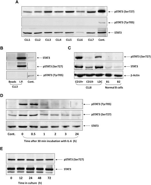
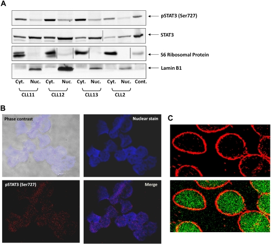
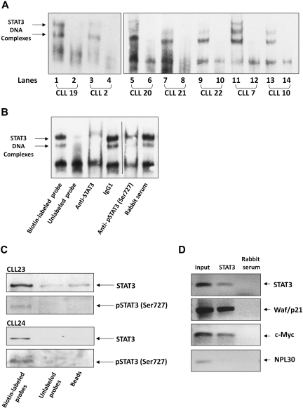
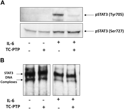
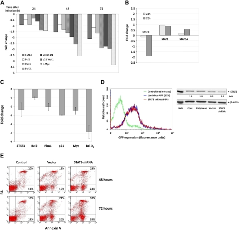
Chronic lymphocytic leukemia (CLL) is the most common leukemia in the Western hemisphere, but its pathogenesis is still poorly understood. Constitutive tyrosine phosphorylation (p) of signal transducer and activator of transcription (STAT) 3 occurs in several solid tumors and hematologic malignancies. In CLL, however, STAT3 is constitutively phosphorylated on serine 727, not tyrosine 705, residues. Because the biologic significance of serine pSTAT3 in CLL is not known, we studied peripheral blood cells of 106 patients with CLL and found that, although tyrosine pSTAT3 was inducible, serine pSTAT3 was constitutive in all patients studied, regardless of blood count, disease stage, or treatment status. In addition, we demonstrated that constitutive serine pSTAT3 translocates to the nucleus by the karyopherin-beta nucleocytoplasmic system and binds DNA. Dephosphorylation of inducible tyrosine pSTAT3 did not affect STAT3-DNA binding, suggesting that constitutive serine pSTAT3 binds DNA. Furthermore, infection of CLL cells with lentiviral STAT3-small hairpin RNA reduced the expression of several STAT3-regulated survival and proliferation genes and induced apoptosis, suggesting that constitutive serine pSTAT3 initiates transcription in CLL cells. Taken together, our data suggest that constitutive phosphorylation of STAT3 on serine 727 residues is a hallmark of CLL and that STAT3 be considered a therapeutic target in this disease.


Figures

Figure 1
Figure 1
STAT3 is constitutively phosphorylated on serine 727 residues in CLL cells. (A) Cell lysates of patients with CLL (CLL 1-7) and of A549 cells (control) were analyzed by Western immunoblotting using antiserine pSTAT3, antityrosine pSTAT3, or anti-STAT3 antibodies. (B) Cell lysates of CLL patient 3 were incubated with rabbit anti–human STAT3 antibodies for immunoprecipitation (I.P.) with protein A-agarose beads. Incubation of cell lysates with beads only was used as a negative control (beads). The immunoprecipitate was analyzed by Western immunoblotting using antiserine pSTAT3, antityrosine pSTAT3, or anti-pSTAT3 antibodies. 3T3 cells were used as a positive control (Cont.). (C) PB cells of CLL patient 8 were fractioned using immunomagnetic beads. CD19+ and CD19 cells were collected. Normal PB CD19+/CD20+ B cells were purified by immunomagnetic beads. Expression of serine pSTAT3 in CLL low-density cells (LDC), CD19+, and CD19 cells and normal donor B cells (B1 and B2) was analyzed by Western immunoblotting using antiserine pSTAT3, anti-STAT3, or anti–β-actin (loading control) antibodies. (D) CLL cells (CLL 9) were stimulated with IL-6 (50 ng/mL, 30 minutes) or left untreated. The cells were harvested at different time points after IL-6 stimulation. Cell lysates were analyzed by Western immunoblotting using antityrosine pSTAT3, antiserine pSTAT3, or anti-STAT3 antibodies. (E) CLL cells (CLL 10) were cultured in medium for 12 to 72 hours, the cells were harvested, and cell lysates were analyzed by Western immunoblotting using antiserine pSTAT3 or anti-STAT3 antibodies. Vertical lines indicate realignment of the same gel's image.
Figure 2
Figure 2
Serine pSTAT3 translocates to the nucleus. (A) Cytoplasmic (Cyt.) and nuclear (Nuc.) fractions were extracted from CLL cells of patients 2, 11, 12, and 13, as described in “Isolation of nuclear and cytoplasmic extracts,” and analyzed by Western immunoblotting using antiserine pSTAT3 and anti-STAT3 antibodies. Adequate fractionation of cytoplasmic and nuclear extracts was confirmed using anti-S6 ribosomal protein and antilamin B1 antibodies. (B) Confocal microscopic images of freshly isolated CLL cells (CLL 14 and 15) that were cytospun and fixed on glass slides, as described in “Confocal microscopy.” As shown (original magnification × 400), the slides were stained with Alexa Fluor 488–mouse antiphosphoserine (serine 727)-STAT3 antibodies (red dots; bottom left corner) and the nuclear stain TOPRO3 (blue; top right corner). Serine pSTAT3 was detected in the nucleus of CLL cells (red dots overlapping blue; bottom right corner). (C) Using a different approach, the slides were stained with antiphosphoserine (serine 727)-STAT3 Alexa Fluor 488–mouse antibodies (green dots) and ribosomal protein S6, followed by phycoerythrin-conjugated rabbit anti–mouse antibodies (red dots). Serine pSTAT3 was detected in the nucleus (green dots) and the cytoplasm (yellow dots represent colocalization of ribosomal protein S6 and serine pSTAT3; X-1000). Vertical lines indicate realignment of the same gel's image. Images were acquired using an Olympus Flowview FV500/X81 system using Flowview software and a 60× oil immersion lens, and cropped and revised using Microsoft Office PowerPoint 2003.
Figure 3
Figure 3
Serine pSTAT3 binds to the nucleocytoplasmic transport proteins importin-β1 and CRM1. Total cell, cytoplasmic (Cyt), and nuclear (Nuc) extracts were obtained from CLL cells (CLL 16 and 17), as described in “Isolation of nuclear and cytoplasmic extracts.” Whole-cell extracts, Cyt, and Nuc fractions were immunoprecipitated (I.P.) with anti-STAT3 (A), -importin-β1 (B), and -CRM1 (C) antibodies using protein A–agarose beads. Incubation with beads only (B) was used as a negative control. The immune complex was separated by SDS-PAGE and analyzed by Western immunoblotting using the indicated antibodies. Expression of STAT3, serine pSTAT3, importin-β1, importin-α1, -α3, -α6, -α5 and 7, and CRM1 in total cell extracts of CLL cells was used as a positive control. (D) Freshly isolated CLL 18 cells were incubated for 3 hours with increasing concentrations (2.5-12.5nM) of the CRM1 inhibitor leptomycin B. Nuclear extracts were fractionated and analyzed by Western immunoblotting using anti-STAT3 and antilamin B1 (nuclear protein control) antibodies. Vertical lines indicate realignment of the same gel's image.
Figure 4
Figure 4
Serine pSTAT3 binds DNA. (A) Nuclear extracts from 7 CLL patients (CLL 2, 7, 10, and 19-22) were analyzed using EMSA, as described in “EMSA.” Binding of STAT3 to biotin-labeled DNA probes is shown (lanes 1, 3, 5, 7, 9, 11, and 13). To compete with the binding, an unlabeled STAT3 binding-site DNA probe was added to the reaction in 100 times molar excess (lanes 2, 4, 6, 8, 10, 12, and 14). The 2 STAT3-DNA complexes are marked by arrows. (B) Elimination of STAT3-DNA binding by antibodies to STAT3 or serine pSTAT3. Nuclear extract of CLL patient 9 was studied by EMSA. The assay was conducted with biotin-labeled STAT3 binding-site DNA probe (lane 1) or with unlabeled probe (lane 2), with the addition of mouse anti–human STAT3 antibodies (lane 3), mouse immunoglobulin G1 isotype control (lane 4), rabbit anti–human serine pSTAT3 (lane 5), or rabbit serum (lane 6). Vertical line indicates realignment of the gel's image. (C) Pull-down of STAT3 by biotin-labeled DNA probe. Nuclear extract from CLL 23 and 24 was incubated with biotin-labeled STAT3 binding-site DNA probe and agarose-conjugated streptavidin beads. The attached proteins were separated by SDS electrophoresis, and STAT3 and serine pSTAT3 were detected by Western blot analysis (Biotin-labeled probe). Incubation of nuclear extracts with unlabeled STAT3 binding-site DNA probes (Unlabeled probe) and agarose-conjugated streptavidin beads only (Beads) were used as negative controls. (D) ChIP assay of CLL cells. CLL cell-derived chromatin was immunoprecipitated with STAT3 or rabbit serum (control). The coimmunoprecipitated DNA was amplified by PCR using upstream promoter constructs of STAT3, waf1/p21, c-Myc, or the control gene RPL30. As shown, STAT3-regulated (STAT3, waf1/p21, and c-Myc), but not control (RPL30), genes were amplified. RPL30 as well as STAT3, waf1/p21, and c-Myc genes were detected in whole cell chromatin-extracted DNA (Input).
Figure 5
Figure 5
TC-PTP dephosphorylates IL-6–induced tyrosine pSTAT3 but does not affect STAT3-DNA binding. CLL cells (CLL 16 and 25) were incubated for 30 minutes, with or without 50 ng/mL IL-6. Nuclear fractions were extracted and treated with TC-PTP for 30 minutes, as described in “EMSA.” After TC-PTP treatment, nuclear extracts were analyzed by (A) Western blot analysis using antityrosine pSTAT3 and antiserine pSTAT3 to detect the corresponding proteins or (B) EMSA to detect binding of STAT3 to the biotin-labeled DNA probe, as described in “EMSA.”
Figure 6
Figure 6
Serine pSTAT3 initiates transcription of STAT3-regulated genes in CLL cells. CLL cells were infected with a lentivirus harboring GFP-STAT3-shRNA or with lentivirus GFP-empty vector. (A) RT-PCR results of mRNA levels of STAT3 and the STAT3-regulated genes Bcl2, Pim1, Bcl-XL, Cyclin D1, p21 (Waf1), and c-Myc in CLL cells (CLL 26) that were infected by lentivirus harboring GFP-STAT3-shRNA for 24, 48, and 72 hours. (B) RT-PCR results of mRNA levels of STAT1, STAT3, and STAT5 in CLL cells (CLL 26) that were infected by lentivirus harboring GFP-STAT3-shRNA for 24 and 72 hours. The RT-PCR results are shown as fold change (decrease or increase) relative to the mRNA levels of the STAT3-regulated genes in CLL cells that were infected with lentiviral GFP-empty vector. (C) RT-PCR results of mRNA levels of the STAT3-regulated genes STAT3, Bcl2, Pim1, Bcl-XL, p21 (Waf1), and c-Myc in CLL cells (CLL 15, 19, 24, and 27). Changes in mRNA levels (mean ± SD) are shown. As in the previous experiment, infection with empty virus did not significantly affect STAT3-regulated gene levels. (D) Left panel: Infection efficiency analyzed by flow cytometry of control and GFP+ empty virus– and STAT3-shRNA–infected CLL cells (CLL 26). Right panel: Western blot of control, empty vector- and STAT3 shRNA-infected CLL cells. Forty-eight hours after infection with STAT3, shRNA down-regulated STAT3 protein levels by 50%. (E) Flow cytometric analysis of PI+/− and annexin V+ cells 48 and 72 hours after infection with GFP-STAT3-shRNA or empty vector. The percentages of early apoptotic cells are shown in the bottom right corners and of late apoptotic (PI and annexin V+) cells in the top right corners.

Comment in

Similar articles

Cited by

References

    1. Yee KW, O'Brien SM. Chronic lymphocytic leukemia: diagnosis and treatment. Mayo Clin Proc. 2006;81(8):1105–1129. - PubMed
    1. Chiorazzi N, Rai KR, Ferrarini M. Chronic lymphocytic leukemia. N Engl J Med. 2005;352(8):804–815. - PubMed
    1. Messmer BT, Messmer D, Allen SL, et al. In vivo measurements document the dynamic cellular kinetics of chronic lymphocytic leukemia B cells. J Clin Invest. 2005;115(3):755–764. - PMC - PubMed
    1. Bueso-Ramos CE, Ferrajoli A, Medeiros LJ, Keating MJ, Estrov Z. Aberrant morphology, proliferation, and apoptosis of B-cell chronic lymphocytic leukemia cells. Hematology. 2004;9(4):279–286. - PubMed
    1. Calin GA, Croce CM. Genomics of chronic lymphocytic leukemia microRNAs as new players with clinical significance. Semin Oncol. 2006;33(2):167–173. - PubMed

Publication types

MeSH terms