. 2010 Apr 8;115(14):2928-37.
doi: 10.1182/blood-2009-06-227629. Epub 2010 Feb 12.

Perturbed hematopoiesis in the Tc1 mouse model of Down syndrome

Affiliations

Perturbed hematopoiesis in the Tc1 mouse model of Down syndrome

Kate A Alford et al. Blood. .

Abstract

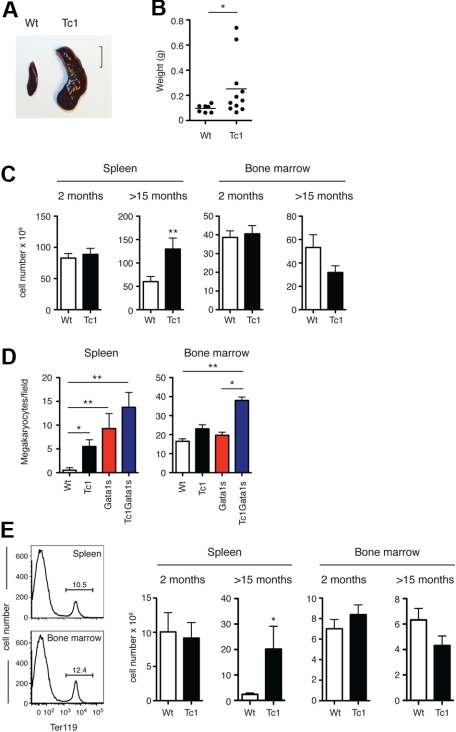
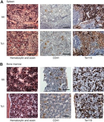
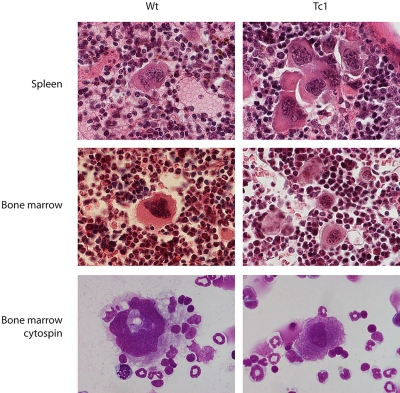
Trisomy of human chromosome 21 (Hsa21) results in Down syndrome (DS), a disorder that affects many aspects of physiology, including hematopoiesis. DS children have greatly increased rates of acute lymphoblastic leukemia and acute megakaryoblastic leukemia (AMKL); DS newborns present with transient myeloproliferative disorder (TMD), a preleukemic form of AMKL. TMD and DS-AMKL almost always carry an acquired mutation in GATA1 resulting in exclusive synthesis of a truncated protein (GATA1s), suggesting that both trisomy 21 and GATA1 mutations are required for leukemogenesis. To gain further understanding of how Hsa21 contributes to hematopoietic abnormalities, we examined the Tc1 mouse model of DS, which carries an almost complete freely segregating copy of Hsa21, and is the most complete model of DS available. We show that although Tc1 mice do not develop leukemia, they have macrocytic anemia and increased extramedullary hematopoiesis. Introduction of GATA1s into Tc1 mice resulted in a synergistic increase in megakaryopoiesis, but did not result in leukemia or a TMD-like phenotype, demonstrating that GATA1s and trisomy of approximately 80% of Hsa21 perturb megakaryopoiesis but are insufficient to induce leukemia.


Figures

Figure 1
Figure 1
Tc1 mice present with macrocytic anemia throughout life. (A) Graphs of mean (± SEM) red blood cell (RBC), white blood cell (WBC), and platelet counts and mean corpuscular volume (MCV) of neonate (< 1 day old) Tc1 and wild-type (Wt) mice (n = 6). (B) Graphs show mean (± SEM) RBC, WBC, and platelet counts and MCV as a function of age of Tc1 and wild-type mice (n = 15). Tc1 mice show decreased RBC counts from 8 to 52 weeks of age and increased MCV from 4 to 52 weeks of age (P < .05). (C) Low-power (left) and high-power (right) views of Wright-Giemsa–stained blood smears from Tc1 mice aged 64 weeks showing clouds of platelets.
Figure 2
Figure 2
Splenomegaly and extramedullary hematopoiesis in Tc1 mice older than 15 months. (A) Photograph shows appearance of an enlarged spleen found in a Tc1 mouse aged 15 months, alongside a spleen from a wild-type (Wt) age-matched control mouse. Size marker indicates 1 cm. (B) Graph shows weights of spleens taken from mice aged 15 months or older. Horizontal bar indicates the mean. (C) Graphs showing mean (± SEM) total number of cells in the spleen and bone marrow of mice of the indicated genotypes and ages (n = 6-11). (D) Graphs showing mean (± SEM) number of megakaryocytes per microscope field (40× objective) in mice aged older than 15 months (n = 5). (E) Histograms of flow cytometric analysis of surface levels of Ter119 expression on spleen and bone marrow cells from a Wt mouse at 2 months of age. Numbers indicate percentage of cells falling into the indicated markers. Graphs show mean (± SEM) numbers of Ter119+ cells in the spleens and bone marrow of mice of the indicated ages and genotypes (2 months, n = 11 [Wt], n = 10 [Tc1]; > 15 months, n = 7 [Wt], n = 11 [Tc1]). *P < .05, **P < .01.
Figure 3
Figure 3
Increased megakaryopoiesis in Tc1 mice older than 15 months. (A) Photomicrographs of sections of spleens from a Tc1 mouse with an enlarged spleen and a wild-type (Wt) control mouse, both aged 15 months. Sections were stained with hematoxylin and eosin, or with antibodies to CD41 or Ter119. The enlarged Tc1 spleen shows an increased number of megakaryocytes seen both in the hematoxylin and eosin and anti-CD41 stains. (B) Bone marrow sections from Wt and Tc1 mice stained as in panel A. Scale bars in panels A and B indicate 100 μm.
Figure 4
Figure 4
Megakaryocytes in Tc1 mice are less mature. Photomicrographs of sections of spleen and bone marrow stained with hematoxylin and eosin and cytospins of bone marrow cells stained with May-Grünwald and Giemsa stains, from Wt and Tc1 mice aged 15 months.
Figure 5
Figure 5
Analysis of hematopoietic progenitors in Tc1 mice. (A) Two-dimensional contour plots of bone marrow cells showing flow cytometric analysis used to identify hematopoietic progenitors. Numbers indicate percentage of cells falling into the gates. Lineage cells were separated by expression of cKit and Sca1, and the earliest progenitors were identified as LineageSca1+cKit+ (LSK). LineageSca1cKit+ cells were further subdivided by expression of FcγR and CD34 into common myeloid progenitors (CMPs, CD34+FcγR), the granulocyte-macrophage progenitors (GMPs, CD34+FcγR+), and the megakaryocyte-erythroid progenitor (MEPs, CD34FcγR). Note that before flow cytometric analysis, Lineage+ cells had been depleted, to enrich for Lineage cells, thus explaining the apparently high percentage of Lineage cells. (B-D) Graphs showing mean (± SEM) percentage of hematopoietic progenitors in the spleen (B-C) and bone marrow (D-E) of Tc1 and wild-type mice aged 2 months (B,D) and older than 15 months (C,E; 2 months, n = 8 [Wt], n = 13 [Tc1]; > 15 months, n = 7 [Wt], n = 11 [Tc1]).
Figure 6
Figure 6
Increased hematopoietic progenitor numbers in the spleen of Tc1 mice. Graphs showing mean (± SEM) frequency of CFU-MK, CFU-E, and CFU-GM progenitors in the spleen (A-B) or bone marrow (C-D) of Tc1 and wild-type mice aged 2 months (A,C) and older than 15 months (B,D; 2 months, n = ≥ 8 [Wt], ≥ 13 [Tc1]; > 15 months, n = 7 [Wt], 11 [Tc1]). *P < .05.
Figure 7
Figure 7
Gata1s expression does not lead to leukemia in Tc1 mice. (A) Graphs show mean (± SEM) red blood cell (RBC), white blood cell (WBC), and platelet counts and mean corpuscular volume (MCV) in neonatal mice of the indicated genotypes (n = 12 [Wt, Tc1], n = 8 [Gata1s, Tc1Gata1s]). (B) Graphs show mean (± SEM) RBC, WBC, and platelet counts and MCV as a function of age in mice of the indicated genotypes (n = 10-15). (C) Graph showing mean (± SEM) splenic weights of 15-month-old mice of the indicated genotypes (n = 12 [Wt], n = 7 [Tc1, Gata1s], n = 9 [Tc1Gata1s]). (D-E) Mean (± SEM) frequencies of CFU-MKs, CFU-Es, and CFU-GMs in the liver of (D) e11.5 and (E) e16.5 fetuses of the indicated genotypes (D: n = 8 [Wt, Tc1Gata1s], n = 10 [Tc1], n = 6 [Gata1s]; E: n = 7 [Wt], n = 6 [Tc1], n = 8 [Gata1s], n = 9 [Tc1Gata1s]). In panels A and C through E, significant differences relative to wild-type are indicated: *P < .05, **P < .01, ***P < .001.

Similar articles

Cited by

References

    1. Antonarakis SE, Lyle R, Dermitzakis ET, Reymond A, Deutsch S. Chromosome 21 and down syndrome: from genomics to pathophysiology. Nat Rev Genet. 2004;5(10):725–738. - PubMed
    1. Henry E, Walker D, Wiedmeier SE, Christensen RD. Hematological abnormalities during the first week of life among neonates with Down syndrome: data from a multihospital healthcare system. Am J Med Genet A. 2007;143(1):42–50. - PubMed
    1. Hord JD, Gay JC, Whitlock JA. Thrombocytopenia in neonates with trisomy 21. Arch Pediatr Adolesc Med. 1995;149(7):824–825. - PubMed
    1. Miller M, Cosgriff JM. Hematological abnormalities in newborn infants with Down syndrome. Am J Med Genet. 1983;16(2):173–177. - PubMed
    1. Roizen NJ, Amarose AP. Hematologic abnormalities in children with Down syndrome. Am J Med Genet. 1993;46(5):510–512. - PubMed

Publication types

MeSH terms