. 2010 Apr;298(4):H1283-9.
doi: 10.1152/ajpheart.01070.2009. Epub 2010 Feb 12.

Novel bradykinin signaling in adult rat cardiac myocytes through activation of p21-activated kinase

Affiliations

Novel bradykinin signaling in adult rat cardiac myocytes through activation of p21-activated kinase

Yunbo Ke et al. Am J Physiol Heart Circ Physiol. 2010 Apr.

Abstract

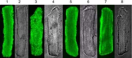
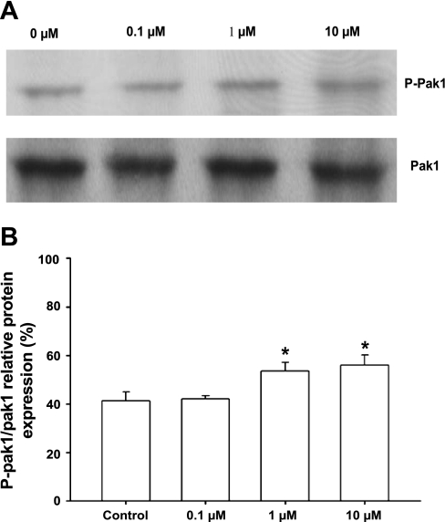
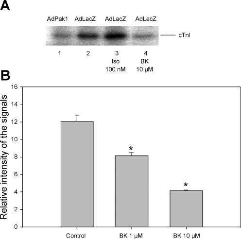
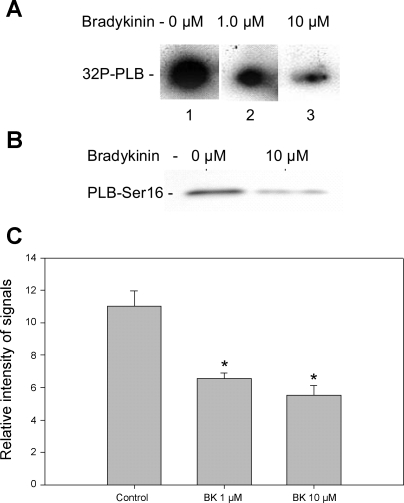
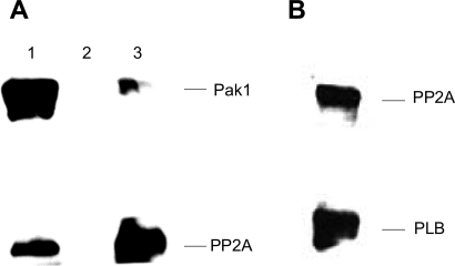
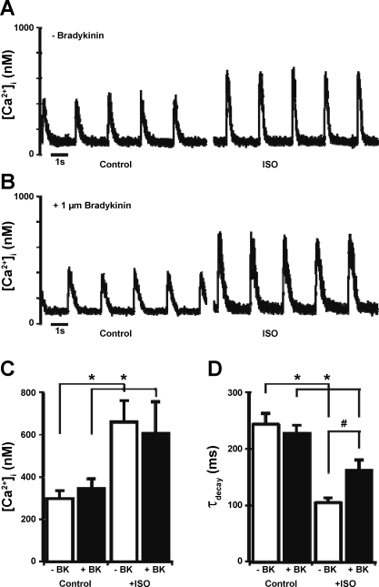
Although bradykinin (BK) is known to exert effects on the myocardium, its intracellular signaling pathways remain poorly understood. Experiments in other cell types indicated that p21-activated kinase-1 (Pak1), a Ser/Thr kinase downstream of small monomeric G proteins, is activated by BK. We previously reported that the expression of active Pak1 in adult cardiac myocytes induced activation of protein phosphatase 2A and dephosphorylation of myofilament proteins (Ke et al. Circ Res 94: 194-200, 2004). In experiments reported here, we tested the hypothesis that BK signals altered protein phosphorylation in adult rat cardiac myocytes through the activation and translocation of Pak1. Treatment of myocytes with BK resulted in the activation of Pak1 as demonstrated by increased autophosphorylation at Thr423 and a diminished striated localization, which is present in the basal state. BK induced dephosphorylation of both cardiac troponin I and phospholamban. Treatment of isolated myocytes with BK also blunted the effect of isoproterenol to enhance peak Ca(2+) and relaxation of Ca(2+) transients. Protein phosphatase 2A was demonstrated to associate with both Pak 1 and phospholamban. Our studies indicate a novel signaling mechanism for BK in adult rat cardiac myocytes and support our hypothesis that Pak 1 is a significant regulator of phosphatase activity in the heart.


Figures

Fig. 1.
Fig. 1.
Intracellular localization of endogenous p21-activated kinase-1 (Pak1) regulated by bradykinin (BK) in rat ventricle myocytes. Lanes 1, 3, 5, and 7: confocal images with immunofluorescence signals for Pak1. Lanes 2, 4, 6, and 8: transmitted light images of the myocytes. Lanes 1 and 2: control myocytes without BK treatment. Lanes 3 and 4: myocytes treated with 0.1 μM BK. Lanes 5 and 6: myocytes treated with 1 μM BK. Lanes 7 and 8: myocytes treated with 10 μM BK. For immunofluorescence, the primary antibody was from Santa Cruz (sc-881). The secondary antibody was FITC-conjugated anti-rabbit IgG from Sigma (F-9887).
Fig. 2.
Fig. 2.
A: Western blot analysis of autophosphorylation of endogenous Pak1 in myocytes treated with BK at different concentrations. A, lanes 1–4: myocytes treated with BK at 0, 0.1, 1, and 10 μM, respectively, for 10 min. Pak1 autophosphorylation was detected by phospho (p)-specific (Pak1 Thr423) antibody. Total Pak1 served as an internal control. B: histogram showing increase of Pak1 Thr423 autophosphorylation normalized to total Pak1. Results shown are representative of 3 independent experiments. p-Pak1 Thr423 levels are expressed as percentage of total Pak1. *P < 0.05 compared with control without BK exposure.
Fig. 3.
Fig. 3.
BK-induced activation of Pak1 associated with dephosphorylation of cardiac troponin I (cTnI). A: inhibition of phosphorylation of cTnI by constitutively active Pak1 and by BK, as detected by [32P] incorporation: myocytes infected with adenovirus Pak1 (AdPak1; lane 1), myocytes infected with AdLacZ (control; lane 2), myocytes infected with AdLacZ and treated with 100 nM isoproterenol (Iso; lane 3), and myocytes infected with AdLacZ and treated with BK at 10 μM (lane 4). B: histogram showing dephosphorylation of cTnI induced by BK at 1 and 10 μM (n = 3). Signal intensity is given in arbitrary units. Results shown are measurements from 4 independent experiments. *P < 0.05 compared with control without exposure to BK.
Fig. 4.
Fig. 4.
Bradykinin induced a reduction in phosphorylation of phospholamban (PLB) in cardiac myocytes. A: [32P] incorporation into PLB from control myocytes (lane 1) and from myocytes treated with 1 μM (lane 2) and 10 μM (lane 3) BK, respectively. Equal amounts of cardiac myocytes were plated in 35-mm culture dish. [32P] incorporation was carried out at room temperature for 30 min. At 30 min of incubation in buffer containing [32P]orthophosphate, BK was added and was incubated for 2 min more. Equal amounts of total protein were loaded in each sample lane as demonstrated by Coommassie blue staining. B: Western blot detection of PLB-Ser16 phosphorylation in control myocytes treated with 100 nM Iso (lane 1) and Western blot of proteins from myocytes treated with 100 nM Iso + 10 μM BK for 2 min (lane 2). C: histogram summarizing the effect of BK effect on PLB phosphorylation corresponding to lanes 1, 2, and 3 of [32P] incorporation shown in A. Results shown are from 3 independent experiments. *P < 0.05 compared with control without exposure to BK.
Fig. 5.
Fig. 5.
Demonstration of protein phosphate 2A (PP2A) association with both Pak1 and PLB by immunoprecipitation (IP). A: PP2A association with Pak1 when Pak1 antibody was used in IP (lane 1), negative control with the same experimental conditions except without the primary antibody (lane 2), and Pak1 association with PP2A when PP2Ac antibody was used in IP. B: Western blotting showing the presence of both PP2Ac and PLB in IP products using a PLB antibody. A control with no PLB or PP2A antibody showed no staining.
Fig. 6.
Fig. 6.
Intracellular Ca2+ concentration ([Ca2+]i) transients recorded from representative field-stimulated single adult rat ventricular myocytes in control conditions and after application of 100 nM Iso in an untreated cell (A) and a cell pretreated for 20 min with 1 μM BK (B) are shown. Summaries of peak [Ca2+]i transients (C) and the [Ca2+]i-transient time constant of decay (τdecay; D) from the same set of records are shown. Data are presented as means ± SE. *P < 0.05 compared with control; #P < 0.05 compared with −BK.

Similar articles

Cited by

References

    1. Arteaga GM, Warren CM, Milutinovic S, Martin AF, Solaro RJ. Specific enhancement of sarcomeric response to Ca2+ protects murine myocardium against ischemia-reperfusion dysfunction. Am J Physiol Heart Circ Physiol 289: H2183–H2192, 2005 - PubMed
    1. Barr E, Carroll J, Kalynych AM, Tripathy SK, Kozarsky K, Wilson JM, Leiden JM. Efficient catheter-mediated gene transfer into the heart using replication-defective adenovirus. Gene Ther 1: 51–58, 1994 - PubMed
    1. Baxter GF, Ebrahim Z. Role of bradykinin in preconditioning and protection of the ischaemic myocardium. Br J Pharmacol 135: 843–854, 2002 - PMC - PubMed
    1. Cannell MB, Cheng H, Lederer WJ. Spatial non-uniformities in [Ca2+]i during excitation-contraction coupling in cardiac myocytes. Biophys J 67: 1942–1956, 1994 - PMC - PubMed
    1. Chen J, Parsons S, Brautigan DL. Tyrosine phosphorylation of protein phosphatase 2A in response to growth stimulation and v-src transformation of fibroblasts. J Biol Chem 269: 7957–7962, 1994 - PubMed

Publication types