. 2010 May;298(5):H1357-64.
doi: 10.1152/ajpheart.00464.2009. Epub 2010 Feb 12.

Ac-SDKP inhibits transforming growth factor-beta1-induced differentiation of human cardiac fibroblasts into myofibroblasts

Affiliations

Ac-SDKP inhibits transforming growth factor-beta1-induced differentiation of human cardiac fibroblasts into myofibroblasts

Hongmei Peng et al. Am J Physiol Heart Circ Physiol. 2010 May.

Abstract

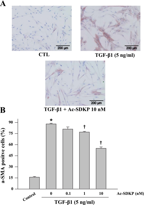
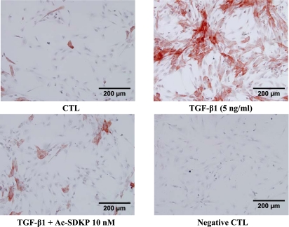
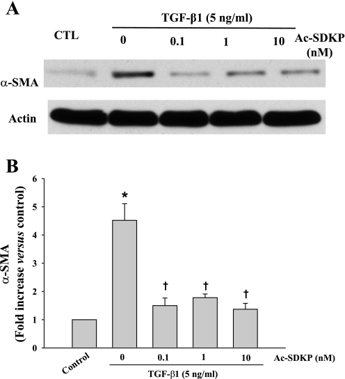
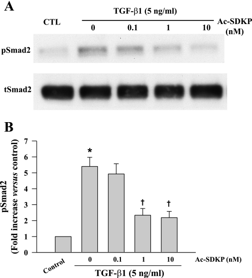
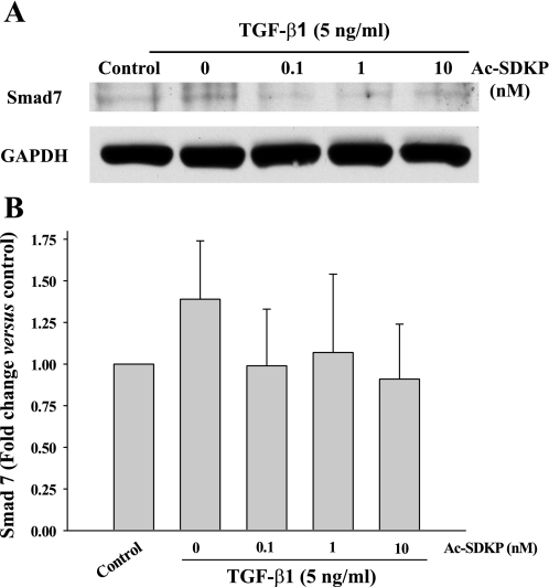
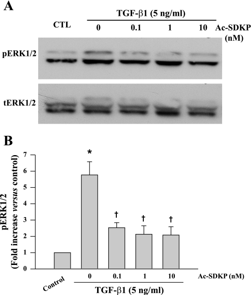
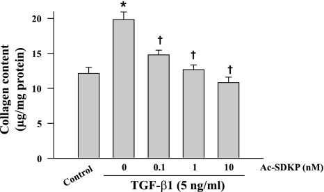
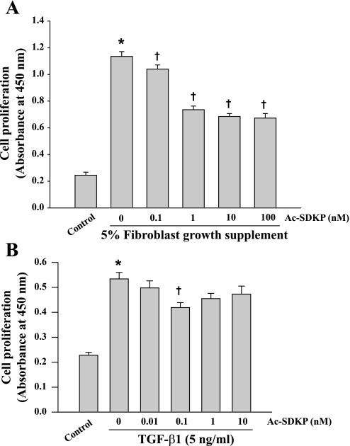
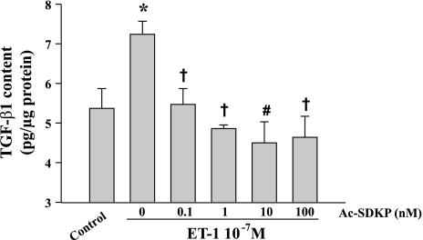
N-acetyl-seryl-aspartyl-lysyl-proline (Ac-SDKP) inhibits collagen production and cell proliferation in cultured rat cardiac fibroblasts, but its effect on differentiation of fibroblasts into myofibroblasts is not known. High amounts of transforming growth factor-beta1 (TGF-beta1) have been found in fibrotic cardiac tissue. TGF-beta1 converts fibroblasts into myofibroblasts, which produce more extracellular matrix proteins than fibroblasts. We hypothesized that 1) Ac-SDKP inhibits TGF-beta1-induced differentiation of fibroblasts into myofibroblasts; and 2) this effect is mediated in part by blocking phosphorylation of small-mothers-against-decapentaplegic (Smad) 2 and extracellular signal-regulated kinase (ERK) 1/2. For this study, we used human fetal cardiac fibroblasts (HCFs), which do not spontaneously become myofibroblasts when cultured at low passages. We investigated the effect of Ac-SDKP on TGF-beta1-induced HCF transformation into myofibroblasts, Smad2 and ERK1/2 phosphorylation, Smad7 expression, cell proliferation, and collagen production. We also investigated TGF-beta1 production by HCFs stimulated with endothelin-1 (ET-1). As expected, HCFs treated with TGF-beta1 transformed into myofibroblasts as indicated by increased expression of alpha-smooth muscle actin and a higher proportion of the embryonic isoform of smooth muscle myosin compared with untreated cells. TGF-beta1 also increased Smad2 and ERK1/2 phosphorylation but did not affect Smad7 expression. In addition, TGF-beta1 stimulated HCF proliferation as indicated by an increase in mitochondrial dehydrogenase activity and collagen production (hydroxyproline assay). Ac-SDKP significantly inhibited all of the effects of TGF-beta1. It also inhibited ET-1-stimulated TGF-beta1 production. We concluded that Ac-SDKP markedly suppresses differentiation of human cardiac fibroblasts into myofibroblasts, probably by inhibiting the TGF-beta/Smad/ERK1/2 signaling pathway, and thus mediating its anti-fibrotic effects.


Figures

Fig. 1.
Fig. 1.
Representative micrographs (A) and quantitative data (B) representing smooth muscle α-actin (α-SMA)-stained cultured fibroblasts treated with transforming growth factor-β1 (TGF-β1, 5 ng/ml for 24 h) in the presence or absence of N-acetyl-seryl-aspartyl-lysyl-proline (Ac-SDKP). Cells were counterstained with hematoxylin to show the nuclei. P = 0.001, control vs. TGF-β1 (*) and Ac-SDKP + TGF-β1 vs. TGF-β1 alone (†) (n = 5 experiments/group).
Fig. 2.
Fig. 2.
Representative micrographs showing the effect of Ac-SDKP on the embryonic isoform of smooth muscle myosin expression in human fetal cardiac fibroblasts treated with TGF-β1 for 24 h. Conditions are the same as in Fig. 1.
Fig. 3.
Fig. 3.
Representative Western blot (A) and quantitative data (B) representing α-SMA expression in fibroblasts treated with TGF-β1 (5 ng/ml) for 24 h in the presence or absence of Ac-SDKP. α-SMA was normalized to actin. *P = 0.004, control vs. TGF-β1; †P < 0.007, Ac-SDKP + TGF-β1 vs. TGF-β1 alone (n = 5/group).
Fig. 4.
Fig. 4.
Representative Western blot (A) and quantitative data (B) representing phosphorylated (p) small-mothers-against-decapentaplegic (Smad) 2 levels in fibroblasts treated with TGF-β1 for 30 min in the presence or absence of Ac-SDKP. pSmad2 was normalized to total (t) Smad2. *P = 0.005, control vs. TGF-β1; †P < 0.05, Ac-SDKP + TGF-β1 vs. TGF-β1 alone (n = 4/group).
Fig. 5.
Fig. 5.
Representative Western blot (A) and quantitative data (B) representing Smad7 levels in fibroblasts treated with TGF-β1 for 30 min in the presence or absence of Ac-SDKP. Smad7 was normalized to glyceraldehydes-3-phosphate dehydrogenase (GAPDH) (n = 4/group).
Fig. 6.
Fig. 6.
Representative Western blot (A) and quantitative data (B) representing phosphorylated extracellular signal-regulated kinase (ERK) 1/2 levels in fibroblasts treated with TGF-β1 for 15 min in the presence or absence of Ac-SDKP. pERK1/2 was normalized to tERK1/2. *P = 0.001, control vs. TGF-β1; †P < 0.02, Ac-SDKP + TGF-β1 vs. TGF-β1 alone (n = 6/group).
Fig. 7.
Fig. 7.
Effects of Ac-SDKP on TGF-β1-stimulated collagen production in cultured fibroblasts (5 ng/ml TGF-β1 for 48 h). *P = 0.002, control vs. TGF-β1; †P = 0.001, Ac-SDKP + TGF-β1 vs. TGF-β1 alone (n = 6/group).
Fig. 8.
Fig. 8.
Effects of Ac-SDKP on proliferation of fibroblasts stimulated with either fibroblast growth supplement (FGS) (A) or TGF-β1 (B). Cell proliferation was measured by measuring mitochondrial dehydrogenase activity (A: n = 28–29/group; B: n = 12/group). Absorbance was recorded using a microtiter plate reader at 450 nm and expressed as arbitrary units (AU). P = 0.001, control vs. TGF-β1 (*) and Ac-SDKP + TGF-β1 vs. TGF-β1 alone (†).
Fig. 9.
Fig. 9.
Effect of Ac-SDKP on endothelin-1 (ET-1)-stimulated TGF-β1 secretion by human cardiac fibroblasts. TGF-β1 was measured with a quantitative sandwich enzyme immunoassay. *P < 0.05, control vs. ET-1; †P < 0.05, Ac-SDKP + ET-1 vs. ET-1 alone; #P = 0.0021, Ac-SDKP + ET-1 vs. ET-1 alone (n = 4/group).

Similar articles

Cited by

References

    1. Azizi M, Rousseau A, Ezan E, Guyene TT, Michelet S, Grognet JM, Lenfant M, Corvol P, Ménard J. Acute angiotensin-converting enzyme inhibition increases the plasma level of the natural stem cell regulator N-acetyl-seryl-aspartyl-lysyl-proline. J Clin Invest 97: 839–844, 1996 - PMC - PubMed
    1. Border WA, Noble NA. Transforming growth factor β in tissue fibrosis. N Engl J Med 331: 1286–1292, 1994 - PubMed
    1. Brooks WW, Conrad CH. Myocardial fibrosis in transforming growth factor β(1)heterozygous mice. J Mol Cell Cardiol 32: 187–195, 2000 - PubMed
    1. Bujak M, Frangogiannis NG. The role of TGF-β signaling in myocardial infarction and cardiac remodeling. Cardiovasc Res 74: 184–195, 2007 - PMC - PubMed
    1. Calderone A, Murphy RJ, Lavoie J, Colombo F, Beliveau L. TGF-β1 and prepro-ANP mRNAs are differentially regulated in exercise-induced cardiac hypertrophy. J Appl Physiol 91: 771–776, 2001 - PubMed

Publication types

MeSH terms