. 2010 May 15;19(10):1883-96.
doi: 10.1093/hmg/ddq064. Epub 2010 Feb 13.

Functional conservation of human Spastin in a Drosophila model of autosomal dominant-hereditary spastic paraplegia

Affiliations

Functional conservation of human Spastin in a Drosophila model of autosomal dominant-hereditary spastic paraplegia

Fang Du et al. Hum Mol Genet. .

Abstract

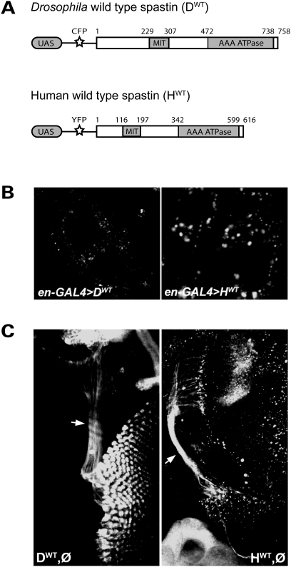
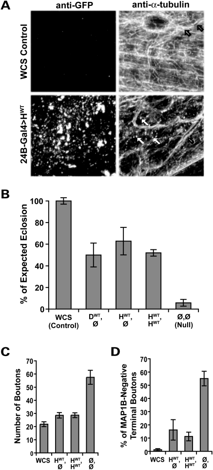
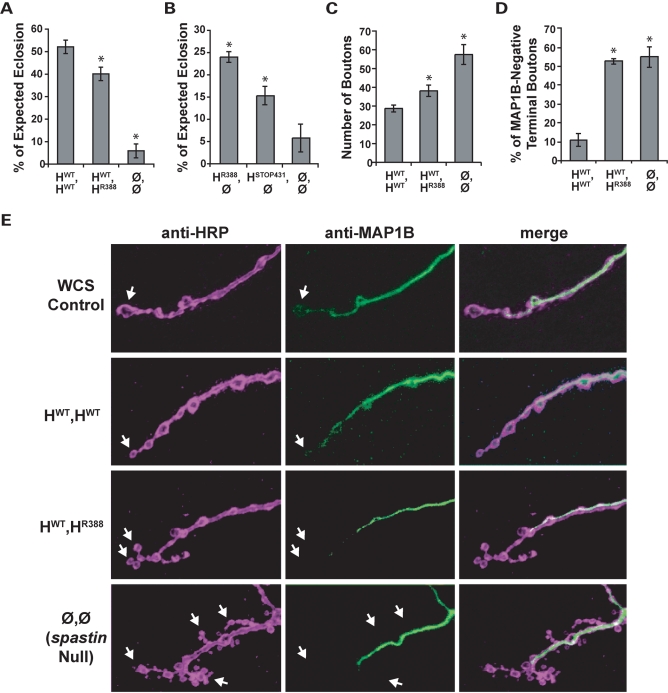
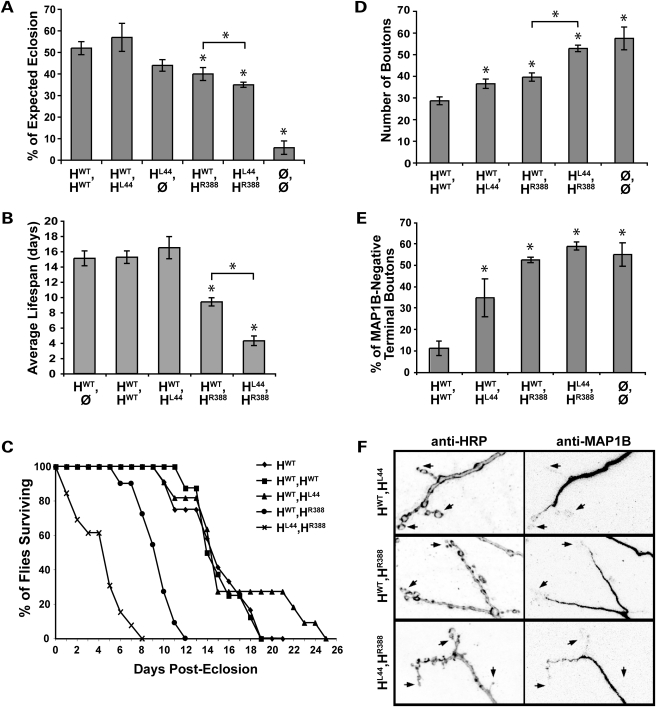
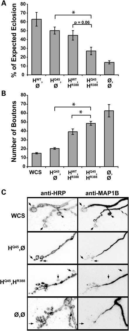
Mutations in spastin are the most frequent cause of the neurodegenerative disease autosomal dominant-hereditary spastic paraplegia (AD-HSP). Drosophila melanogaster lacking spastin exhibit striking behavioral similarities to human patients suffering from AD-HSP, suggesting conservation of Spastin function between the species. Consistent with this, we show that exogenous expression of wild-type Drosophila or human spastin rescues behavioral and cellular defects in spastin null flies equivalently. This enabled us to generate genetically representative models of AD-HSP, which arises from dominant mutations in spastin rather than a complete loss of the gene. Flies co-expressing one copy of wild-type human spastin and one encoding the K388R catalytic domain mutation in the fly spastin null background, exhibit aberrant distal synapse morphology and microtubule distribution, similar to but less severe than spastin nulls. R388 or a separate nonsense mutation act dominantly and are furthermore sufficient to confer partial rescue, supporting in vitro evidence for additional, non-catalytic Spastin functions. Using this model, we tested the observation from human pedigrees that S44L and P45Q are trans-acting modifiers of mutations affecting the Spastin catalytic domain. As in humans, both L44 and Q45 are largely silent when heterozygous, but exacerbate mutant phenotypes when expressed in trans with R388. These transgenic 'AD-HSP' flies therefore provide a powerful and tractable model to enhance our understanding of the cellular and behavioral consequences of human spastin mutations and test hypotheses directly relevant to the human disease.


Figures

Figure 1.
Figure 1.
Fly and human spastin transgenes have similar subcellular distributions. (A) Constructs containing GAL4 binding sites (UAS) followed by genes encoding a fluorescent tag (CFP or Venus YFP) and either the spastin full-length wild-type fly genomic (DWT) or human cDNA (HWT) sequence were generated using the Drosophila Gateway system. Transgenic flies bearing either construct could then be used to express fluorescently tagged fly or human Spastin under the spatiotemporal control of a promoter-GAL4 line. Numbers denote amino acid position. (B) en-GAL4 was used to drive expression of the constructs in larval epidermal cells to reveal their subcellular distribution. Anti-GFP immunostaining shows, in both cases, diffuse cytoplasmic signal with scattered aggregates. No expression is detected in the nucleus. (C) A single copy of wild-type Drosophila or human spastin driven by GS-elav-GAL4 in the spastin null background (genotypes DWT,Ø and HWT,Ø, respectively) induced pan-neuronal spastin transgene expression. Anti-GFP reveals cytoplasmic expression of both proteins in neuronal cell bodies as well as their processes, including the long photoreceptor axons (arrows).
Figure 2.
Figure 2.
Human and Drosophila Spastin are functionally equivalent. (A) Microtubules in the body wall muscles of wild-type third-instar Drosophila larvae were visualized using an antibody against α-tubulin, revealing a dense filamentous mesh throughout the tissue (top right). Microtubules in the walls of the trachea overlying the muscle are also seen (black arrows). Muscle-specific overexpression of human wild-type (HWT) Spastin revealed by anti-GFP (bottom left) causes an overall reduction in the intensity of α-tubulin staining (bottom right), as well as a profusion of punctae (white arrows). The punctate microtubule signal is distinct from the pattern of GFP expression, and likely reflects fragments resulting from the severing activity of the overexpressed human Spastin. (B) Pan-neuronal expression of fly or human spastin in the fly spastin null background rescues eclosion rates equivalently. Flies deleted for spastin (Ø,Ø) eclose successfully only ∼6% of the time, compared with WCS control flies, which nearly always eclose. GS-elav-GAL4-driven expression of a single copy of the fly or human spastin transgene (genotypes DWT,Ø and HWT,Ø, respectively) restores eclosion to over 50%, as does two copies of the human transgene (HWT,HWT; P < 5 × 10⁻⁶ for all groups compared with nulls, P > 0.4 between all transgenic lines). Similar results were observed for multiple independent insertions of the HWT transgene (Supplementary Material, Table S1). (C) At the cellular level, increased synaptic bouton number caused by loss of spastin is almost fully rescued by neuronal expression of the wild-type human transgene. Bouton number is 2.5-fold greater in spastin nulls (Ø,Ø) compared with WCS controls, and doubled compared with animals expressing human spastin (see also Fig. 3E). (D) Correspondingly, expression of human spastin significantly restores the penetration of microtubules into terminal synaptic boutons, such that only 16% (HWT,Ø) and 11% (HWT,HWT) are devoid of detectable MAP1B staining compared with >50% in nulls (P < 2 × 10⁻⁴ for either rescued genotype). WCS animals have <2% of terminal boutons lacking microtubules. Averages in this and subsequent figures are mean ± s.e.; P-values were calculated by one-way ANOVA; N's are detailed in the Methods.
Figure 3.
Figure 3.
Heterozygous expression of R388 catalytic mutant Spastin causes mild loss of function phenotypes. (A) Flies mimicking the AD-HSP genotype, HWT,HR388, eclose less frequently compared with ‘wild-type’ HWT,HWT flies, but are still much healthier than nulls (Ø,Ø). *, P < 2 × 10⁻³ relative to wild-type. (B) Spastin retains significant function even in the likely absence of ATPase activity. Four times as many flies survive to adulthood when the R388 mutant protein is expressed alone in the spastin null background (HR388,Ø; P < 6 × 10⁻⁵ compared with nulls). STOP431, a mutant lacking over half of the catalytic domain, also confers significant rescue of eclosion to over 2-fold more than nulls (P < 8 × 10⁻³). (C) Larval NMJ bouton number is increased in R388 heterozygous mutants compared with wild-type controls (P < 0.02), although not as severely as in nulls (P < 2 × 10⁻⁴). (D) Microtubule distribution in terminal boutons is depleted in both HWT,HR388 heterozygotes and spastin nulls (P < 6 × 10⁻⁶ compared with HWT,HWT controls). (E) Representative synaptic bouton arbors from muscle 4 of third-instar larvae immunostained with anti-HRP (left, purple), which delineates the neuronal cell membrane, and anti-Futsch (center, green), which recognizes the Drosophila ortholog of MAP1B. WCS control boutons are linearly arrayed and MAP1B signal is typically detected throughout the arbor, including into terminal boutons where the microtubules form bundled loops (arrow). Boutons in animals deleted for spastin (‘Null’), in contrast, are highly clustered, smaller and more numerous, and in most cases lack distal MAP1B-positive microtubules. Expression of wild-type human spastin in the spastin null background (HWT,HWT) restores bouton size, linearity and microtubule penetration towards the WCS control phenotype. Expression of one copy each of wild-type and R388 mutant spastin (HWT,HR388) causes a mild loss of function phenotype, with more clustered boutons that often lack MAP1B signal in comparison to controls.
Figure 4.
Figure 4.
The amino-terminus S44L mutation exacerbates R388 catalytic domain mutant phenotypes but is itself only mildly deleterious. (A) L44 expressed heterozygously (HWT,HL44) or alone (HL44,Ø) rescues eclosion as effectively as one or two copies of the wild-type transgene, HWT (P > 0.1). Co-expression of R388 and L44 slightly but significantly lowers eclosion compared with the HWT,HR388 genotype, consistent with L44 being largely silent in combination with wild-type Spastin, but deleterious when in trans with a catalytic domain mutant in Spastin (P < 0.02). (B, C) Post-eclosion, survival of adult flies is dramatically affected by Spastin genotype. Compared with controls, HWT,HR388 heterozygotes survive on average only about half as long, and HL44,HR388 compound heterozygotes survive less than half as well as those (P < 9 × 10⁻⁶). No deleterious effect of the L44 allele is seen in HWT,HL44 heterozygotes compared with wild-type, however. (D, E) HWT,HL44 larvae show moderate but significant increases in bouton number and the percentage of terminal boutons lacking MAP1B compared with HWT,HWT controls, indicating that L44 is somewhat deleterious at the cellular level (P < 0.01 for both parameters). When in trans with R388, however, L44 strongly enhances the R388 loss of function phenotype, to levels equivalent to spastin nulls (P > 0.3). Bouton number is significantly greater in the compound versus single mutants (P < 6 × 10⁻⁷), although both genotypes are deficient in terminal bouton microtubules, similar to nulls. (F) Representative synaptic bouton arbors with their MAP1B-positive microtubule distribution are shown for the different ‘AD-HSP’ genotypes. Presynaptic arbors in HWT,HL44 animals resemble wild-type, with large, round terminal boutons (e.g. arrows) sometimes penetrated by microtubules. HWT,HR388 larvae exhibit smaller, more clustered boutons often devoid of microtubule staining, and HL44,HR388 synapses are similarly, but much more severely, affected.
Figure 5.
Figure 5.
Like L44, Q45 enhances R388 mutant effects in trans. (A) Expression of the Q45 mutation alone in spastin null flies (HQ45,Ø) rescues eclosion to the same level as HWT (P > 0.2). In contrast, ∼40% fewer HQ45,HR388 compound heterozygotes eclose compared with HWT,HR388 single mutants, although for the numbers tested this effect did not achieve a P-value of <0.05 (P = 0.06). Q45 may therefore be a weaker intragenic modifier of catalytic mutations compared with L44. (B, C) Bouton number in HQ45,HR388 compound heterozygotes is increased, similar to nulls (P > 0.07), and significantly greater than in single heterozygotes (HWT,HR388 or HQ45,Ø; P < 0.03). Expression of the HQ45 mutation alone in the null background restores bouton number and microtubule distribution to near wild-type levels. Arrows in (C) denote examples of terminal boutons.

Similar articles

Cited by

References

    1. Fink J.K. Hereditary spastic paraplegia. Neurol. Clin. 2002;20:711–726. - PubMed
    1. Beetz C., Nygren A., Schickel J., Auer-Grumbach M., Burk K., Heide G., Kassubek J., Klimpe S., Klopstock T., Kreuz F., et al. High frequency of partial SPAST deletions in autosomal dominant hereditary spastic paraplegia. Neurology. 2006;67:1926–1930. - PubMed
    1. Fink J.K., Rainier S. Hereditary spastic paraplegia: spastin phenotype and function. Arch. Neurol. 2004;61:830–833. - PubMed
    1. Salinas S., Carazo-Salas R.E., Proukakis C., Schiavo G., Warner T.T. Spastin and microtubules: Functions in health and disease. J. Neurosci. Res. 2007;85:2778–2782. - PubMed
    1. Roll-Mecak A., Vale R.D. The Drosophila homologue of the hereditary spastic paraplegia protein, spastin, severs and disassembles microtubules. Curr. Biol. 2005;15:650–655. - PubMed

Publication types

MeSH terms