

Ventral lateral geniculate input to the medial pons is necessary for visual eyeblink conditioning in rats
- PMID: 20154353
- PMCID: PMC2825698
- DOI: 10.1101/lm.1572710
Ventral lateral geniculate input to the medial pons is necessary for visual eyeblink conditioning in rats
Abstract
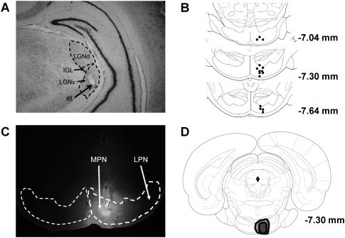
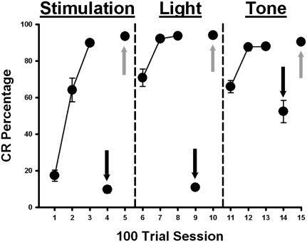
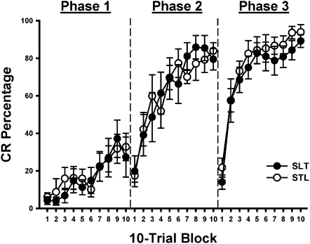
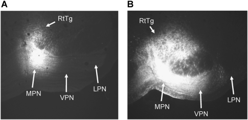
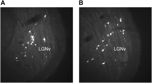
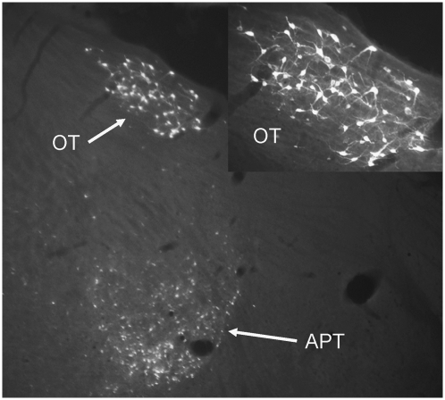
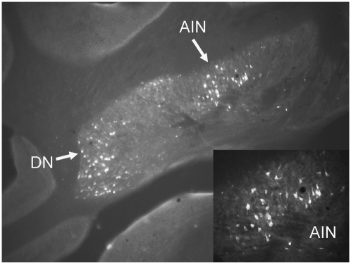
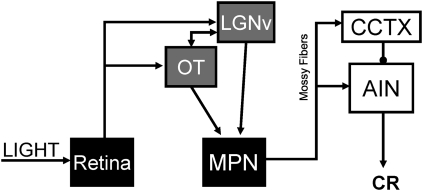
The conditioned stimulus (CS) pathway that is necessary for visual delay eyeblink conditioning was investigated in the current study. Rats were initially given eyeblink conditioning with stimulation of the ventral nucleus of the lateral geniculate (LGNv) as the CS followed by conditioning with light and tone CSs in separate training phases. Muscimol was infused into the medial pontine nuclei (MPN) after each training phase to examine conditioned response (CR) retention to each CS. The spread of muscimol infusions targeting the MPN was examined with fluorescent muscimol. Muscimol infusions into the MPN resulted in a severe impairment in retention of CRs with the LGNv stimulation and light CSs. A less severe impairment was observed with the tone CS. The results suggest that CS information from the LGNv and light CSs is relayed to the cerebellum through the MPN. Retrograde tracing with fluoro-gold (FG) showed that the LGNv and nucleus of the optic tract have ipsilateral projections to the MPN. Unilateral inputs to the MPN from the LGNv and nucleus of the optic tract may be part of the visual CS pathway that is necessary for visual eyeblink conditioning.
Figures
Similar articles
-
Inactivation of the ventral lateral geniculate and nucleus of the optic tract impairs retention of visual eyeblink conditioning.Behav Neurosci. 2013 Oct;127(5):690-3. doi: 10.1037/a0033729. Epub 2013 Aug 26. Behav Neurosci. 2013. PMID: 23978151 Free PMC article.
-
Medial auditory thalamic input to the lateral pontine nuclei is necessary for auditory eyeblink conditioning.Neurobiol Learn Mem. 2010 Jan;93(1):92-8. doi: 10.1016/j.nlm.2009.08.008. Epub 2009 Aug 23. Neurobiol Learn Mem. 2010. PMID: 19706335 Free PMC article.
-
Stimulation of the lateral geniculate, superior colliculus, or visual cortex is sufficient for eyeblink conditioning in rats.Learn Mem. 2009 Apr 24;16(5):300-7. doi: 10.1101/lm.1340909. Print 2009 May. Learn Mem. 2009. PMID: 19395671 Free PMC article.
-
Neural circuitry and plasticity mechanisms underlying delay eyeblink conditioning.Learn Mem. 2011 Oct 3;18(10):666-77. doi: 10.1101/lm.2023011. Print 2011. Learn Mem. 2011. PMID: 21969489 Free PMC article. Review.
-
Modulation of eyeblink conditioning through sensory processing of conditioned stimulus by cortical and subcortical regions.Behav Brain Res. 2019 Feb 1;359:149-155. doi: 10.1016/j.bbr.2018.10.035. Epub 2018 Oct 29. Behav Brain Res. 2019. PMID: 30385367 Review.
Cited by
-
Optogenetic stimulation of mPFC pyramidal neurons as a conditioned stimulus supports associative learning in rats.Sci Rep. 2015 May 14;5:10065. doi: 10.1038/srep10065. Sci Rep. 2015. PMID: 25973929 Free PMC article.
-
Reevaluating the ability of cerebellum in associative motor learning.Sci Rep. 2019 Apr 15;9(1):6029. doi: 10.1038/s41598-019-42413-5. Sci Rep. 2019. PMID: 30988338 Free PMC article.
-
Use of a tissue clearing technique combined with retrograde trans-synaptic viral tracing to evaluate changes in mouse retinorecipient brain regions following optic nerve crush.Neural Regen Res. 2023 Apr;18(4):913-921. doi: 10.4103/1673-5374.353852. Neural Regen Res. 2023. PMID: 36204863 Free PMC article.
-
Learning-related neuronal activity in the ventral lateral geniculate nucleus during associative cerebellar learning.J Neurophysiol. 2014 Nov 1;112(9):2234-50. doi: 10.1152/jn.00185.2013. Epub 2014 Aug 13. J Neurophysiol. 2014. PMID: 25122718 Free PMC article.
-
Classical eyeblink conditioning using electrical stimulation of caudal mPFC as conditioned stimulus is dependent on cerebellar interpositus nucleus in guinea pigs.Acta Pharmacol Sin. 2012 Jun;33(6):717-27. doi: 10.1038/aps.2012.32. Epub 2012 May 7. Acta Pharmacol Sin. 2012. PMID: 22562015 Free PMC article.
References
-
- Bao S, Chen L, Thompson RF. Learning-and cerebellum-dependent neuronal activity in the lateral pontine nucleus. Behav Neurosci. 2000;114:254–261. - PubMed
-
- Bloedel JR, Courville L. Cerebellar afferent systems. In: Brookhart JM, editor. Handbook of physiology, section I: The nervous system. II. Williams and Wilkins; Baltimore, MD: 1981. pp. 735–830.
-
- Brodal A. Neurological anatomy. Oxford University Press; New York: 1981.
Publication types
MeSH terms
Grants and funding
LinkOut - more resources
Full Text Sources