. 2010 May 14;285(20):15027-15037.
doi: 10.1074/jbc.M109.064790. Epub 2010 Feb 13.

Protein complex of Drosophila ATRX/XNP and HP1a is required for the formation of pericentric beta-heterochromatin in vivo

Affiliations

Protein complex of Drosophila ATRX/XNP and HP1a is required for the formation of pericentric beta-heterochromatin in vivo

Alexander V Emelyanov et al. J Biol Chem. .

Abstract

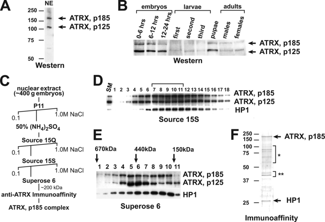
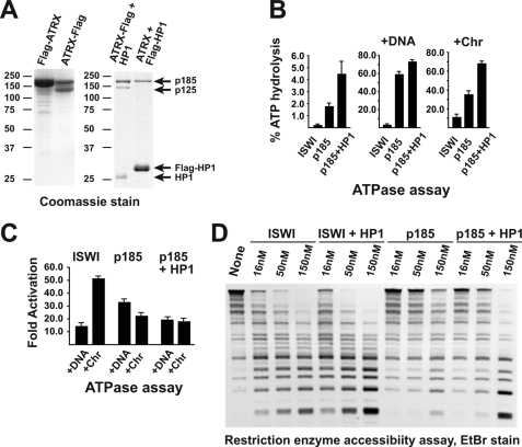
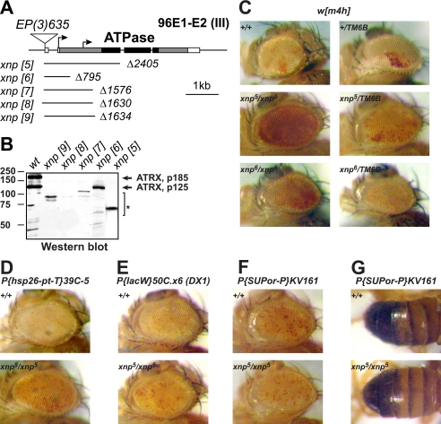
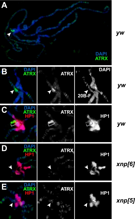
ATRX belongs to the family of SWI2/SNF2-like ATP-dependent nucleosome remodeling molecular motor proteins. Mutations of the human ATRX gene result in a severe genetic disorder termed X-linked alpha-thalassemia mental retardation (ATR-X) syndrome. Here we perform biochemical and genetic analyses of the Drosophila melanogaster ortholog of ATRX. The loss of function allele of the Drosophila ATRX/XNP gene is semilethal. Drosophila ATRX is expressed throughout development in two isoforms, p185 and p125. ATRX185 and ATRX125 form distinct multisubunit complexes in fly embryo. The ATRX185 complex comprises p185 and heterochromatin protein HP1a. Consistently, ATRX185 but not ATRX125 is highly concentrated in pericentric beta-heterochromatin of the X chromosome in larval cells. HP1a strongly stimulates biochemical activities of ATRX185 in vitro. Conversely, ATRX185 is required for HP1a deposition in pericentric beta-heterochromatin of the X chromosome. The loss of function allele of the ATRX/XNP gene and mutant allele that does not express p185 are strong suppressors of position effect variegation. These results provide evidence for essential biological functions of Drosophila ATRX in vivo and establish ATRX as a major determinant of pericentric beta-heterochromatin identity.


Figures

FIGURE 1.
FIGURE 1.
Purification of the native form of ATRX/XNP. A, Western blot analysis of Drosophila nuclear extract (NE) is shown. Antibodies against a C-terminal epitope of Drosophila ATRX recognize two polypeptide bands with apparent molecular masses of 185 and 125 kDa (arrows). B, developmental analysis of ATRX/XNP expression is shown. Whole-animal lysates of various developmental stages of Drosophila were analyzed by a Western blot with anti-ATRX antibodies. Uniformity of the total protein loading was controlled by Coomassie staining of similarly loaded SDS-PAGE gels (not shown). C, shown is a chromatographic scheme for purification of the native form of ATRX185 from Drosophila embryos. D, source 15S chromatography is shown. The peak ATRX (p185 and p125) fractions from the Source 15Q step were applied to the Source 15S column. The column fractions were analyzed by Western blot with antibodies against Drosophila ATRX and HP1a. ATRX/XNP and HP1a co-fractionate with a peak in fractions 7–11 (bracket, of a total of 45 gradient fractions). E, size-exclusion chromatography is shown. The peak fractions from the Source 15S step were applied to the Superose 6 column, and the column fractions were analyzed by Western blot. Approximate molecular masses of the native proteins were estimated based on the peaks of fractionation of molecular mass markers (arrows). ATRX125 fractionates with a peak in fractions 5–7 (estimated molecular mass, ∼500 kDa), whereas p185 fractionates with two approximately equivalently abundant peaks in fractions 5–7 and 8–10 (estimated molecular mass, ∼200 kDa). HP1a co-fractionates with the second peak of p185. F, the native ATRX185 complex comprises ATRX185 and HP1a. The second peak (fraction 9) of ATRX185 from Superose 6 step (∼200 kDa) was applied to the anti-ATRX immunoaffinity column, and the protein was eluted with a low pH buffer and analyzed by SDS-PAGE and silver staining. The identities of the protein bands were determined by mass spectometry sequencing (arrows and brackets). Brackets: *, proteolysis products of ATRX/XNP; **, yolk proteins.
FIGURE 2.
FIGURE 2.
Drosophila ATRX185 physically and functionally interacts with HP1a. A, ATRX185 specifically binds HP1a. N-terminal or C-terminal FLAG-tagged Drosophila ATRX proteins were expressed alone (left panel) or in combination with Drosophila HP1a protein (wild-type or N-terminal FLAG-tagged) in Sf9 cells using baculovirus expression system. The protein complexes were purified by FLAG affinity chromatography. The complex purified through FLAG-HP1a does not contain untagged ATRX125 (right panel, right lane). ATRX185 appears substoichiometric due to more efficient expression of FLAG-HP1a. The positions and sizes (kDa) of molecular mass markers are indicated on the left of the panels. B, ATPase activities of recombinant Drosophila ISWI, ATRX185, and ATRX185·HP1a complex are shown. The indicated proteins (∼1.3 pmol) were analyzed in ATPase reactions in the absence or presence of 260 ng (∼0.13 pmol) of plasmid DNA or equimolar amounts of reconstituted oligonucleosomes (∼2.6 pmol of nucleosomes; Chr). All reactions were performed in triplicate. Error bars represent S.D. C, DNA and nucleosomes stimulate the ATPase activity of ATRX. The indicated proteins were analyzed in ATPase assay reactions in the presence of DNA or oligonucleosomes as in B. D, stimulation of nucleosomal array remodeling activity of Drosophila ATRX by HP1a is shown. Chromatin template (∼0.2 pmol and ∼4 pmol of nucleosomes) was digested with HaeIII in the absence or presence increasing amounts (∼0.3, ∼1, ∼3 pmol) of the indicated remodeling enzymes. Where indicated, double equimolar amounts of HP1a were added to the reactions. Deproteinated DNA fragments were resolved by agarose gel electrophoresis and stained with ethidium bromide. Recombinant HP1a alone does not possess appreciable nucleosome remodeling activity (not shown).
FIGURE 3.
FIGURE 3.
Mutations of xnp/atrx suppress heterochromatic silencing. A, Drosophila ATRX genomic region and xnp/atrx imprecise excision alleles are shown. ATRX/XNP introns are represented by solid lines, and exons are represented by rectangles. The coding sequence is shaded, and its portion that codes for the ATPase domain is painted black. Arrows indicate alternative translation start methionine codons of the native ATRX/XNP. Solid lines under the gene schematic indicate the extent and breakpoints of the xnp deficiency alleles. Triangle, the insertion site of P{EP}xnp[EP635]; scale bar, 1 kbp. B, shown is Western analysis of xnp/atrx alleles. Wild-type (wt) or homozygous mutant embryos of indicated alleles were used to prepare protein lysates and analyzed on a Western blot with anti-ATRX/XNP antibody. The positions of native Drosophila ATRX polypeptides are indicated by arrows. The molecular masses of markers are shown in kDa on the left of the panel. *, truncated polypeptide products of the gene that are recognized by the C-terminal specific antibody. C–G, shown is the effect of xnp/atrx mutations on variegation of heterochromatin-silenced genes. xnp/atrx suppresses PEV in pericentric X chromatin of the In(1)w[m4h] rearrangement (C). It also suppresses silencing of the transgene insertion 39C-5 in 2L subtelomeric region (D) and tandem array of transgenes DX1 in 2R euchromatin (E). However, it does not suppress PEV in pericentric insertion KV161 on the second chromosome (F and G). Alleles and crosses are described under “Experimental Procedures.” Variegated expression of w (C–F) or y (G) transgenes was assayed.
FIGURE 4.
FIGURE 4.
Drosophila ATRX facilitates deposition of HP1a into pericentric beta-heterochromatin of the X chromosome. A, the majority of Drosophila ATRX localizes to a pericentric region of polytene chromosomes. Polytene chromosomes from salivary glands of wild-type (y w) L3 larvae were stained with 4′,6-diamidino-2-phenylindole (DAPI, blue) and anti-XNP/ATRX antibodies (green). Drosophila ATRX localizes to multiple sites in euchromatic arms; however, the major signal is observed in the pericentric region (arrowhead). B, the major locus of ATRX staining in pericentric heterochromatin corresponds to beta-heterochromatin of the proximal X (cytogenetic region 20B-F, arrowheads). Polytene chromosomes were stained as in A. C, pericentric staining of ATRX/XNP co-localizes with weak HP1a staining of beta-heterochromatin. Red, HP1a. Brackets, 20B-F cytogenetic region. D, ATRX185 is specific to the X beta-heterochromatin and is required for HP1a loading in this region. Polytene chromosomes from salivary glands of atrx[6] L3 larvae were stained with 4′,6-diamidino-2-phenylindole (blue), anti-HP1a (red), and anti-ATRX/XNP (green) antibodies. In the xnp[6] allele, which does not express p185, ATRX/XNP and HP1a staining is missing in 20B-F region (arrowheads). E, the loss-of function mutation of xnp/atrx results in elimination of HP1a from pericentric beta-heterochromatin of X. Polytene chromosomes from salivary glands of xnp[5] L3 larvae were stained with 4′,6-diamidino-2-phenylindole (blue), anti-HP1a (red), and anti-ATRX/XNP (green) antibodies. ATRX and HP1a staining is missing in 20B-F region (arrowheads). Residual ATRX/XNP staining of the truncated protein is apparent in euchromatic regions.

Similar articles

Cited by

References

    1. Wolffe A. P. (1995) Curr. Biol. 5, 452–454 - PubMed
    1. Luger K., Mäder A. W., Richmond R. K., Sargent D. F., Richmond T. J. (1997) Nature 389, 251–260 - PubMed
    1. Eissenberg J. C., Reuter G. (2009) Int. Rev. Cell Mol. Biol. 273, 1–47 - PubMed
    1. de Wit E., van Steensel B. (2009) Chromosoma 118, 25–36 - PubMed
    1. Fanti L., Pimpinelli S. (2008) Curr. Opin. Genet. Dev. 18, 169–174 - PubMed

Publication types

LinkOut - more resources