

EBV positive mucocutaneous ulcer--a study of 26 cases associated with various sources of immunosuppression
- PMID: 20154586
- PMCID: PMC6437677
- DOI: 10.1097/PAS.0b013e3181cf8622
EBV positive mucocutaneous ulcer--a study of 26 cases associated with various sources of immunosuppression
Abstract
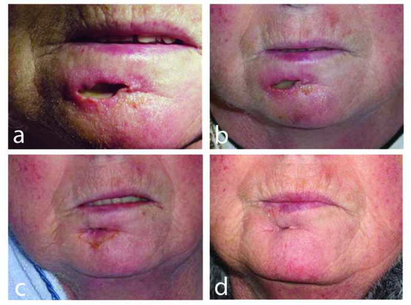
We describe a series of Epstein Barr virus (EBV)-positive circumscribed, ulcerative lesions associated with various types of immunosuppression (IS). The study group (26 patients) comprised 10 males and 16 females, median age 77 years (range 42 to 101). IS in 9 cases included azathioprine (AZA), methotrexate (MTX) or cyclosporin-A (CyA). Seventeen patients had age-related immunosenescence. Patients presented with isolated sharply circumscribed ulcers involving oropharyngeal mucosa (16), skin (6), and gastrointestinal tract (4). Lesions were histologically characterized by a polymorphous infiltrate and atypical large B-cell blasts often with Hodgkin/Reed-Sternberg (HRS) cell-like morphology. The B cells showed strong CD30 and EBER positivity, some with reduced CD20 expression, in a background of abundant T cells. CD15 was positive in 43% of cases (10/23). The pathologic features were identical regardless of the anatomic site or cause of IS. Polymerase chain reaction revealed 39% (7/18) clonal Ig gene rearrangements with 38% (6/16) and 31% (5/16) clonal and restricted T-cell patterns, respectively. Twenty-five percent of patients (5/20) received standard chemotherapy and/or radiotherapy. Forty-five percent (9/20) regressed spontaneously with no treatment and 15% (3/20) were characterized by a relapsing and remitting course. All of the iatrogenic lesions (6/6) with available follow-up responded to reduction of IS. All patients achieved complete remission with no disease-associated deaths over a median follow-up period of 22 months (range 3 to 72). We propose EBV-positive mucocutaneous ulcer as a newly recognized clinicopathologic entity with Hodgkin-like features and a self-limited, indolent course, generally responding well to conservative management. Association with various forms of IS implies a common pathogenetic mechanism. The localized nature of the disease may be owing to a minimal and localized lapse in immunosurveillance over EBV.
Figures


References
-
- WHO Classification of Tumours of Haematopoietic and Lymphoid Tissue. Lyon: IARC; 2008.
-
- Asano N, Yamamoto K, Tamaru J, et al. Age-related Epstein-Barr virus (EBV)-associated B-cell lymphoproliferative disorders: comparison with EBV-positive classic Hodgkin lymphoma in elderly patients. Blood. 2009;113:2629–36. - PubMed
-
- Au WY, Ma ES, Choy C, et al. Therapy-related lymphomas in patients with autoimmune diseases after treatment with disease-modifying anti-rheumatic drugs. Am J Hematol. 2006;81:5–11. - PubMed
-
- Beaty MW, Toro J, Sorbara L, et al. Cutaneous lymphomatoid granulomatosis: correlation of clinical and biologic features. Am J Surg Pathol. 2001;25:1111–20. - PubMed
-
- Bharadwaj M, Burrows SR, Burrows JM, et al. Longitudinal dynamics of antigen-specific CD8+ cytotoxic T lymphocytes following primary Epstein-Barr virus infection. Blood. 2001;98:2588–9. - PubMed
Publication types
MeSH terms
Substances
Grants and funding
LinkOut - more resources
Full Text Sources
Other Literature Sources
Medical
