. 2010 Mar;6(3):244.
doi: 10.1038/nchembio0310-244c.

Aminoglycoside activity observed on single pre-translocation ribosome complexes

Aminoglycoside activity observed on single pre-translocation ribosome complexes

Michael B Feldman et al. Nat Chem Biol. 2010 Mar.

Abstract

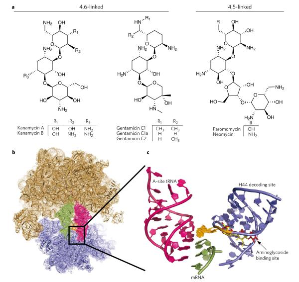
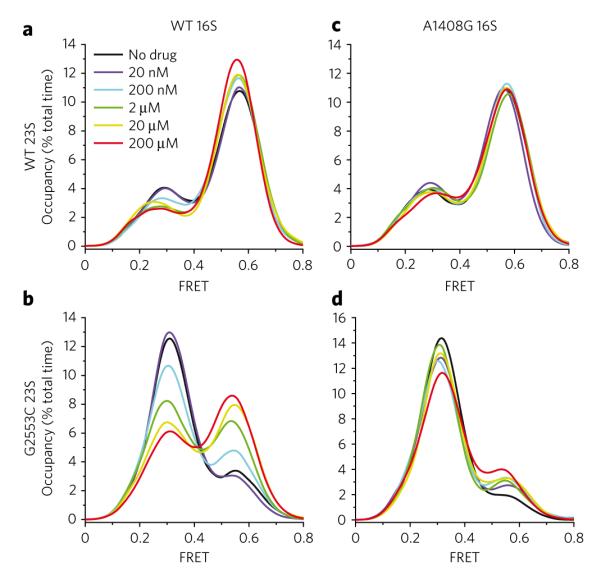
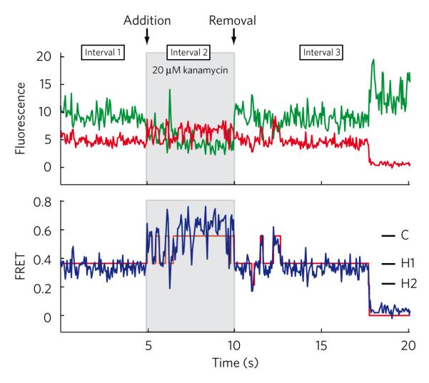
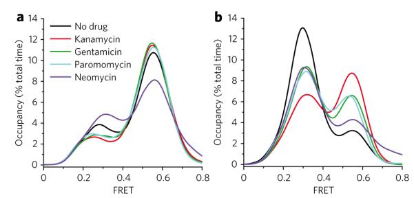
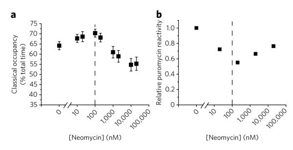
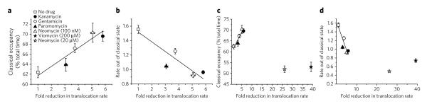
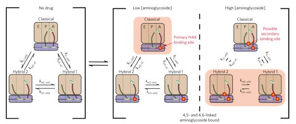
Aminoglycoside-class antibiotics bind directly to ribosomal RNA, imparting pleiotropic effects on ribosome function. Despite in-depth structural investigations of aminoglycoside–RNA oligonucleotide and aminoglycoside-ribosome interactions, mechanisms explaining the unique ribosome inhibition profiles of chemically similar aminoglycosides remain elusive. Here, using single-molecule fluorescence resonance energy transfer (smFRET) methods, we show that high-affinity aminoglycoside binding to the conserved decoding site region of the functional pre-translocation ribosome complex specifically remodels the nature of intrinsic dynamic processes within the particle. The extents of these effects, which are distinct for each member of the aminoglycoside class, strongly correlate with their inhibition of EF-G–catalyzed translocation. Neomycin, a 4,5-linked aminoglycoside, binds with lower affinity to one or more secondary binding sites, mediating distinct structural and dynamic perturbations that further enhance translocation inhibition. These new insights help explain why closely related aminoglycosides elicit pleiotropic translation activities and demonstrate the potential utility of smFRET as a tool for dissecting the mechanisms of antibiotic action.


Figures

Figure 1
Figure 1. Aminoglycoside-induced restructuring of the ribosome 16S decoding site
(a) Chemical structures of kanamycin, gentamicin, paromomycin and neomycin. (b) Overview of the 70S prokaryotic ribosome structure composed of the large 50S subunit (tan) and the small 30S subunit (blue). The decoding site is boxed and contains the aminoglycoside binding portion of the 16S rRNA helix 44. (c) Enlarged view of the decoding site 16S rRNA (blue) with bound aminoglycoside antibiotics. The A1492-A1493 bases (orange) ‘turn out’ in the presence of aminoglycosides or upon cognate codon-anticodon pairing and interact with the tRNA (magenta) and mRNA (green) at the site of codon-anticodon interaction. Gentamicin (yellow) and neomycin (red) are shown bound in the decoding site. Crystal structures were constructed from Protein Data Bank entries 2J00 and 2J01 (ref. 4),with aminoglycosides mapped from 2QB9 and 2QAL (ref. 35) using PyMol software (Delano Scientific).
Figure 2
Figure 2. Kanamycin binding to the decoding site increases time-averaged occupancy of specific ribosome conformations
The time-averaged occupancy (Supplementary Fig. 1) in all nonzero FRET states was measured for the surface immobilized population of intact 70S E. coli ribosomes at increasing concentrations of kanamycin. The FRET values between P-site tRNAfMet(Cy3-s4U8) and A-site fMet-Phe-tRNAPhe(Cy5-acp3U47) report directly on three specific conformational intermediates in the translocation pathway: the classical state (~0.55 FRET), the hybrid 1 (~0.39 FRET) state and the hybrid 2 state (~0.24 FRET). (ad) Histograms are shown for wild-type (a), G2553C 23S (b), A1408G 16S (c) and G2553C 23S + A1408G 16S (d) ribosomal complexes in the presence of increasing kanamycin concentrations (black to red lines). Histograms were constructed by summing FRET values over the first 50 frames of each acquired movie over all nonzero FRET states and plotting the spline-interpolated data points.
Figure 3
Figure 3. Changes in population time-averaged state occupancy are caused by drug-induced changes in single ribosomes
Single-molecule fluorescence traces were collected for surface-immobilized ribosomes containing the G2553C mutation over three 5-s intervals: (i) in the absence of drug, (ii) following a 2-min incubation in 20 μM kanamycin and (iii) following removal of kanamycin with drug-free buffer for 2 min. Shown are donor (green line) and acceptor (red line) fluorescence traces (top panel), along with calculated FRET trajectory (blue) and idealization (red line).
Figure 4
Figure 4. Alterations in tRNA dynamics are a common effect of aminoglycoside binding to the ribosome
(a,b) Population time-averaged state occupancy was measured for wild-type ribosomes (a) and ribosomes containing the large subunit G2553C mutation (b) in the presence of 20 μM aminoglycosides representing both the 4,6-linked (kanamycin, gentamicin) and the 4,5-linked (paromomycin, neomycin) structural families.
Figure 5
Figure 5. Neomycin alters tRNA dynamics on the ribosome in a bimodal fashion
(a) smFRET traces obtained in the presence of increasing neomycin concentrations were idealized to a four-state hidden Markov model (Methods). Time-averaged classical-state occupancy is shown versus neomycin concentration along with standard errors calculated from 1,000 bootstrap samples. (b) Puromycin was delivered to ribosomal complexes containing tRNAfMet in the P-site and Cy3-DSP-Met-Phe-tRNAPhe in the A-site in the presence of increasing concentrations of neomycin. Fluorescence intensity decays with time as fluorescently labeled peptide is released from the surface (Methods; Supplementary Fig. 7). Decreased rates of puromycin reactivity indicate decreased hybrid-state occupancy. Mean relative rates of puromycin reactivity (n = 2, s.d. = 0.05) calculated from the total normalized change in fluorescence over a 10-min period are shown versus neomycin concentration.
Figure 6
Figure 6. Stabilization of the classical state is strongly correlated with inhibition of translocation by decoding site–binding aminoglycosides
(a) Correlation of time-averaged classical-state occupancy versus the fold-reduction of the single-step translocation rate of wild-type ribosomes in the presence of drug (20 μM, unless otherwise noted) revealed a strong correlation with classical-state occupancy. (b) A strong correlation is also observed between translocation rates and the rate constant of transitioning from the classical to hybrid states, as calculated using hidden Markov modeling (Methods). (c,d) Translocation inhibition for 20 μM neomycin (c) and 200 μM viomycin (d) is shown on the expanded axis and was similar to that observed for the no EF-G control. Error bars on translocation rates represent the s.d. of triplicate experiments. Error bars on state occupancy and transition rates were calculated in each experiment from 1,000 bootstrap samples of smFRET traces.
Figure 7
Figure 7. Kinetic scheme of aminoglycoside-induced changes in tRNA dynamics
This scheme describes the changes in the rates of transitions between the classical state, the hybrid 1 state and the hybrid 2 state following aminoglycoside binding to the ribosome complex. 50S (tan) and 30S (blue) subunits are shown, as well as the A-site tRNA (red) and the P-site tRNA (green). At low concentrations, aminoglycosides bind with high affinity to the primary h44 binding site. High concentrations of aminoglycosides can lead to binding at a low-affinity second binding site. Arrow lengths represent the rate of transition relative to the drug-free system, and the predominantly stabilized state is highlighted (pink box).

References

    1. Rodnina MV, Gromadski KB, Kothe U, Wieden HJ. Recognition and selection of tRNA in translation. FEBS Lett. 2005;579:938–942. - PubMed
    1. Shoji S, Walker SE, Fredrick K. Ribosomal translocation: one step closer to the molecular mechanism. ACS Chem. Biol. 2009;4:93–107. - PMC - PubMed
    1. Korostelev A, Trakhanov S, Laurberg M, Noller HF. Crystal structure of a 70S ribosome-tRNA complex reveals functional interactions and rearrangements. Cell. 2006;126:1065–1077. - PubMed
    1. Selmer M, et al. Structure of the 70S ribosome complexed with mRNA and tRNA. Science. 2006;313:1935–1942. - PubMed
    1. Yusupov MM, et al. Crystal structure of the ribosome at 5.5 angstrom resolution. Science. 2001;292:883–896. - PubMed