. 2010 Mar;12(3):286-93.
doi: 10.1038/ncb2029. Epub 2010 Feb 14.

TGF-beta-mediated phosphorylation of hnRNP E1 induces EMT via transcript-selective translational induction of Dab2 and ILEI

Affiliations

TGF-beta-mediated phosphorylation of hnRNP E1 induces EMT via transcript-selective translational induction of Dab2 and ILEI

Arindam Chaudhury et al. Nat Cell Biol. 2010 Mar.

Abstract

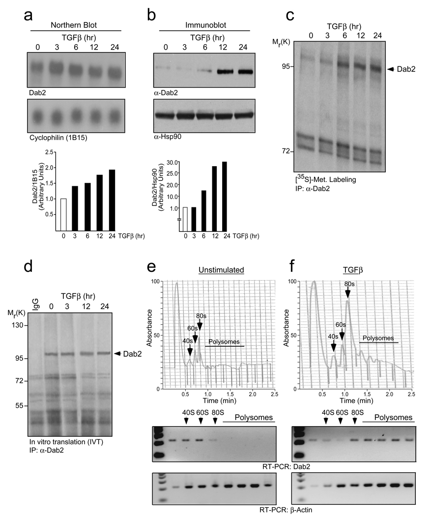
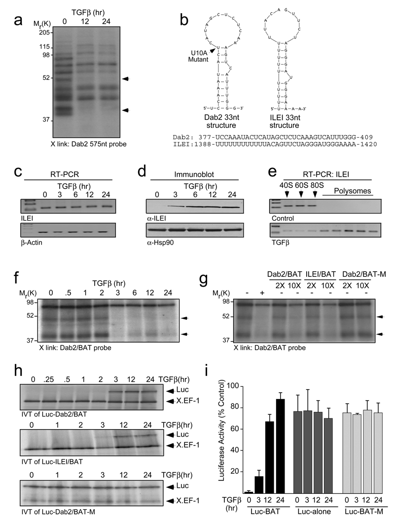
Transforming growth factor-beta (TGF-beta) induces epithelial-mesenchymal transdifferentiation (EMT) accompanied by cellular differentiation and migration. Despite extensive transcriptomic profiling, the identification of TGF-beta-inducible, EMT-specific genes has met with limited success. Here we identify a post-transcriptional pathway by which TGF-beta modulates the expression of EMT-specific proteins and of EMT itself. We show that heterogeneous nuclear ribonucleoprotein E1 (hnRNP E1) binds a structural, 33-nucleotide TGF-beta-activated translation (BAT) element in the 3' untranslated region of disabled-2 (Dab2) and interleukin-like EMT inducer (ILEI) transcripts, and represses their translation. TGF-beta activation leads to phosphorylation at Ser 43 of hnRNP E1 by protein kinase Bbeta/Akt2, inducing its release from the BAT element and translational activation of Dab2 and ILEI messenger RNAs. Modulation of hnRNP E1 expression or its post-translational modification alters the TGF-beta-mediated reversal of translational silencing of the target transcripts and EMT. These results suggest the existence of a TGF-beta-inducible post-transcriptional regulon that controls EMT during the development and metastatic progression of tumours.


Figures

Figure 1
Figure 1
TGFβ translationally up-regulates Dab2 expression. (a) Northern blot analysis examining Dab2 expression levels in NMuMG cells treated with TGFβ for the times indicated. Lower panel represents the quantification of band intensities analyzed by NIH Image J software. Dab2 band intensity was normalized to cyclophilin (1B15), then normalized to the t=0 unstimulated. (b) Immunoblot (IB) analysis examining Dab2 protein levels in NMuMG cells treated with TGFβ for the indicated times. Lower panel represents the quantification of band intensities analyzed by NIH Image J software. Dab2 band intensity was normalized to Hsp90, then normalized to the t=0 unstimulated. (c) Metabolic labeling with [35S]-methionine analyzing the de novo rate of Dab2 synthesis post-TGFβ stimulation. (d) Dab2 mRNA stability analysis by in vitro translation (IVT) of total RNA isolated from NMuMG cells treated with TGFβ for the times indicated followed by immunoprecipitation (IP) with α-Dab2 antibody and mouse IgG. (e) & (f) Translocation of Dab2 mRNA from the non-polysomal to polysomal pool was analyzed by semi-quantitative RT-PCR of RNA isolated from each fraction following polysome profiling. Full scans of (a), (b), (c) and (d) are shown in SI Fig. 7.
Figure 2
Figure 2
The 3’-UTR of Dab2 mRNA contains a cis regulatory (BAT) element, which is also present in ILEI mRNA. (a) UV crosslinking (X-link) analysis to characterize regulatory element(s) in the 3’-UTR of Dab2 mRNA using [α-32P]-labeled Dab2 3’-UTR 575-nt probe (10 fmol) and S100 cytosolic extract from NMuMG cells treated with TGFβ for the times indicated. (b) Secondary structure of the mouse Dab2/BAT (dG = −5.0 Kcal/mol) and ILEI/BAT (dG = −2.5 Kcal/mol) elements as predicted by the Mfold algorithm. Substituted nucleotide (U10A), indicated in boldface, represents a mutant form. ILEI/BAT element was folded under (F 5 0 2)/ (F 9 0 2)/ (P 11 0 2) constraints. (c) Semi-quantitative RT-PCR and (d) IB analyses examining mRNA and protein expression levels of ILEI in NMuMG cells treated with TGFβ. (e) Translocation of ILEI mRNA from the non-polysomal to polysomal pool was analyzed by semi-quantitative RT-PCR of RNA isolated from each fraction following polysome profiling. (f) X-link analysis was performed with [α-32P]-labeled Dab2/BAT probe (10 fmol) and S100 cytosolic extract from NMuMG cells treated with TGFβ. The arrows indicate the positions of two proteins that fail to bind the probe following TGFβ treatment. (g) Specificity of the BAT element was examined by decoy X-link using [α-32P]-labeled Dab2/BAT probe and a 2- or 10-fold molar excess (2X or 10X) of unlabeled Dab2/BAT, ILEI/BAT, and mutant (U10A) Dab2/BAT-M cRNA. (h) IVT analyses with chimeric Luc-Dab2/BAT, Luc-ILEI/BAT and Luc-Dab2/BAT-M shows that TGFβ treatment relieves translational silencing conferred by the WT and not the mutant BAT element following 3 hr of TGFβ treatment. (i) Dual-luciferase assay examining the in vivo translational silencing activity conferred by the BAT element by co-transfecting with wild-type, mutant (Luc-BAT, Luc-BAT-M) or luciferase alone (Luc-alone) and CMV-driven renilla luciferase constructs. The firefly luciferase values were normalized to renilla luciferase values (which were checked for uniformity to monitor equal transfection efficiency). Results are shown as means ± s.d. for three independent sets of experiments (n=3), each experiment done in triplicates. Full scans of (a), (d), (f), (g) and (h) are shown in SI Fig. 7.
Figure 3
Figure 3
hnRNP E1 is an integral functional component of the mRNP complex. (a) IVT assay for translation inhibitory activity of chimeric Dab2/BAT-Luc cRNA using size exclusion chromatographic fractions. (b) Chromatographic fractions (# 36–38) harboring translational silencing activity were subjected to pull-down with Dab2/BAT cRNA bound to cyanogens bromide (CNBr)-activated sepharose beads after pre-clearing with U10A Dab2/BAT-M cRNA. Precipitated mRNP complex was visualized by silver staining (left panel) and the band (arrowhead) which migrated similarly to the band that does not bind the BAT element after TGFβ treatment (shown by arrowhead in right panel) was analyzed by LC-MS. (c) IB analysis of chromatographic fractions with α-hnRNP E1 antibody exclusively detected hnRNP E1 in fractions harboring translational silencing activity. (d) RNA affinity pull-down and IB analyses using S100 cytosolic extracts for the times indicated to define the temporal association of hnRNP E1 with the Dab2 and ILEI BAT element. (e) RNA affinity pull-down IB analyses using BAT, BAT-M and DICE cRNAs of unstimulated and TGFβ-treated S100 cytosolic extracts examining the selective binding of hnRNP E1, and not hnRNP K to the BAT element. (f), (g) & (h) hnRNP E1 interacts with the BAT element in vivo. Immunoprecipitation with α-hnRNP E1 (f) or mouse IgG (g) of cytosolic extracts from NMuMG cells treated with TGFβ for the times indicated followed by semi-quantitative RT-PCR (using Dab2, ILEI, and β-actin specific primers) analyses of RNA isolated from the immunoprecipitates to examine in vivo association of hnRNP E1 with the BAT element. RNA isolated from input extracts were also analyzed by semi-quantitative RT-PCR (h). Full scans of (a), (b), (c), (d) and (e) are shown in SI Fig. S7.
Figure 4
Figure 4
Phosphorylation of hnRNP E1 at serine-43 by TGFβ-mediated activation of Akt2 disrupts its binding to the BAT element and activates translation of Dab2 and ILEI. (a) IB analysis of immunoprecipitates derived from NMuMG WCLs with α-phospho-serine (p-ser) antibody (top panel) and α-hnRNP E1 antibody (bottom panel) to examine TGFβ-dependent hnRNP E1 phosphorylation. (b) IB analysis of WCLs to examine TGFβ-mediated Akt activation, revealed by phospho-Akt (pS473). (c) IB analysis of α-hnRNP E1 immunoprecipitates derived from NMuMG WCLs were probed with the phospho-Akt substrate antibody that recognizes the RXRXXpS/pT motif. (d) IB analysis of α-hnRNP E1 immunoprecipitates from LY294002 treated and untreated WCLs with α-phospho-serine (p-ser) antibody (top panel) and α-hnRNP E1 antibody (bottom panel) to confirm Akt as the kinase. (e) RNA affinity pull-down and IB analysis of cytosolic extracts from unstimulated and LY294002-treated cells to examine temporal association of hnRNP E1 and the BAT element. (f) Phosphorylated hnRNP E1 does not bind the BAT element. Increasing amounts of phosphorylated-GST-hnRNP E1 protein was subjected to pull-down with Dab2/BAT cRNA. The precipitates and the supernatants post pull-down were analyzed by IB. (j) Akt phosphorylates hnRNP E1 at Ser43. Activated kinases were recovered by anti-p-Akt (pSer473) or PAK1 immunoprecipitation and incubated with 5 µg of GST-hnRNP E1 or serine-43-alanine (S43A) mutant GST-hnRNP E1 in the presence of [γ-32P]-ATP. The kinase reaction products were detected by autoradiography. (g) IB analysis of WCLs derived from NMuMG cells post insulin and TGFβ stimulation to examine insulin and TGFβ-mediated Akt activation (top panel). IB analysis of immunoprecipitates derived from NMuMG WCLs with α-phospho-serine (p-ser) antibody (bottom panel) to examine insulin and TGFβ-dependent hnRNP E1 phosphorylation. (h) IB analysis of Akt1 and Akt2 immunoprecipitates derived from NMuMG WCLs with α-phospho-Akt (pS473) antibody to examine insulin and TGFβ-dependent isoform specific Akt activation. (i) TGFβ activated Akt2 specifically phosphorylates hnRNP E1. Activated Akt1 or Akt2 was recovered by anti-Akt1 or anti-Akt2 immunoprecipitation following TGFβ stimulation and incubated with 5 µg of GST-hnRNP E1 in the presence of [γ-32P]-ATP. Scans of (a), (b), (c), (d), (e), (f), (g), (h) and (i) are shown in SI Fig. S7.
Figure 5
Figure 5
Modulation of hnRNP E1 expression or its posttranslational modification alters translation of Dab2 and ILEI and sensitivity of NMuMG cells to TGFβ-induced EMT. (a) Phase contrast images of unstimulated and TGFβ-treated (24 hr) WT, E23 and E2KD cells examining morphological changes post TGFβ-stimulation. Images were taken at 10× magnification. (b) IB analysis monitoring Dab2, ILEI, N-cadherin, vimentin and b-actin protein levels in WT, E23 and E2KD cells treated with TGFβ for the times indicated. (c) In vivo validation of Ser43 as the hnRNP E1 phosphorylation site. WCLs derived from NMuMG, KIM2 and KIWT6 cells were immunoprecipitated with α-hnRNP E1 antibody and analyzed by IB with α-phospho serine antibody (top panel) and α-hnRNP E1 antibody (second panel). TGFβ-dependent Akt activation analyzed by IB analysis of WCLs derived from NMuMG, KIWT6 and KIM2 cells treated with TGFβ for the times indicated (third and bottom panel). (d) IB analysis examining Dab2, ILEI, N-cadherin, vimentin and β-actin protein levels in cells treated with TGFβ for the times indicated. (e) & (f) IVT and RNA pull-down assays with cytosolic extracts from SH14, KIWT6 and KIM2 cells treated with TGFβ for the times indicated to examine translational silencing of chimeric Luc-Dab2/BAT cRNA (d) and temporal association of the modified hnRNP E1 with the Dab2/BAT cRNA (e). (g) Role of hnRNP E1 on EMT is mediated by Dab2 and ILEI. IB analysis of WCLs derived from SH14 cells, un-transfected or transiently transfected with ILEI, Dab2 or control-A siRNA to confirm knockdown of Dab2 and ILEI, respectively (first and second panel, respectively). IB analysis examining N-cadherin, vimentin and Hsp90 protein levels in these cells (third, fourth and bottom panel, respectively). Full scans of (b), (c), (d), (e), (f) and (g) are shown in SI Fig. S7.

Similar articles

Cited by

References

    1. Massague J. TGFβeta in Cancer. Cell. 2008;134:215–230. - PMC - PubMed
    1. Bierie B, Moses HL. TGF-beta and cancer. Cytokine Growth Factor Rev. 2006;17:29–40. - PubMed
    1. Derynck R, Akhurst RJ, Balmain A. TGF-beta signaling in tumor suppression and cancer progression. Nature Genet. 2001;29:117–129. - PubMed
    1. Zavadil J, Bottinger EP. TGF-beta and epithelial-to-mesenchymal transitions. Oncogene. 2005;24:5764–5774. - PubMed
    1. Thiery JP, Sleeman JP. Complex networks orchestrate epithelial-mesenchymal transitions. Nature Rev. Mol. Cell. Biol. 2006;7:131–142. - PubMed

Publication types

MeSH terms

Substances