. 2010 Mar;17(3):379-88.
doi: 10.1038/nsmb.1771. Epub 2010 Feb 14.

Mechanism of error-free and semitargeted mutagenic bypass of an aromatic amine lesion by Y-family polymerase Dpo4

Affiliations

Mechanism of error-free and semitargeted mutagenic bypass of an aromatic amine lesion by Y-family polymerase Dpo4

Olga Rechkoblit et al. Nat Struct Mol Biol. 2010 Mar.

Abstract

The aromatic amine carcinogen 2-aminofluorene (AF) forms covalent adducts with DNA, predominantly with guanine at the C8 position. Such lesions are bypassed by Y-family polymerases such as Dpo4 via error-free and error-prone mechanisms. We show that Dpo4 catalyzes elongation from a correct 3'-terminal cytosine opposite [AF]G in a nonrepetitive template sequence with low efficiency. This extension leads to cognate full-length product, as well as mis-elongated products containing base mutations and deletions. Crystal structures of the Dpo4 ternary complex, with the 3'-terminal primer cytosine base opposite [AF]G in the anti conformation and with the AF moiety positioned in the major groove, reveal both accurate and misalignment-mediated mutagenic extension pathways. The mutagenic template-primer-dNTP arrangement is promoted by interactions between the polymerase and the bulky lesion rather than by a base pair-stabilized misaligment. Further extension leads to semitargeted mutations via this proposed polymerase-guided mechanism.


Figures

Figure 1
Figure 1
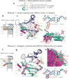
Structure of the [AF]G•A-1 Dpo4 extension ternary complex. (a) Chemical formula of 2-aminofluorene-C8-guanine, [AF]G, and 2-acetylaminofluorene-C8-guanine, [AAF]G, adducts. (b) Schematic of the expected pairing of the [AF]G-modified 19-mer template with the 13-mer primer, ending with a 2′,3′-dideoxy-A, and dGTP in the extension ternary complex with Dpo4. The insertion position at the Dpo4 active site is denoted by (0), and the post-insertion position is denoted by (–1). (c) Schematic of the observed base pairing arrangement within the Dpo4 active site. (d) Overall structure of the [AF]G•A-1 complex. (e) Structure of the active site of the [AF]G•A-1 complex. [AF]G(syn) at the (–1) position is opposite disordered 3′-terminal A14 base of the primer strand. The next template base C5 is paired with an incoming dGTP at the (0) position of the active site. The first Ca2+, cation A, is coordinated by invariant D7, D105, and E106 residues. The second Ca2+, cation B, is chelated by the phosphate groups of the incoming dGTP. Simulated annealing Fo-Fc omit map contoured at 3σ level and colored in blue (2.96 Å resolution) is shown for [AF]G, A14 and Arg336 residues. (f) [AF]G(syn) opposite A14 of the primer strand. [AF]G in the anti conformation (black lines) does not fit the map. Only the phosphate group of A14 has well-ordered density. (g) The N2 group of modified-G(syn) forms hydrogen bonds with the phosphate oxygens of C5. (h) Base stacking arrangement of the [AF]G(syn) and neighboring base pairs. The intercalated AF-moiety leaves no room for the disordered base of A14 within the template/primer helix.
Figure 2
Figure 2
Structure of the [AF]G•C-1 Dpo4 extension ternary complex containing two distinct molecules per asymmetric unit (AU), with ‘correct’ and ‘mutagenic’ alignment for extension from the [AF]G(anti)•C base pair. (a) Schematic of the expected pairing of the [AF]G-template with the 13-mer primer, ending with a 2′,3′-dideoxy-C, and dGTP. (b) Schematic of the observed base pairing arrangement within the Dpo4 active site of molecule 1. (c) Structure of the active site of molecule 1. [AF]G(anti) at the (–1) position is opposite the primer C14 base. The simulated annealing Fo-Fc omit map contoured at 3σ level is colored in blue (2.70 Å resolution). (d) Watson-Crick base pair between the [AF]G(anti) and C14(anti) at the (–1) position. (e) Base stacking arrangement of the [AF]G(anti) opposite C14 and neighboring base pairs. The little finger domain residues Leu293, Arg331 and Arg 332 contact the AF-moiety. (f) Schematic of the observed base pairing arrangement within the Dpo4 active site of molecule 2. (g) Structure of the active site of molecule 2. The [AF]G(anti) opposite the primer C14 base are shifted to the (–2) position. Arg336 has repositioned to stack with the ‘bottom’ face of the AF-moiety. Simulated annealing Fo-Fc omit map contoured at 3σ level is colored in blue (2.70 Å resolution). The loop connecting β2 and β3 of the Dpo4 finger domain (residues Phe33 to Ala42) is highlighted by the shaded area. (h) Accommodation of the AF-moiety in a pocket on the surface of the little finger domain. (i) Watson-Crick base pair between the [AF]G(anti) and C14(anti) at the (–2) position.
Figure 3
Figure 3
Interactions of the Dpo4 little finger and thumb domains with template/primer DNA in extension and post-extension [AF]G-modified Dpo4 ternary complexes. The backbone DNA phosphate groups in contact with Dpo4 are labeled in color and are shown by CPK spheres. The template strand is colored in cyan, the primer strand is in green. (a) The [AF]G•A-1 extension complex has a ‘ternary complex-like’ Dpo4/DNA interaction pattern. The little finger domain contacts the phosphate groups of template C5-A9 and of primer A7-G9 positions (top panel). The thumb domain contacts the phosphates of the template A12 and primer A12-G13 positions (bottom panel). The DNA segment, containing template C5-A16, primer T4-A14 bases and dGTP, is shown. (b) Molecule 1 of the [AF]G•C-1 complex with ‘correct’ template/primer-dNTP pairing alignment has a ‘binary complex-like’ Dpo4/DNA contact pattern. The little finger domain contacts the phosphate groups of template C5-A9 and primer A7-G9 positions (top panel), the thumb domain is translocated compared to the position observed in the [AF]G•A-1 complex and contacts the phosphate groups of the template C11 and primer G13-C14 positions (bottom panel). The DNA segment, containing template C5-A16, primer T4-C14 bases and dGTP is shown. (c) Molecule 2 of the [AF]G•C-1 complex with ‘mutagenic’ template/primer-dNTP pairing alignment has a ‘ternary complex-like’ Dpo4/DNA contact pattern. The little finger domain is translocated relative to the position observed in molecule 1; it contacts the phosphate groups of template [AF]G6-T8 (the C5 base is looped out and the phosphate of A4 is disordered) and primer T8-G10 positions (top panel), and the thumb domain interacts with the phosphate groups of the template C11 and primer G13-C14 positions (bottom panel). The DNA segment containing template A4-A16, primer T4-C14 bases and dGTP is shown. (d) The [AF]G•C-2 post-extension complex has a ‘ternary complex-like’ Dpo4/DNA contact pattern. The little finger domain contacts the phosphate groups of template A4-T8 and primer T8-G10 positions (top panel), and the thumb domain interacts with the phosphate groups of the template C11 and primer G13-C14 positions (bottom panel). The DNA segment containing template A4-A16, primer T4-C14 bases and dGTP is shown. The [AF]G•A-2 post-extension complex has a similar Dpo4/DNA interaction pattern to that of [AF]G•C-2.
Figure 4
Figure 4
Structure of the [AF]G•C-2 Dpo4 post-extension ternary complex. (a) Schematic of the expected pairing of the [AF]G-template with the 13-mer primer, ending with a 2′,3′-dideoxy-G, and added dTTP. (b) Schematic of the observed base pairing arrangement within the Dpo4 active site. (c) Structure of the active site. Simulated annealing Fo-Fc omit map contoured at 3σ level is colored in blue (2.0 Å resolution). (d) Watson-Crick base pair between the [AF]G(anti) and C14(anti) at the (–2) position. (e) Accommodation of the AF-moiety in a pocket on the surface of the little finger domain.
Figure 5
Figure 5
Structure of the [AF]G•A-2 Dpo4 post-extension ternary complex containing two molecules per AU with different positions of the partner A14 base. (a) Schematic of the expected template/primer-dTTP pairing. (b) Schematic of the observed base pairing arrangement within the Dpo4 active site of molecule 1, with the A14 apparently oriented outside the helix. (c) The active site of molecule 1. [AF]G(anti) at the (–2) position opposite primer A14 base. Simulated annealing Fo-Fc omit map contoured at 3σ level is colored in blue and at 2σ level is colored in gray (2.10 Å resolution). (d) The A14 base positioned opposite [AF]G(anti), appears to be oriented outside the helix on the minor groove side. (e) Schematic of the observed base pairing arrangement within the Dpo4 active site of molecule 2 with A14 apparently oriented inside the helix. (f) The active site of molecule 2. Simulated annealing Fo-Fc omit map contoured at 3σ level is colored in blue and at 2σ level it is colored in gray. (g) The A14 base positioned opposite [AF]G(anti), appears to be oriented inside the helix.
Figure 6
Figure 6
Efficiency and fidelity of base incorporation and extension of primers bound to unmodified-G and [AF]G-template by Dpo4. (a) Time course of extension of 32P 5′-endlabeled 13-mer primers bound to 19-mer templates in the presence of all four dNTPs. The 3′-end of the 13-mer primer was paired with the template base on the 3′-side of the unmodified-G, or [AF]G; the 3′-end primer base of the 14-mer is paired with G or [AF]G. In the case of the [AF]G-template, additional 15-, 16-, 17- and 18-mers bands that migrate with different mobilities than the correctly elongated bands arising from the unmodified template strand, are detected, thus indicating mutagenic extension. The green triangles represent the correctly extended products; the magenta triangles represent mutagenic extension. In the case of the [AF]G template, the fully extended 19-mer and the shorter 18-mer products comprise ∼22% and ∼20% of the overall extended and unextended primer strands, respectively, observed after a 20 min incubation time (lane 10). (b) dCTP, dATP, dGTP or dTTP single nucleotide insertion. (c) Nucleotide insertion frequencies normalized relative to the insertion of a C base opposite the unmodified-G. The Michaelis-Menten kinetic data are from the Supplementary Table 1. (d) Efficiency and fidelity of extension from a C base opposite the unmodified-G and [AF]G- by Dpo4. Lanes 1-2 demonstrate extension in the presence of a mixture of dGTP, dATP and dTTP. Lanes 3-6 and 7-10 show extension by a single nucleotide at a time (see labels), and lanes 11-16 show extension in the presence of two nucleotides at a time (see labels). The 15-mer to 18-mer primers containing mutated bases are indicated by the magenta triangles (e) Relative extension frequencies from G•C and [AF]G•C pairs in the presence of dGTP. The Michaelis-Menten kinetic data are from Supplementary Table 1. (f) Profile analysis of P-signal intensities shown in lanes 1 and 2 of panel d.
Figure 7
Figure 7
Misalignment-mediated replication errors and proposed mechanism for the [AF]G-induced semi-targeted mutagenesis. (a) Misalignment-mediated DNA synthesis errors on lesion-modified DNA. The intermediates are stabilized by Watson-Crick base-pairing. The schematics are adapted from,. The template bases colored in dark red are distinct from the DNA sequence in the present study. (b) Proposed mechanism for [AF]G-induced semi-targeted mutagenesis in the Dpo4 active site. The misaligned intermediates are stabilized by interactions with the DNA polymerase. Left panel: Schematics of base pairing alignment and the Dpo4 little finger (purple) and thumb domain (red) contacts with the ‘top’ portion of the template/primer DNA in an unmodified ternary complex. State 1, molecule 1 of [AF]G•C-1: C5 stacks above the AF-moiety, the base of dGTP is shifted toward the middle of the template/primer helix and the phosphate groups adopt a ‘goat-tail’ conformation. In this state the thumb domain of Dpo4 is translocated relative to the template/primer DNA compared to the unmodified ternary complex, denoted by a red arrow. The State 1 complex has a ‘binary complex-like’ DNA/Dpo4 interaction pattern that reduces Dpo4 elongation efficiency and allows time for further realignment. State 2, molecule 2 of [AF]G•C-1: The little finger, palm and finger domains are translocated so the [AF]G(anti)•C base pair is now at the (–2) position, the AF-moiety is in a pocket on the surface of the Dpo4 little finger domain. The complex has a ‘ternary complex-like’ interaction pattern. Translocation of the little finger domain is denoted by a purple arrow, and of the finger and palm domains by a blue arrow. The unpaired dGTP can be interchanged with another dNTP. The equilibria between State 2 and State 1 would allow bringing the C5 back into the double helix for correct extension. State 3 (hypothesized): Covalent bond formation following State 2 would result in C5 base deletion, and misinsertion of G opposite template base A4. Binding of the next correct dTTP opposite A3 does not require a change in Dpo4/DNA contacts. The complex now resembles the catalytically competent [AF]G•C-2 post-extension complex.

Similar articles

Cited by

References

    1. Kadlubar FF. DNA adducts of carcinogenic aromatic amines. IARC Sci Publ. 1994;199216 - PubMed
    1. Turesky RJ. Formation and biochemistry of carcinogenic heterocyclic aromatic amines in cooked meats. Toxicol Lett. 2007;168:219–27. - PubMed
    1. Clapp RW, Jacobs MM, Loechler EL. Environmental and occupational causes of cancer: new evidence 2005-2007. Rev Environ Health. 2008;23:1–37. - PMC - PubMed
    1. Heflich RH, Neft RE. Genetic toxicity of 2-acetylaminofluorene, 2-aminofluorene and some of their metabolites and model metabolites. Mutat Res. 1994;318:73–114. - PubMed
    1. Shibutani S, Suzuki N, Tan X, Johnson F, Grollman AP. Influence of flanking sequence context on the mutagenicity of acetylaminofluorene-derived DNA adducts in mammalian cells. Biochemistry. 2001;40:3717–22. - PubMed

Publication types