. 2010 Mar;17(3):365-72.
doi: 10.1038/nsmb.1769. Epub 2010 Feb 14.

A PP4 phosphatase complex dephosphorylates RPA2 to facilitate DNA repair via homologous recombination

Affiliations

A PP4 phosphatase complex dephosphorylates RPA2 to facilitate DNA repair via homologous recombination

Dong-Hyun Lee et al. Nat Struct Mol Biol. 2010 Mar.

Abstract

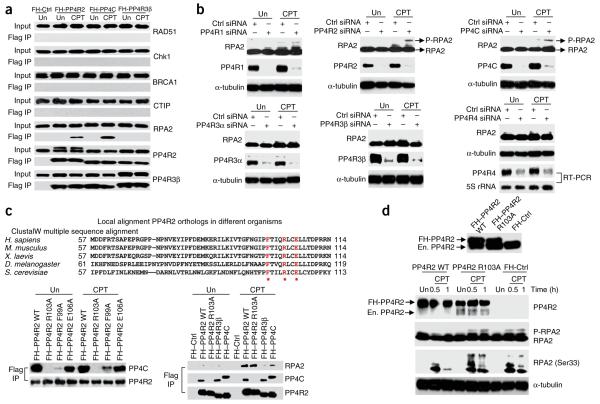
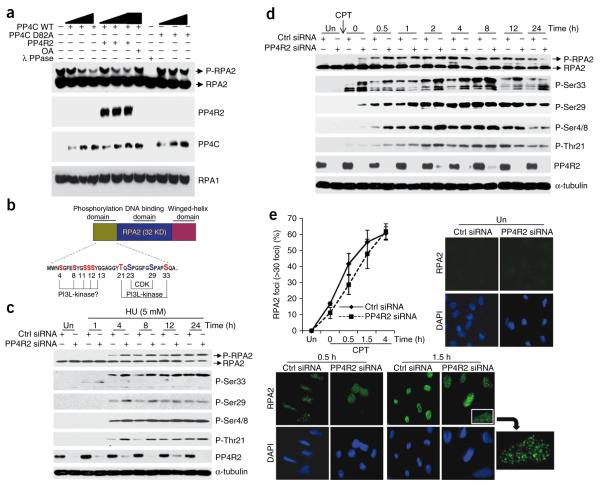
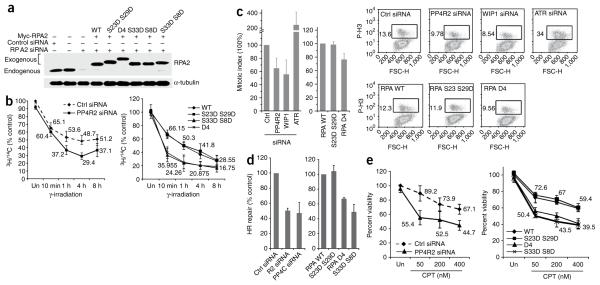
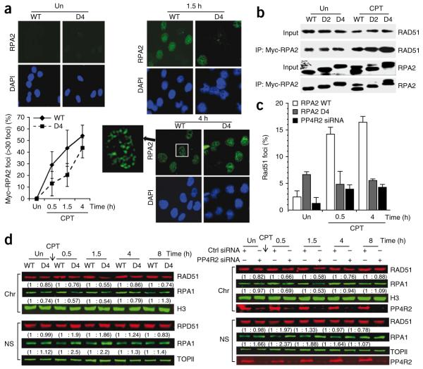
Double-stranded DNA breaks (DSBs) induce a phosphorylation-mediated signaling cascade, but the role of phosphatases in this pathway remains unclear. Here we show that human protein phosphatase 4 (PP4) dephosphorylates replication protein A (RPA) subunit RPA2, regulating its role in the DSB response. PP4R2, a regulatory subunit of PP4, mediates the DNA damage-dependent association between RPA2 and the PP4C catalytic subunit. PP4 efficiently dephosphorylates phospho-RPA2 in vitro, and silencing PP4R2 in cells alters the kinetics and pattern of RPA2 phosphorylation. Depletion of PP4R2 impedes homologous recombination (HR) via inefficient loading of the essential HR factor RAD51, causing an extended G2-M checkpoint and hypersensitivity to DNA damage. Cells expressing phosphomimetic RPA2 mutants have a comparable phenotype, suggesting that PP4-mediated dephosphorylation of RPA2 is necessary for an efficient DNA-damage response. These observations provide new insight into the role and regulation of RPA phosphorylation in HR-mediated repair.


Figures

Figure 1
Figure 1
A PP4 complex interacts with RPA2 in the context of DNA damage, and silencing the PP4 complex enhances RPA2 phosphorylation. (a) Immunoprecipitation from HeLa S3 cells where the indicated FH-tagged PP4 subunits were stably expressed. Anti-Flag antibody was used for immunoprecipitation from untreated (Un) or CPT-treated cells and the western blot probed with indicated antibodies. (b) Silencing PP4R2 and PP4C enhances phospho-RPA2 levels. All known PP4 subunits in U2OS cells were depleted by siRNA transfection, and the phosphorylation of RPA2 was checked by immunoblotting after CPT treatment. Owing to lack of a commercially available antibody, the expression of the newly discovered PP4R4 (ref. 43) subunit was determined by RT-PCR. α-tubulin served as loading control. (c) PP4R2 mediates the interaction of PP4C with RPA2. Top, alignment of the N-terminal region of PP4R2 across different organisms. The highlighted and starred residues represent conserved residues which were consequently mutated to alanine for study. Bottom, HeLa S3 cells stably expressing FH-tagged wild-type (WT) or mutant PP4R2 proteins were subjected to immunoprecipitation after CPT treatment. Immunoprecipitation with FH-PP4C– and FH-PP4R3β–expressing cells were performed as additional controls. (d) Interaction of PP4C and PP4R2 is necessary for dephosphorylation of RPA2. Endogenous PP4R2 was depleted in HeLa S3 cells expressing either FH–PP4R2 WT or FH–PP4R2 R103A by siRNAs targeting the 3′ UTR of PP4R2. Endogenous (En.) and FH-PP4R2 can be distinguished by mobility shift on immunoblot (upper panel). The phosphorylation of RPA2 was observed at indicated times after CPT treatment. Antibody against a RPA2 epitope with phosphorylated Ser33 was used for the immunoblot. α-tubulin served as loading control.
Figure 2
Figure 2
PP4 dephosphorylates RPA2 in vitro and influences the kinetics and pattern of RPA2 phosphorylation in cells. (a) PP4 dephosphorylates RPA2 in vitro. Wild-type PP4C, mutant PP4C (PP4C D82A) and PP4R2 were purified using the baculoviral system and were serially diluted in the phosphatase reaction. PP4C dephosphorylates phospho-RPA2 in a dose-dependent manner. Phosphatase reactions were probed with indicated antibodies. RPA1 serves as a loading control. OA, okadaic acid. (b) Schematic representation of RPA2 with an expanded view of the N-terminal phosphorylation domain. Serine and threonine residues highlighted with red are confirmed or potential DNA damage–responsive PI3-like kinase sites, and blue represents the cell cycle–dependent CDK sites. (c,d) Time course and pattern of RPA2 phosphorylation in PP4R2-depleted U2OS cells after hydroxyurea (HU; c) or CPT (d) treatment using antibodies against specific RPA2 phosphoresidues; untreated (Un) cells serve as controls. In c, cells were incubated in media containing 5 mM hydroxyurea for indicated time periods. In d, cells after CPT treatment were washed and incubated for indicated time points. Elevated levels of hyperphosphorylated RPA2 were detected in PP4R2-silenced cells, a difference found to be more pronounced at early times after DNA damage. (e) Delayed RPA2 focus formation after CPT treatment in PP4R2-depleted U2OS cells. Cells were stained for RPA2 and 4′,6-diamidino-2-phenylindole (DAPI), and images were captured by fluorescence microscopy. The RPA2 focus–positive cells (>30 foci) were quantified manually by comparison with DAPI images (~300 cells total). A magnified image of a RPA2 focus–positive cell is shown.
Figure 3
Figure 3
Hyperphosphorylation of RPA2 affects HR-mediated repair of DSBs and the DNA-damage response. (a) Endogenous RPA2 was replaced with phosphomimetic mutants. Myc-tagged RPA2, wild-type (WT) and indicated RPA2 mutants were expressed in cells depleted of endogenous RPA2. α-tubulin served as loading control. (b) Radioresistant DNA synthesis is inhibited by hyperphosphorylated RPA2. Following silencing of PP4R2 (left) or replacement of endogenous RPA2 with phosphomimetic RPA2 mutants (right), U2OS cells were incubated with 14C, exposed to γ-radiation (undamaged controls, Un), and treated with 3H for indicated time periods. (c) Hyperphosphorylation of RPA2 affects the G2-M checkpoint. U2OS cells similar to those described in b were irradiated and released in medium containing nocodazole, and mitotic cells evaluated by flow cytometry. ATR- and WIP1-silenced cells served as controls. Representative flow cytometry images are shown on the right, and the results from three independent experiments are graphically represented on the left. (d) Measurement of HR-mediated repair of an I-SceI–induced DSB. U2OS cells carrying a single copy of the recombination substrate were transfected with control siRNA, PP4R2 siRNA or PP4C siRNA (left). In a parallel experiment, endogenous RPA2 was replaced with RPA2 WT or indicated mutants. The I-SceI expression plasmid was transfected, and green fluorescent protein–positive cells were measured by flow cytometry. (e) Hyperphosphorylation of RPA2 influences sensitivity to DNA damage. Cells depleted of PP4R2 or expressing RPA2- WT or indicated mutants were incubated with CPT at different concentration.
Figure 4
Figure 4
Premature formation of hyperphosphorylated RPA2 impedes recruitment of RPA and RAD51 to chromatinized DNA damage–induced foci. (a) RPA2 focus formation after CPT or mock (represented as Un) treatment of U2OS cells expressing RPA2 WT or RPA2 D4. Cells were stained for RPA2 and DAPI, and images were captured by fluorescence microscopy. The RPA2 focus–positive cells (>30 foci) were quantified manually by comparison with DAPI images (~300 cells total). Representative images are shown on the left. (b) Interaction of Myc-tagged RPA2 WT, RPA2 D2 or RPA2 D4 with RAD51 after CPT treatment. Cells were subjected to immunoprecipitation after CPT treatment using an anti-Myc and probed for RAD51. (c) Reduced nuclear staining of RAD51 in damaged cells lacking PP4R2 or replaced with RPA2 D4. Following replacement of endogenous RPA2 with phosphomimetic RPA2 mutants or depletion of PP4R2, U2OS cells were mock- or CPT-treated. Cells were then extracted to remove soluble RAD51, stained with DAPI and anti-RAD51, and imaged by epifluorescence microscopy using identical exposure times. RAD51 nuclear staining was quantified and plotted. (d) Nuclear localization of RAD51 is altered by hyperphosphorylated RPA2. RAD51 localization after CPT treatment of U2OS cells, either where endogenous RPA2 was replaced by RPA2 WT or RPA2 D4 (left) or where PP4R2 was silenced (right). Nuclei were biochemically fractionated, and nuclear soluble (NS) and chromatin-bound (Chr) fractions were probed for RAD51. Topoisomerase II (TOPII) and histone H3 (H3) was probed for loading and fractionation controls, respectively. The relative amounts of RAD51 and RPA1 are shown in parentheses.

Similar articles

Cited by

References

    1. Pandey AV, Mellon SH, Miller WL. Protein phosphatase 2A and phosphoprotein SET regulate androgen production by P450c17. J. Biol. Chem. 2003;278:2837–2844. - PubMed
    1. Xu X, Stern DF. NFBD1/KIAA0170 is a chromatin-associated protein involved in DNA damage signaling pathways. J. Biol. Chem. 2003;278:8795–8803. - PubMed
    1. Lal A, et al. miR-24-mediated downregulation of H2AX suppresses DNA repair in terminally differentiated blood cells. Nat. Struct. Mol. Biol. 2009;16:492–498. - PMC - PubMed
    1. Matsuoka S, et al. ATM and ATR substrate analysis reveals extensive protein networks responsive to DNA damage. Science. 2007;316:1160–1166. - PubMed
    1. Chowdhury D, et al. γ-H2AX dephosphorylation by protein phosphatase 2A facilitates DNA double-strand break repair. Mol. Cell. 2005;20:801–809. - PubMed

Publication types

MeSH terms