. 2010 Mar;17(3):289-93.
doi: 10.1038/nsmb.1755. Epub 2010 Feb 14.

The structures of the anti-tuberculosis antibiotics viomycin and capreomycin bound to the 70S ribosome

Affiliations

The structures of the anti-tuberculosis antibiotics viomycin and capreomycin bound to the 70S ribosome

Robin E Stanley et al. Nat Struct Mol Biol. 2010 Mar.

Abstract

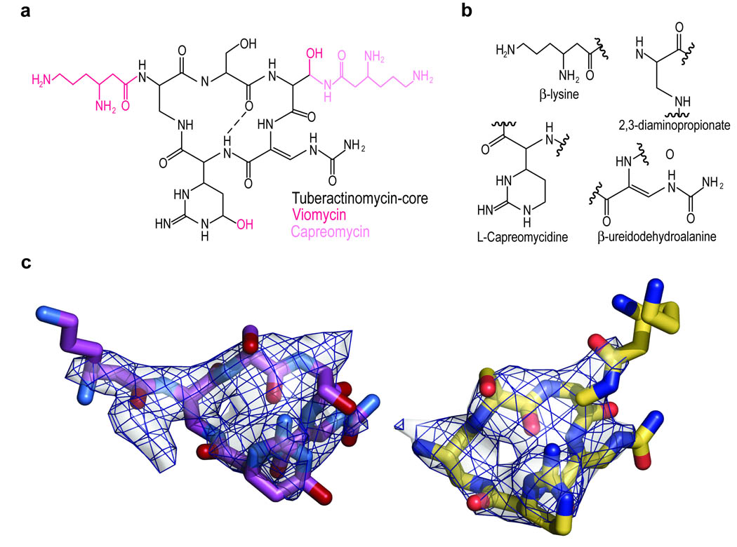
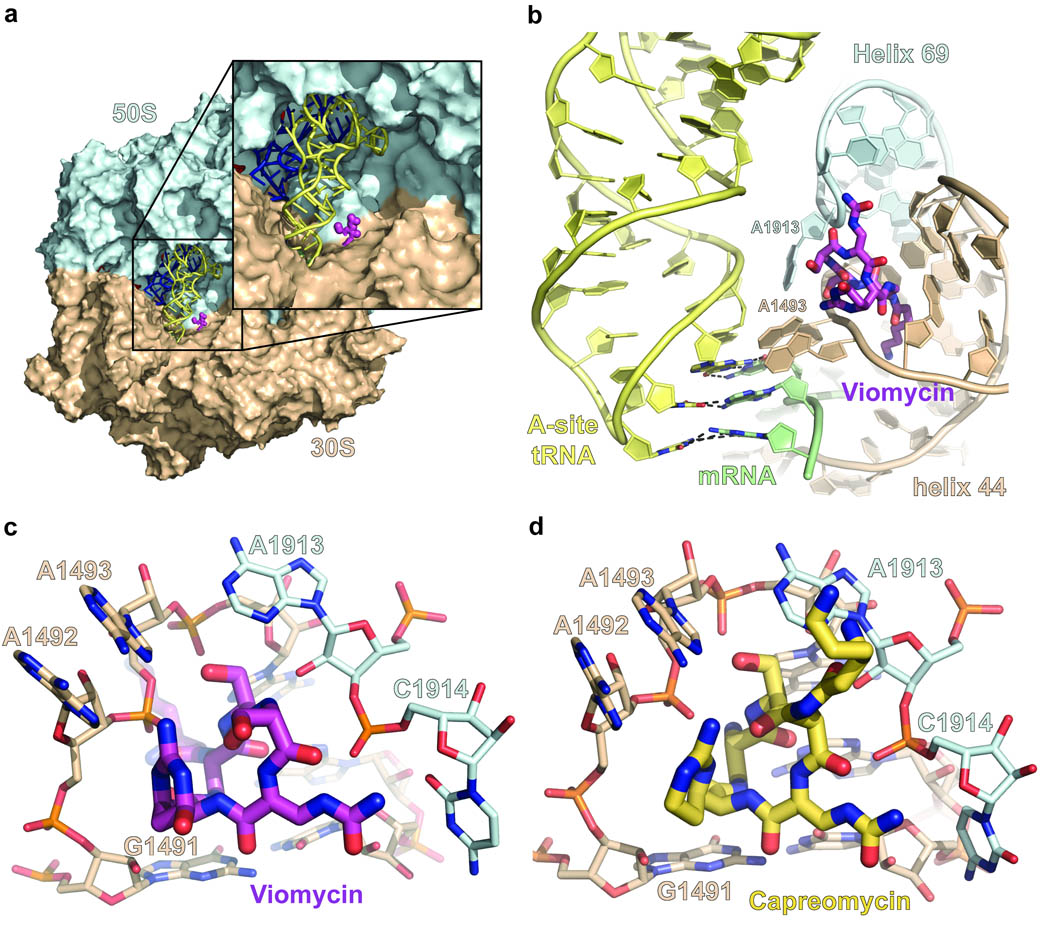
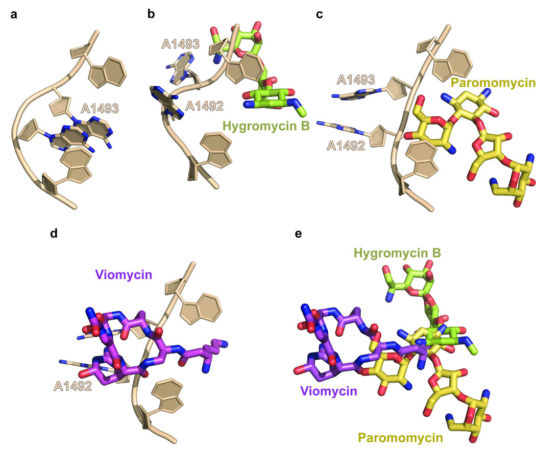
Viomycin and capreomycin belong to the tuberactinomycin family of antibiotics, which are among the most effective antibiotics against multidrug-resistant tuberculosis. Here we present two crystal structures of the 70S ribosome in complex with three tRNAs and bound to either viomycin or capreomycin at 3.3- and 3.5-A resolution, respectively. Both antibiotics bind to the same site on the ribosome, which lies at the interface between helix 44 of the small ribosomal subunit and helix 69 of the large ribosomal subunit. The structures of these complexes suggest that the tuberactinomycins inhibit translocation by stabilizing the tRNA in the A site in the pretranslocation state. In addition, these structures show that the tuberactinomycins bind adjacent to the binding sites for the paromomycin and hygromycin B antibiotics, which may enable the development of new derivatives of tuberactinomycins that are effective against drug-resistant strains.


Figures

Figure 1
Figure 1
Chemical Structure of the tuberactinomycins. (a) The chemical structures of viomycin and capreomycin IA. For simplicity only capreomycin IA is displayed. The other isoforms of capreomycin contain fewer functional groups (supplemental fig.1). The distinctive features of viomycin are in magenta and those of capreomycin are in pink. (b) Chemical structures of the unusual amino acids, from which the tuberactinomycins are built and which are referred to in the text. (c) Unbaised Fo-Fc difference Fourier map for viomycin and capreomyicn bound to a complex of T. thermophilus 70S, mRNA and tRNAs. The difference electron densities are contoured at 2.8 and 3.0 σ for viomycin (left) and capreomycin (right), and the refined models of viomycin (magenta) and capreomyicn (pink) are superimposed.
Figure 2
Figure 2
Binding site of Viomycin and Capreomycin. (a) Overview of the entire structure of the ribosome complex with viomycin. Both the 50S ribosomal subunit (cyan) and the 30S ribosomal subunit (light brown) are shown in surface representation. The A-site tRNA (yellow) and the P-site tRNA (blue) are shown in ribbon, while viomycin (magenta) is shown as a spacefilled model. (b) Close up view of the viomycin binding site as shown in (a). The codon:anticodon base pairing between the A-site tRNA (yellow) and the mRNA (green) is indicated as black dashed lines. (c) Detailed view of the binding site of viomycin (magenta) illustrating its close proximity to A1492, A1493, and G1491 of h44 and A1913 and C1914 of H69. (d) Detailed view of the binding site of capreomycin (yellow) illustrating its close proximity to A1492, A1493, and G1491 of h44 and A1913 and G1941 of H69.
Figure 3
Figure 3
Overlapping Binding Sites. (a) The region around 1492 and 1493 of h44 showing their base stacking in the apo 70S ribosome of E. coli (PDB ID 2AW4) . The nitrogens of the base of A1492 and A1493 are in blue and all other atoms are in light brown. (b) The binding site of hygromycin B showing the “flipped out” orientations of 1492 and 1493. The carbons of hygromycin are in green, the nitrogens in blue and the oxygens in red. The colour coding of the 16S rRNA is the same in (a). The coordinates were taken from PDB IDs 3DF1 . (c) The binding site of paromomycin (yellow). The coordinates were taken from PDB ID 2WDG . (d) The binding site of viomycin (magenta) showing the same “flipped out” orientation of 1492 and 1493 as in (c). (e) Overlap of the binding sites of hygromycin B, paromomycin and viomycin. The superpositioning was based on h44 of the 16S rRNA with the exception of nucleotides A1492 and A1493.

Similar articles

Cited by

References

    1. Guy ES, Mallampalli A. Managing TB in the 21st century: existing and novel drug therapies. Ther Adv Respir Dis. 2008;2:401–408. - PubMed
    1. Hugonnet JE, Tremblay LW, Boshoff HI, Barry CE, 3rd, Blanchard JS. Meropenem-clavulanate is effective against extensively drug-resistant Mycobacterium tuberculosis. Science. 2009;323:1215–1218. - PMC - PubMed
    1. Makarov V, et al. Benzothiazinones kill Mycobacterium tuberculosis by blocking arabinan synthesis. Science. 2009;324:801–804. - PMC - PubMed
    1. WHO. Global Tuberculosis Control: Epidemology, Strategy, Financing: WHO report 2009. Geneva, Switzerland: World Health Organization; 2009. p. 314.
    1. Yamada T, Mizugichi Y, Nierhaus KH, Wittmann HG. Resistance to viomycin conferred by RNA of either ribosomal subunit. Nature. 1978;275:460–461. - PubMed

Methods

    1. Mayer C, RajBhandary UL. Conformational change of Escherichia coli initiator methionyl-tRNA(fMet) upon binding to methionyl-tRNA formyl transferase. Nucleic Acids Res. 2002;30:2844–2850. - PMC - PubMed
    1. Blaha G, Stanley RE, Steitz TA. Formation of the first peptide bond: the structure of EF-P bound to the 70S ribosome. Science. 2009;325:966–970. - PMC - PubMed
    1. Perona JJ, Swanson R, Steitz TA, Soll D. Overproduction and purification of Escherichia coli tRNA(2Gln) and its use in crystallization of the glutaminyl-tRNA synthetase-tRNA(Gln) complex. Journal of Molecular Biology. 1988;202:121–126. - PubMed
    1. Kabsch W. XDS. In: Rossmann MG, Arnold E, editors. International Tables in Crystallography. Vol. F. Dordrecht, Netherlands: Published for the International Union of Crystallography by Springer; 2005. pp. 730–734.
    1. McCoy AJG-K, R.W., Adams PD, Winn MD, Storoni LC, Read RJ. Phaser crystallographic software. J Appl Cryst. 2007;40:658–674. - PMC - PubMed

Publication types