

Transforming growth factor-beta1 upregulates the expression of CXC chemokine receptor 4 (CXCR4) in human breast cancer MCF-7 cells
- PMID: 20154716
- PMCID: PMC4002409
- DOI: 10.1038/aps.2009.204
Transforming growth factor-beta1 upregulates the expression of CXC chemokine receptor 4 (CXCR4) in human breast cancer MCF-7 cells
Abstract
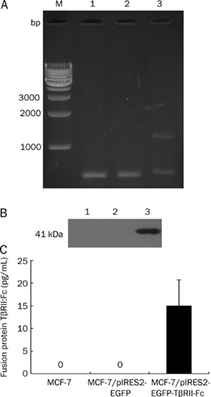
Aim: To investigate whether rhTGF-beta1 or a recombinant vector encoding a fusion protein comprising an extracellular domain of TGF-beta receptor II and an IgG Fc fragment) affects the regulation of CXC chemokine receptor 4 (CXCR4) expression in MCF-7 human breast cancer cells.
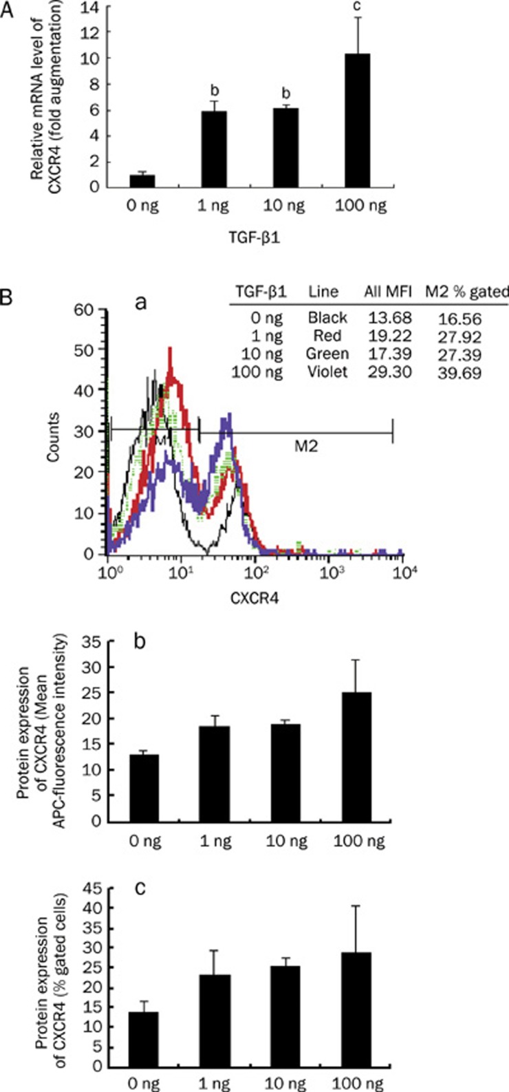
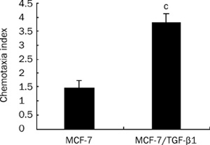
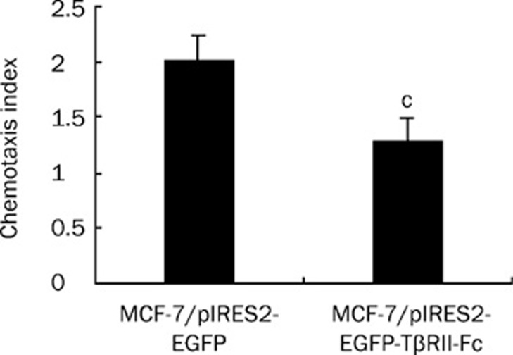
Methods: MCF-7 breast cancer cells were treated with rhTGF-beta1 or transfected with a recombinant vector, pIRES2-EGFP-TbetaRII-Fc. Expression of CXCR4 in these cells was then analyzed at the mRNA and protein levels by quantitative RT-PCR and flow cytometry assay, respectively. A transwell assay was used to measure the chemotactic response of these cells to SDF-1alpha.
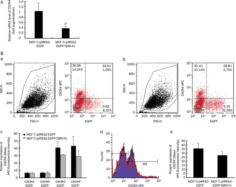
Results: CXCR4 mRNA and protein expression were upregulated in TGF-beta1-treated MCF-7 cells. These cells also demonstrated an enhanced chemotactic response to SDF-1alpha. In MCF-7 cells transiently transfected with pIRES2-EGFP-TbetaRII-Fc, a fusion protein named TbetaRII-Fc (approximately 41 kDa) was produced and secreted. In these transfected cells, there was a reduction in CXCR4 expression and in the SDF-1alpha-mediated chemotactic response.
Conclusion: TGF-beta1 upregulated CXCR4 expression in MCF-7 cells, which subsequently enhanced the SDF-1alpha-induced chemotactic response. The results suggest a link between TGF-beta1 and CXCR4 expression in MCF-7 human breast cancer cells, which may be one of the mechanisms of TGF-beta1-mediated enhancement of metastatic potential in breast cancer cells.
Figures
Similar articles
-
TLR4 ligand/H₂O₂ enhances TGF-β1 signaling to induce metastatic potential of non-invasive breast cancer cells by activating non-Smad pathways.PLoS One. 2013 May 29;8(5):e65906. doi: 10.1371/journal.pone.0065906. Print 2013. PLoS One. 2013. PMID: 23734265 Free PMC article.
-
Induction of chemokine receptor CXCR4 expression by transforming growth factor-β1 in human basal cell carcinoma cells.J Dermatol Sci. 2013 Nov;72(2):123-33. doi: 10.1016/j.jdermsci.2013.06.011. Epub 2013 Jul 1. J Dermatol Sci. 2013. PMID: 23856244
-
Expression of transforming growth factor-beta receptor type II and tumorigenicity in human breast adenocarcinoma MCF-7 cells.J Cell Physiol. 1998 Aug;176(2):424-34. doi: 10.1002/(SICI)1097-4652(199808)176:2<424::AID-JCP21>3.0.CO;2-1. J Cell Physiol. 1998. PMID: 9648930
-
SDF-1α reduces fibronectin expression in rat mesangial cells induced by TGF-β1 and high glucose through PI3K/Akt pathway.Exp Cell Res. 2013 Jul 15;319(12):1796-1803. doi: 10.1016/j.yexcr.2013.03.030. Epub 2013 Apr 6. Exp Cell Res. 2013. PMID: 23567184
-
[Construction of recombinant adenovirus vector expressing extracellular domain of TbetaR-II-RANTES fusion gene and its anti-tumor effects].Zhonghua Zhong Liu Za Zhi. 2007 Jun;29(6):405-10. Zhonghua Zhong Liu Za Zhi. 2007. PMID: 17974270 Chinese.
Cited by
-
CXCR7 promotes migration and invasion in head and neck squamous cell carcinoma by upregulating TGF-β1/Smad2/3 signaling.Sci Rep. 2019 Dec 2;9(1):18100. doi: 10.1038/s41598-019-54705-x. Sci Rep. 2019. PMID: 31792315 Free PMC article.
-
CXCL12 chemokine and GABA neurotransmitter systems crosstalk and their putative roles.Front Cell Neurosci. 2014 Apr 28;5:115. doi: 10.3389/fncel.2014.00115. eCollection 2014. Front Cell Neurosci. 2014. PMID: 24808825 Free PMC article. Review.
-
Local CXCR4 Upregulation in the Injured Arterial Wall Contributes to Intimal Hyperplasia.Stem Cells. 2016 Nov;34(11):2744-2757. doi: 10.1002/stem.2442. Epub 2016 Jul 17. Stem Cells. 2016. PMID: 27340942 Free PMC article.
-
Immunohistochemical expression of CXCR4 on breast cancer and its clinical significance.Anal Cell Pathol (Amst). 2015;2015:891020. doi: 10.1155/2015/891020. Epub 2015 Jun 16. Anal Cell Pathol (Amst). 2015. PMID: 26161302 Free PMC article. Review.
-
A Novel Necroptosis-Associated lncRNA Signature Can Impact the Immune Status and Predict the Outcome of Breast Cancer.J Immunol Res. 2022 May 5;2022:3143511. doi: 10.1155/2022/3143511. eCollection 2022. J Immunol Res. 2022. PMID: 35578667 Free PMC article.
References
-
- Derynck R, Akhurst RJ, Balmain A. TGF-β signaling in tumor suppression and cancer progression. Nat Genet. 2001;29:117–29. - PubMed
-
- Müller A, Homey B, Soto H, Ge N, Catron D, Buchanan ME, et al. Involvement of chemokine receptors in breast cancer metastasis. Nature. 2001;410:50–6. - PubMed
-
- Bleul CC, Farzan M, Choe H, Parolin C, Clark-Lewis I, Sodroski J, et al. The lymphocyte chemoattractant SDF-1 is a ligand for LESTR/fusin and blocks HIV-1 entry. Nature. 1996;382:829–33. - PubMed
-
- Zlotnik A, Yoshie O. Chemokines: a new classification system and their role in immunity. Immunity. 2000;12:121–7. - PubMed
-
- Balkwill F. Cancer and the chemokine network. Nat Rev Cancer. 2004;4:540–50. - PubMed
Publication types
MeSH terms
Substances
LinkOut - more resources
Full Text Sources
Medical
