. 2010 Apr 29;29(17):2488-98.
doi: 10.1038/onc.2010.7. Epub 2010 Feb 15.

Notch-1 stimulates survival of lung adenocarcinoma cells during hypoxia by activating the IGF-1R pathway

Affiliations

Notch-1 stimulates survival of lung adenocarcinoma cells during hypoxia by activating the IGF-1R pathway

S Eliasz et al. Oncogene. .

Abstract

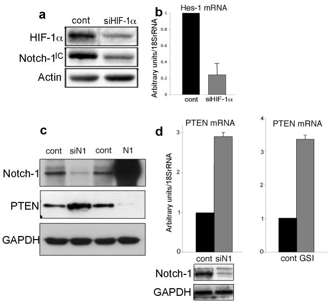
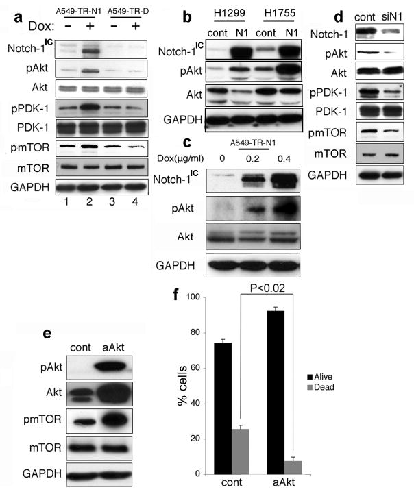
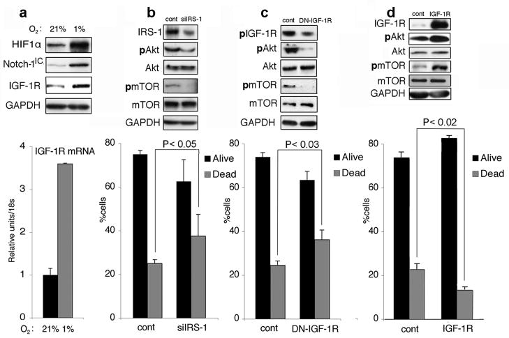
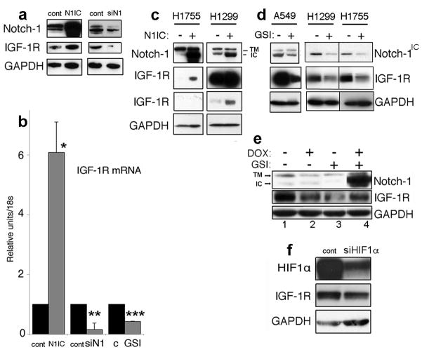
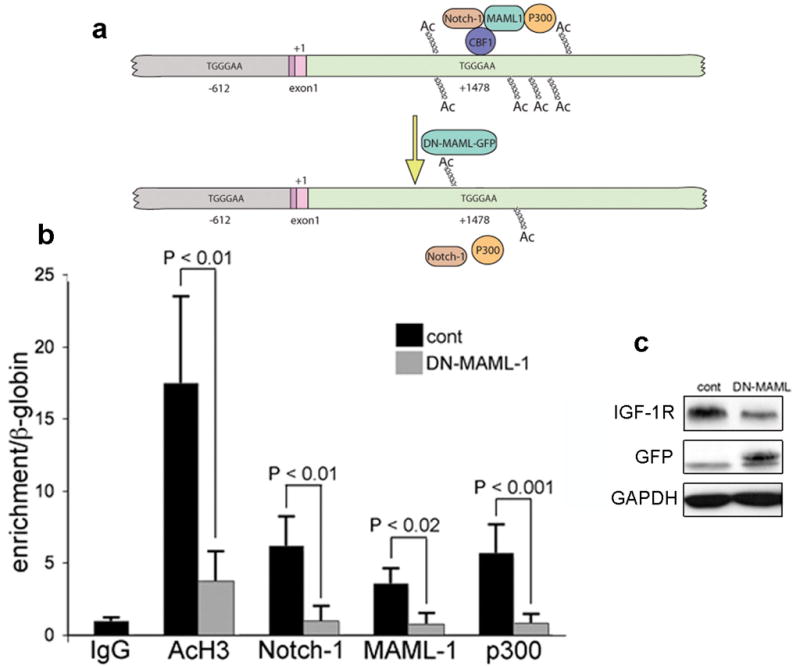
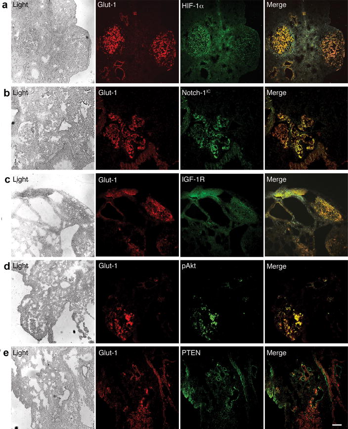
Hypoxic microenvironment supports cancer stem cell survival, causes poor response to anticancer therapy and tumor recurrence. Inhibition of Notch-1 signaling in adenocarcinoma of the lung (ACL) cells causes apoptosis specifically under hypoxia. Here, we found that Akt-1 activation is a key mediator of Notch-1 pro-survival effects under hypoxia. Notch-1 activates Akt-1 through repression of phosphatase and tensin (PTEN) homolog expression and induction of the insulin-like growth factor 1 receptor (IGF-1R). The latter seems to be the major determinant of Akt-1 stimulation, as Notch-1 signaling affects Akt-1 activation in PTEN(-/-) ACL cells. Both downregulation of insulin receptor substrate 1 (IRS-1) and dominant-negative IGF-1R sensitized ACL cells to gamma-secretase inhibitor (GSI)-induced apoptosis. Conversely, overexpression of IGF-1R protected ACL cells from GSI toxicity. Inhibition of Notch-1 caused reduced IGF-1R expression, whereas forced Notch-1 expression yielded opposite effects. Chromatin immunoprecipitation experiments suggested Notch-1 direct regulation of the IGF-1R promoter. Experiments in which human ACL cells were injected in mice confirmed elevated and specific co-expression of Notch-1(IC), IGF-1R and pAkt-1 in hypoxic tumor areas. Our data provide a mechanistic explanation for Notch-1-mediated pro-survival function in hypoxic ACL tumor microenvironment. The results identify additional targets that may synergize with Notch-1 inhibition for ACL treatment.


Conflict of interest statement

Conflict of interest. The authors declare no conflict of interest.

Figures

Figure 1
Figure 1
Notch-1 signaling is dependent on HIF-1α and negatively regulates PTEN expression in ACL cells. (a) Representative Western blot analysis of A549 cells transfected with either a control siRNA (cont) or with a siRNA targeting the HIF-1α mRNA (siHIF-1α). Note decreased Notch-1IC expression when HIF-1α was artificially downregulated. (b) Quantitative RT-PCR of the HES-1 mRNA in A549 transfected with either a control siRNA (cont, black column) or with a siRNA targeting the HIF-1α mRNA (siHIF-1α, gray column). mRNA levels were normalized for 18S rRNA expression levels. Columns represent the average of three independent experiments. Error bars represent SD. (c) Western blot analysis of the indicated proteins in H1299 cells transfected with a control siRNA (cont, left), with a siRNA targeting the Notch-1 mRNA (siN1), with a control plasmid (cont, right) or with a plasmid expressing Notch-1IC (N1). (d) Both siRNA to Notch-1 and GSI treatment increase the PTEN mRNA expression levels. H1299 cells were transfected with either a control siRNA (cont, left) or with a siRNA targeting the Notch-1 mRNA (siN1). Alternatively, H1299 cells were treated with either DMSO (cont, right) or with 20 μM MRK-003 for 48 hr (GSI). The mRNA levels were measured using Q-RT-PCR and normalized for 18S rRNA content. Note that both genetic and chemical inhibition of Notch-1 causes increased PTEN mRNA expression. Columns represent average of three independent experiments. Error bars represent SD. We obtained similar results in A549 and H1755 cells (one experiment for each cell line).
Figure 2
Figure 2
Notch-1 regulates Akt-1 phosphorylation in ACL cells under hypoxia; Akt-1 activation protects ACL cells from apoptosis triggered by Notch inhibition under hypoxia. (a) A549 cells were transduced with an empty lentiviral vector (A549-TR-D) or with a lentivirus in which Notch-1IC was under the control of a doxycycline-inducible promoter (A549-TR-N1). Upon doxycycline induction of Notch-1IC, increased phosphorylation of Akt-1, PDK-1 and mTOR was observed (compare lanes 2 and 4). (b) Transient transfection of ACL cells with a plasmid expressing Notch-1IC causes Akt-1 phosphorylation in ACL cells. Western blot analysis of the indicated proteins; cont, ACL cells transfected with pCDNA3; N1, ACL cells transfected with a plasmid expressing Notch-1IC. (c) A549-TR-N1 cells were treated with the indicated concentrations of doxycycline. Notch-1 appeared to stimulate Akt-1 phosphorylation in a dose-dependent fashion. (d) Notch-1 artificial downregulation obtained using a siRNA to Notch-1 caused decreased activation of PDK-1, Akt-1 and mTOR. A549 cells were transfected with either a control siRNA (cont) or a siRNA targeting the Notch-1 mRNA (siN1). 48 hr after transfection cells were assayed by Western blot. Similar results were obtained in H1299 and H1755 cells (not shown). Constitutively active Akt-1 protects ACL cells from GSI-induced cell death under hypoxia. (e) Western blot analysis of the indicated proteins in A549 cells transfected with either a control vector (cont) or with a vector expressing an NH2-terminal myristoylatable Akt-1 (constitutively active Akt, or aAkt). (f) Annexin V/7-AAD staining of A549 cells transfected with either a control plasmid (cont) or with aAkt, and exposed to 20 μM MRK-003 for 48 hr. The histogram represents the average of three independent experiments. Error bars represent SD. Black columns: alive cells; gray columns: dead cells (annexin V positive). Of note, A549 cells grown under hypoxia (with no additional manipulations) have 6.42 ± 2.11% annexin V positive cells. This number is not significantly different from cells transfected with aAkt-1 and treated with MRK-003.
Figure 3
Figure 3
Notch-1 modulates the activation levels of PDK-1, Akt-1 and mTOR in PTEN null ACL cells. Western blot analysis of the indicated proteins. H1650 cells were transfected with either a control siRNA (cont), with a siRNA targeting the Notch-1 mRNA (siN1), with a control plasmid (pcDNA) or with a plasmid expressing Notch-1IC (N1IC). Cells were analyzed 48 hr after transfection. Note that artificial downregulation of Notch-1 depresses the activation of the PDK-1/Akt-1/mTOR axis, while forced expression of Notch-1IC elicits opposite effects in ACL cells lacking PTEN.
Figure 4
Figure 4
IGF-1R signaling affects ACL susceptibility to GSI treatment. (a) A549 cells cultured in a standard incubator or under hypoxia. Top, representative Western blot analysis of the indicated proteins; bottom, quantitative RT-PCR of the IGF-1R mRNA in A549 grown either in normoxia (21% O2, black column) or under hypoxia (1% O2, gray columns). Messenger RNA levels were normalized for 18S rRNA content. Columns represent the average of three independent experiments. Error bars represent SD. (b) siRNA to IRS-1 sensitizes ACL cells to MRK-003 induced cell death under hypoxia. A549 cells were transfected with either a control siRNA (cont) or with a siRNA targeting the IRS-1 mRNA. Top, representative Western blot analysis of the indicated proteins; bottom, annexin V/7AAD staining of cells transfected with siRNA and exposed to 20 μM MRK-003 under hypoxia for 48 hr. Black column, alive cells; gray column, annexin V positive cells. Columns represent the average of three independent experiments. Error bars representing SD. P value is indicated. (c) Dominant negative IGF-1R sensitizes ACL cells to MRK-003 induced cell death under hypoxia. A549 cells were transfected with either a control plasmid (cont) or with a plasmid expressing a dominant-negative IGF-1R. Top, representative Western blot analysis of the indicated proteins; bottom, annexin V/7AAD staining of cells transfected with plasmids and exposed to 20 μM MRK-003 under hypoxia for 48 hr. Black column, alive cells; gray column, annexin V positive cells. Columns represent the average of three independent experiments. Error bars represent SD. P value is indicated. (d) Transfection of IGF-1R under the control of a CMV promoter protects ACL cells from MRK-003 induced cell death under hypoxia. A549 cells were transfected with either a control plasmid (cont) or with a plasmid expressing full-length IGF-1R. Top, representative Western blot analysis of the indicated proteins; bottom, annexin V/7AAD staining of cells transfected with plasmids and exposed to 20 μM MRK-003 under hypoxia for 48 hr. Black column, alive cells; gray column, annexin V positive cells. Columns represent the average of three independent experiments. Error bars represent SD. P value is indicated. As all experiments where the oxygen concentration is not indicated, the analyses in (b), (c) and (d) were performed under hypoxia.
Figure 5
Figure 5
Notch-1 regulates IGF-1R expression. (a) Representative Western blot of the indicated proteins in A549 cells trasfected with a control plasmid (cont, left) or with a plasmid expressing Notch-1IC (N1IC), or transfected with a control siRNA (cont, right) or with a siRNA targeting the Notch-1 mRNA (siN1). (b) quantitative RT-PCR of the IGF-1R mRNA in A549 transfected with the nucleic acids described above. Additionally, A549 were exposed to DMSO (C) or with 20 μM MRK-003 (GSI) for 48 hr under hypoxia. mRNA values were normalized for 18S rRNA expression levels. *: P< 0.001; **: P< 0.001; ***: P< 0.01. Columns represent the average of three independent experiments. Error bars represent SD. (c) Western blot analysis of the indicated proteins. H1755 and H1299 cells were either transfected with a control plasmid (N1IC −) or with a plasmid expressing Notch-1IC (N1IC +). The two panels for IGF-1R represent two different exposure times of the same gel. Note increased IGF-1R expression when Notch-1IC was overexpressed. Additional abbreviations: TM, transmembrane, uncleaved Notch-1; IC, Notch-1IC. (d) Western blot analysis of the indicated proteins of different ACL lines (indicated) exposed to 20 μM MRK-003 (GSI+) or to vehicle alone (GSI−) for 48 hr. (e) A549-TR-DEST (lanes 1 and 2) and A549-TR-N1 (lanes 3 and 4) were exposed to either 1 μg/ml doxycycline (DOX), 20 μM MRK-003 (GSI) or both for 48 hr. Note that doxycycline treatment alone reduced the IGF-1R expression levels. A more marked effect was elicited by MRK-003 treatment on the expression of the IGF-1R. However, induction (obtained through doxycycline treatment) of Notch-1IC led to a 4.8-fold increase in the IGF-1R expression levels (compare lanes 3 and 4). Notch-1IC failed to restore the IGF-1R expression levels completely possibly because of the compounding doxycycline effects (compare lanes 1 and 2). (f) Artificial downregulation of HIF-1α causes reduced expression of the IGF-1R in ACL cells under hypoxia. A549 cells were transfected with either a control siRNA or with a siRNA targeting the HIF-1α mRNA. 48 hr after transfection cells were assayed by Western blot analysis for the indicated proteins.
Figure 6
Figure 6
Notch-1 associates with the +1478 region of the IGF-1R gene alongside MAML-1 and p300; transfection of ACL cells with dominant negative MAML-1 disrupts such association. (a) Schematic representation of the region of the IGF-1R that we analyzed by ChIP for the indicated proteins. We focused our attention to the two DNA fragments containing canonical CBF-1 binding sites (indicated). Gray: region upstream of the initiation of transcription; darker pink: 5′ UTR; lighter pink: first fragment of the CDS (+1, initiation of translation); green: 5′ region of the first intron. Top; at the +1478 region we detected association of Notch-1, MAML-1 and p300. Upon transfection with dominant negative MAML-1 (DN-MAML-GFP) (bottom) these proteins are no longer associated to this DNA region, and the amount of acetylated histone H3 is significantly reduced (see panel b). (b) Quantitative ChIP of the +1478 region (62 bp-long TaqMan fragment) after immunoprecipitation of the indicated proteins in A549 cells transfected with a control plasmid (black columns) or with a plasmid coding for dominant negative MAML-1 (gray columns). The IgG column represents the average of three different pre-immune IgGs. Each column represents the average of four (black columns) or five (gray columns) independent experiments (IgG colum represents 9 independent immunoprecipitations). Error bars represent SD. (c) Representative Western blot analysis of A549 cells transfected with either control plasmid or the plasmid expressing dominant negative (dn) MAML-1 (visualized with an antibody against GFP). Note that upon dnMAML-1 expression (24 hr after transfection), the expression levels of IGF-1R are substantially decreased. All the experiments shown in this figure were conducted 24 hr after transfection. This was because dnMAML-1 appeared to be toxic to ACL cells for longer time-points after transfection.
Figure 7
Figure 7
Notch-1IC is coexpressed in hypoxic tumor cells together with IGF-1R and phosphorylated Akt-1. Coimmunofluorescence stainings of the indicated proteins on 8 μm-thick slides obtained from tumor-bearing lungs of SCID mice injected with A549 cells through the tail vein. (a) Co-expression of GLUT-1 and HIF-1α. (b) Co-expression of GLUT-1 and Notch-1IC. (c) Expression of IGF-1R is maximal in GLUT-1 expressing tumor cells. (d) Phosphorylated (S473) Akt-1 is detected in GLUT-1 expressing (hypoxic) tumor cell only. (e) PTEN is expressed in GLUT-1 negative tumor cell only. Note that GLUT-1 and PTEN positive cells appear to be contiguous. This is likely an event due to the lateral, asymmetric nature of Notch signaling. Bar, 20 μm. Similar results were obtained in mice injected with H1299 and H1755.

Similar articles

Cited by

References

    1. Adams TE, Epa VC, Garrett TP, Ward CW. Structure and function of the type 1 insulin-like growth factor receptor. Cell Mol Life Sci. 2000;57:1050–1093. - PMC - PubMed
    1. Alessi DR, James SR, Downes CP, Holmes AB, Gaffney PR, Reese CB, et al. Characterization of a 3-phosphoinositide-dependent protein kinase which phosphorylates and activates protein kinase Balpha. Curr Biol. 1997;7:261–269. - PubMed
    1. Artavanis-Tsakonas S, Rand MD, Lake RJ. Notch signaling: cell fate control and signal integration in development. Science. 1999;284:770–776. - PubMed
    1. Baserga R, Hongo A, Rubini M, Prisco M, Valentinis B. The IGF-I receptor in cell growth, transformation and apoptosis. Biochim Biophys Acta. 1997;1332:F105–F126. - PubMed
    1. Behrooz A, Ismail-Beigi F. Stimulation of Glucose Transport by Hypoxia: Signals and Mechanisms. News Physiol Sci. 1999;14:105–110. - PubMed

Publication types