Review
. 2010 Mar;119(3):277-90.
doi: 10.1007/s00401-010-0652-7. Epub 2010 Feb 14.

Vascular pathology in the aged human brain

Affiliations
Review

Vascular pathology in the aged human brain

Lea Tenenholz Grinberg et al. Acta Neuropathol. 2010 Mar.

Abstract

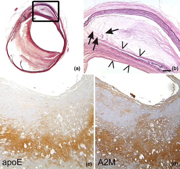
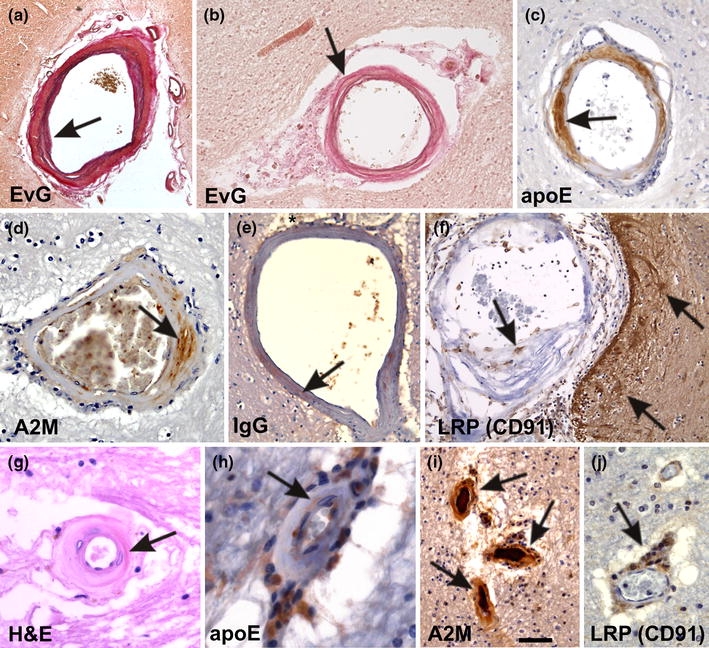
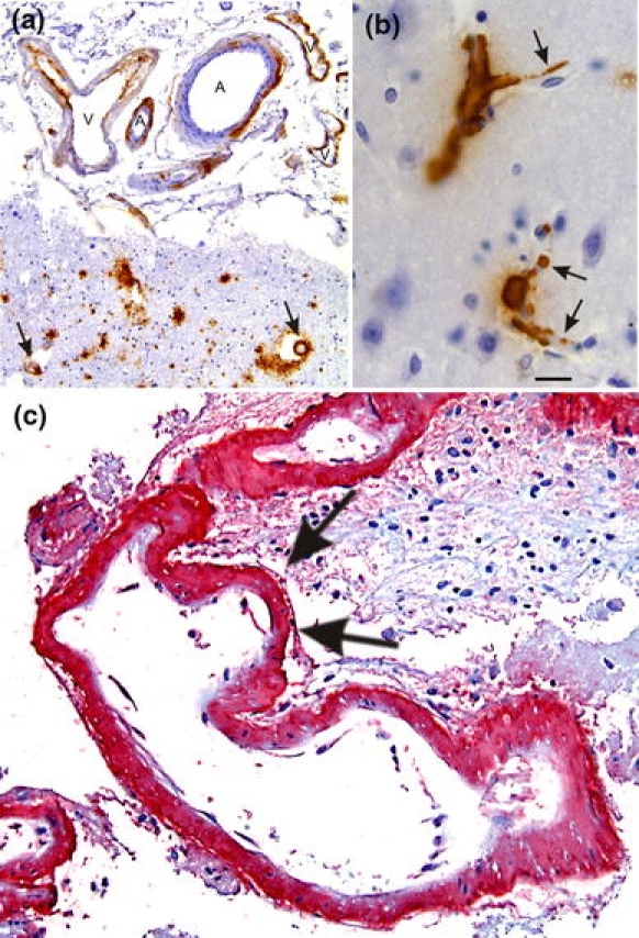
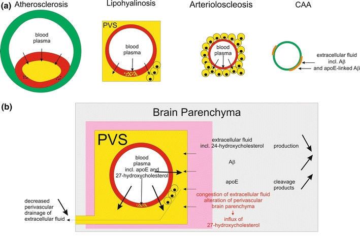
Cerebral atherosclerosis (AS), small vessel disease (SVD), and cerebral amyloid angiopathy (CAA) are the most prevalent arterial disorders in the aged brain. Pathogenetically, AS and SVD share similar mechanisms: plasma protein leakage into the vessel wall, accumulation of lipid-containing macrophages, and fibrosis of the vessel wall. CAA, on the other hand, is characterized by the deposition of the amyloid beta-protein in the vessel wall. Despite these differences between CAA, AS and SVD, apolipoprotein E (apoE) is involved in all three disorders. Such a pathogenetic link may explain the correlations between AS, SVD, CAA, and Alzheimer's disease in the brains of elderly individuals reported in the literature. In addition, AS, SVD, and CAA can lead to tissue lesions such as hemorrhage and infarction. Moreover, intracerebral SVD leads to plasma protein leakage into the damaged vessel wall and into the perivascular space resulting in a blood-brain barrier (BBB) dysfunction. This SVD-related BBB dysfunction is considered to cause white matter lesions (WMLs) and lacunar infarcts. In this review, we demonstrate the relationship between AS, SVD, and CAA as well as their contribution to the development of vascular tissue lesions and we emphasize an important role for apoE in the pathogenesis of vessel disorders and vascular tissue lesions as well as for BBB dysfunction on WML and lacunar infarct development.


Figures

Fig. 1
Fig. 1
a, b The left internal carotid artery of a 79-year-old man exhibits severe atherosclerotic changes (Type 5 according to Stary [119]). There is a thinning of the lamina media, proliferation and lipid accumulation in the intima including cholesterol clefts (arrows). The necrotic core is covered by a fibromuscular tissue layer (arrowheads) indicative for Stary Type 5 lesions. b Corresponds to a high magnification view of the boxed area in (a). ApoE (c) and A2M (d) occur in the plaque core of an AS plaque. Staining in (a, b) Elastica van Gieson (EVG), c anti-apoE [Covance (Dedham, USA), D6E10, 1/500, formic acid and microwave pretreatment], d anti-A2M [BioMac (Germany, Leipzig), polyclonal rabbit, 1/5,000]. The calibration bar in (b) corresponds to: a 400 μm, b 90 μm, c, d 70 μm
Fig. 2
Fig. 2
Small vessel disease-related changes. a A leptomeningeal artery shows intima proliferation and a splitting of the internal elastic lamina (arrow). These changes are related to small vessel arteriosclerosis/atherosclerosis. b A white matter artery exhibits fibrosis, lipohyalinosis of the vessel wall, and fibrinoid necrosis (arrow). Lipohyalinosis affected vessels exhibit the plasma proteins apoE (c), A2M (d), and IgG (e) within the vessel wall (arrows in ce) indicating the leakage of plasma proteins into the vessel wall and into the perivascular space (asterisk in e). f Macrophages within the lipohyalinotic lesions and perivascular astrocytes strongly exhibit the A2M and apoE receptor LRP (CD91) (arrows) indicating that these cells are capable of taking up A2M and apoE. g Arteriolosclerosis of a white matter artery shows severe hyalinization (arrow) of the vessel wall. hj ApoE and A2M were observed within the vessel wall of arteriolosclerotic vessels (arrows in h, i). Within the enlarged perivascular spaces high numbers of apoE (h), A2M (i), and LRP-positive cells (arrow in j) were observed indicating that these perivascular cells accumulate apoE and A2M due to an insufficient perivascular drainage. These perivascular macrophages are often Prussian blue negative and do not necessarily represent hemorrhagic residues [129]. Stainings in aj as indicated. Anti-apoE and anti-A2M staining was performed as indicated in Fig. 1. For anti-IgG and anti-LRP immunohistochemistry the following antibodies were used [anti-IgG: polyclonal goat; Biomeda, Foster City, CA; 1/100; microwave pretreatment; anti-LRP (anti-CD91): α2-M-R-II2C7; BioMac, Leipzig, Germany; 1/150; microwave and protease pretreatment]. The calibration bar in i corresponds to: a 300 μm, b 80 μm, c 40 μm, d, j 35 μm, e, f 60 μm, g 20 μm, h 16 μm, i 50 μm. a and b are reproduced from Thal et al. 2003 [129] with kind permission
Fig. 3
Fig. 3
Cerebral amyloid angiopathy (CAA). a Aβ deposition in the vessel wall of leptomeningeal arteries (A) and veins (V) as well as in cortical arteries (arrows). b Capillary CAA is characterized by Aβ deposits at the basement membrane of cortical capillaries (arrows). c Severe CAA in a case of CAA-related hemorrhage. The CAA-affected artery exhibits multiple aneurysmal dilations of the vessel wall as indicated by arrows. Aβ deposits are stained in red (permanent red; DAKO, Glostrup, Denmark) with an antibody against Aβ17–24 (4G8, Covance, Dedham, USA, 1/5,000, pretreatment with formic acid). The same antibody was also used in figures a and b but 3,3-diaminobencidine–HCl was used as chromogen. The calibration bar in b corresponds to: a 85 μm, b, c 20 μm
Fig. 4
Fig. 4
Plasma protein leakage induced by vessel disorders and its relation to perivascular alterations of the brain parenchyma. a This schematic representation shows that plasma proteins occur (1) in the plaque cores of AS plaques, (2) in the vessel wall of lipohyalinotic vessels as well as in the perivascular space and in macrophages within the perivascular space, and (3) in the vessel wall of arteriolosclerotic vessels as well as in accompanying macrophages. CAA, on the other hand, is characterized by the deposition of proteins of the extracellular fluid of the brain, i.e. Aβ [15] and apoE [133]. b Impact of plasma protein leakage into the brain. Physiologically, extracellular fluid is drained into the perivascular space and along the vascular basement membranes [16, 60, 155]. In the event of SVD, there is plasma protein leakage into the vessel wall and into the perivascular space [139] resulting in (1) a competition between leaking plasma and extracellular fluid from the brain for perivascular drainage and (2) the congestion of extracellular fluid leading to the accumulation and/or alternative processing of proteins of the extracellular fluid, and 3) the influx of the peripheral cholesterol metabolite 27-hydroxycholesterol into the brain [46, 127, 139]. The influx of 27-hydroxycholesterol into the brain is accompanied by decreased levels of brain derived 24-hydroxycholesterol indicating a reduction in the cerebral 24-hydroxycholesterol production [46]

Similar articles

Cited by

References

    1. Alafuzoff I, Adolfsson R, Grundke-Iqbal I, Winblad B. Perivascular deposits of serum proteins in cerebral cortex in vascular dementia. Acta Neuropathol. 1985;66:292–298. - PubMed
    1. Alafuzoff I, Thal DR, Arzberger T, Bogdanovic N, Al-Sarraj S, Bodi I, Boluda S, Bugiani O, Duyckaerts C, Gelpi E, Gentleman S, Giaccone G, Graeber M, Hortobagyi T, Hoftberger R, Ince P, Ironside JW, Kavantzas N, King A, Korkolopoulou P, Kovacs GG, Meyronet D, Monoranu C, Nilsson T, Parchi P, Patsouris E, Pikkarainen M, Revesz T, Rozemuller A, Seilhean D, Schulz-Schaeffer W, Streichenberger N, Wharton SB, Kretzschmar H. Assessment of beta-amyloid deposits in human brain: a study of the BrainNet Europe Consortium. Acta Neuropathol. 2009;117:309–320. - PMC - PubMed
    1. Attems J. Sporadic cerebral amyloid angiopathy: pathology, clinical implications, and possible pathomechanisms. Acta Neuropathol. 2005;110:345–359. - PubMed
    1. Attems J, Jellinger KA. Only cerebral capillary amyloid angiopathy correlates with Alzheimer pathology—a pilot study. Acta Neuropathol (Berl) 2004;107:83–90. - PubMed
    1. Beach TG, Wilson JR, Sue LI, Newell A, Poston M, Cisneros R, Pandya Y, Esh C, Connor DJ, Sabbagh M, Walker DG, Roher AE. Circle of Willis atherosclerosis: association with Alzheimer’s disease, neuritic plaques and neurofibrillary tangles. Acta Neuropathol. 2007;113:13–21. - PubMed

Publication types

Substances