

Differential proteomic analysis of a highly metastatic variant of human breast cancer cells using two-dimensional differential gel electrophoresis
- PMID: 20155427
- PMCID: PMC11828232
- DOI: 10.1007/s00432-010-0812-0
Differential proteomic analysis of a highly metastatic variant of human breast cancer cells using two-dimensional differential gel electrophoresis
Abstract
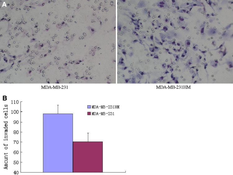
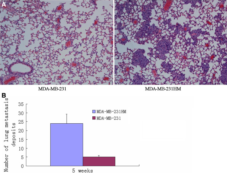
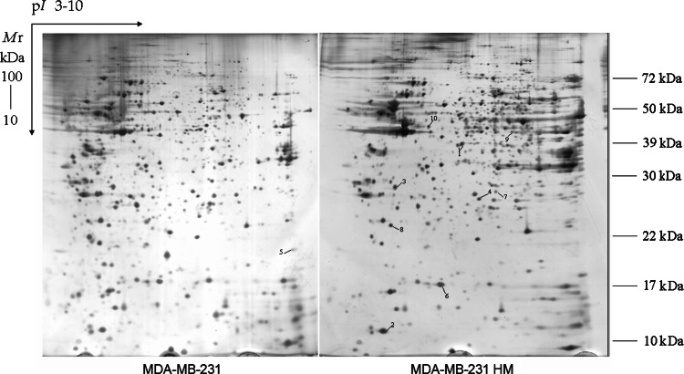
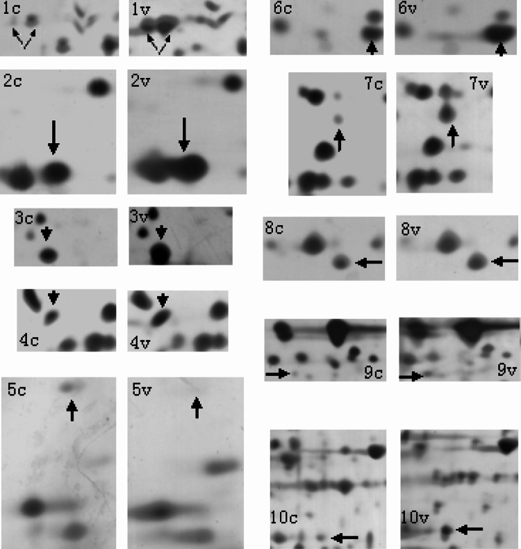
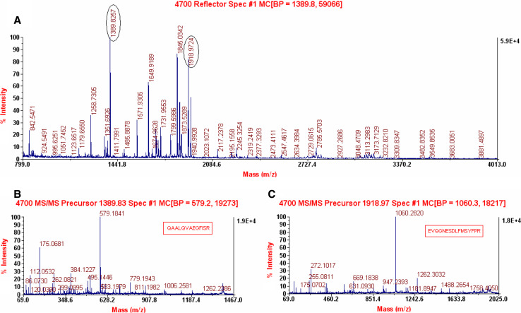
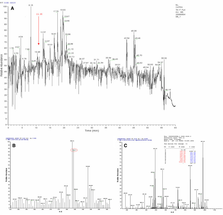
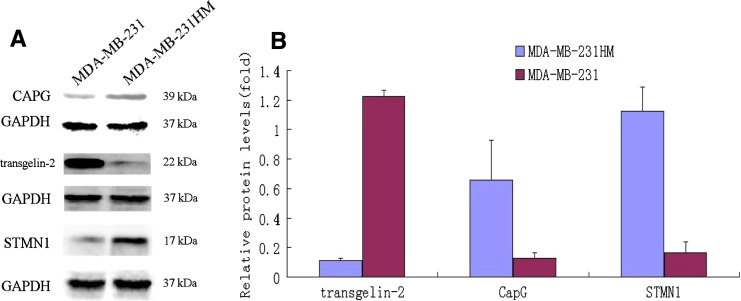
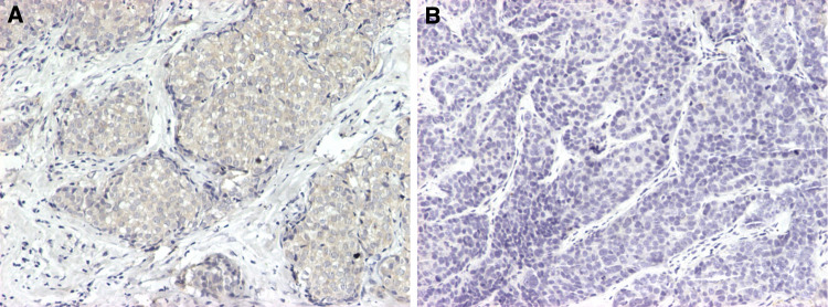
Distant metastasis represents the major lethal cause of breast cancer. To understand the molecular mechanisms of breast cancer metastasis and identify markers with metastatic potential, we established a highly metastatic variant of parental MDA-MB-231 cells (MDA-MB-231HM). Using two-dimensional electrophoresis (2-DE), we performed a proteomic comparison of the two kinds of cells. As much as 51 protein spots were differentially expressed between the selected variant and its parental counterpart in at least 3 experiments. Ten unique proteins were identified using matrix-assisted laser desorption/ionization-time of flight (MALDI-TOF) mass spectrometry (MS), liquid chromatography-electrospray ionization-tandem mass spectrometry (LC-ESI-MS/MS), and database searching software. Among them, nine proteins were up-regulated in MDA-MB-231HM cells, including Macrophage-capping protein (CapG), Galectin-1, Chloride intracellular channel protein 1, Endoplasmic reticulum protein ERp29 precursor, Stathmin-1 (STMN1), Isoform 1 of uridine-cytidine kinase 2(UCK2), Rho GDP-dissociation inhibitor 2 (ARHGDIB), isocitrate dehydrogenase [NADP] cytoplasmic (IDH1), and N-myc downstream regulated gene 1 (NDRG1) protein. Only transgelin-2 was down-regulated. Differential expression was confirmed for three proteins including CapG, STMN1, and transgelin-2 by Western blotting analysis. Transgelin-2 was chosen for further verification by immunohistochemistry. The results suggested that 2-DE would be an efficient way to screen the proteins responsible for specific biological function. Furthermore, the findings imply that different proteins may be involved in the metastatic process in breast carcinomas.
Figures
Similar articles
-
Identification of transgelin as a potential novel biomarker for gastric adenocarcinoma based on proteomics technology.J Cancer Res Clin Oncol. 2008 Nov;134(11):1219-27. doi: 10.1007/s00432-008-0398-y. Epub 2008 Apr 30. J Cancer Res Clin Oncol. 2008. PMID: 18446369 Free PMC article.
-
Differential proteomic analysis of human colorectal carcinoma cell lines metastasis-associated proteins.J Cancer Res Clin Oncol. 2007 Oct;133(10):771-82. doi: 10.1007/s00432-007-0222-0. Epub 2007 May 15. J Cancer Res Clin Oncol. 2007. PMID: 17503081 Free PMC article.
-
Comparative proteomic analysis of paclitaxel sensitive A549 lung adenocarcinoma cell line and its resistant counterpart A549-Taxol.J Cancer Res Clin Oncol. 2011 Mar;137(3):521-32. doi: 10.1007/s00432-010-0913-9. Epub 2010 May 25. J Cancer Res Clin Oncol. 2011. PMID: 20499251 Free PMC article.
-
Breast cancer proteomics: a review for clinicians.J Cancer Res Clin Oncol. 2011 Jun;137(6):915-25. doi: 10.1007/s00432-011-0978-0. Epub 2011 Apr 5. J Cancer Res Clin Oncol. 2011. PMID: 21465318 Free PMC article. Review.
-
A systematic review of evidence on malignant spinal metastases: natural history and technologies for identifying patients at high risk of vertebral fracture and spinal cord compression.Health Technol Assess. 2013 Sep;17(42):1-274. doi: 10.3310/hta17420. Health Technol Assess. 2013. PMID: 24070110 Free PMC article.
Cited by
-
Cross-Species Proteomics Identifies CAPG and SBP1 as Crucial Invasiveness Biomarkers in Rat and Human Malignant Mesothelioma.Cancers (Basel). 2020 Aug 27;12(9):2430. doi: 10.3390/cancers12092430. Cancers (Basel). 2020. PMID: 32867073 Free PMC article.
-
Far upstream element-binding protein 1 is a prognostic biomarker and promotes nasopharyngeal carcinoma progression.Cell Death Dis. 2015 Oct 15;6(10):e1920. doi: 10.1038/cddis.2015.258. Cell Death Dis. 2015. PMID: 26469968 Free PMC article.
-
Placenta-specific novel splice variants of Rho GDP dissociation inhibitor β are highly expressed in cancerous cells.BMC Res Notes. 2012 Dec 3;5:666. doi: 10.1186/1756-0500-5-666. BMC Res Notes. 2012. PMID: 23206989 Free PMC article.
-
Superior Therapeutic Efficacy of Nanoparticle Albumin Bound Paclitaxel Over Cremophor-Bound Paclitaxel in Experimental Esophageal Adenocarcinoma.Transl Oncol. 2018 Apr;11(2):426-435. doi: 10.1016/j.tranon.2018.01.022. Epub 2018 Feb 20. Transl Oncol. 2018. PMID: 29475139 Free PMC article.
-
Stathmin decreases cholangiocarcinoma cell line sensitivity to staurosporine-triggered apoptosis via the induction of ERK and Akt signaling.Oncotarget. 2017 Feb 28;8(9):15775-15788. doi: 10.18632/oncotarget.15005. Oncotarget. 2017. PMID: 28178656 Free PMC article.
References
-
- Ahn J, Murphy M, Kratowicz S, Wang A, Levine AJ, George DL (1999) Down-regulation of the stathmin/Op18 and FKBP25 genes following p53 induction. Oncogene 18:5954–5958 - PubMed
-
- Albini A, Iwamoto Y, Kleinman HK, Martin GR, Aaronson SA, Kozlowski JM, McEwan RN (1987) A rapid in vitro assay for quantitating the invasive potential of tumor cells. Cancer Res 47:3239–3245 - PubMed
-
- Baldassarre G, Belletti B, Nicoloso MS, Schiappacassi M, Vecchione A, Spessotto P, Morrione A, Canzonieri V, Colombatti A (2005) p27(Kip1)-stathmin interaction influences sarcoma cell migration and invasion. Cancer Cell 7:51–63 - PubMed
-
- Belmont LD, Mitchison TJ (1996) Identification of a protein that interacts with tubulin dimers and increases the catastrophe rate of microtubules. Cell 84:623–631 - PubMed
-
- Blackstock WP, Weir MP (1999) Proteomics: quantitative and physical mapping of cellular proteins. Trends Biotechnol 17:121–127 - PubMed
MeSH terms
Substances
LinkOut - more resources
Full Text Sources
Other Literature Sources
Medical
Research Materials
Miscellaneous
