Review
. 2010 Jun;67(11):1799-815.
doi: 10.1007/s00018-010-0276-z. Epub 2010 Feb 14.

Spontaneous arterial dissection: phenotype and molecular pathogenesis

Affiliations
Review

Spontaneous arterial dissection: phenotype and molecular pathogenesis

Caspar Grond-Ginsbach et al. Cell Mol Life Sci. 2010 Jun.

Abstract

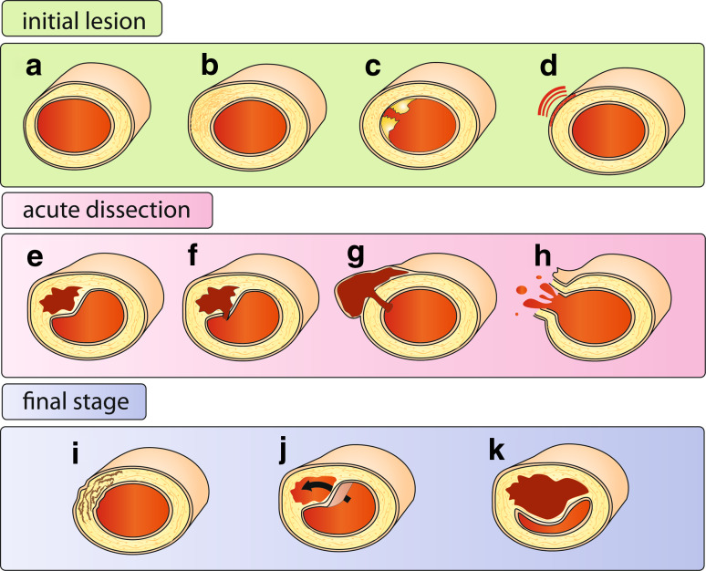
Arterial dissection (AD) is defined as the longitudinal splitting up of the arterial wall caused by intramural bleeding. It can occur as a spontaneous event in all large and medium sized arteries. The histological hallmark of AD is medial degeneration. Histological investigations, gene expression profiling and proteome studies of affected arteries reveal disturbances in many different biological processes including inflammation, proteolytic activity, cell proliferation, apoptosis and smooth muscle cell (SMC) contractile function. Medial degeneration can be caused by various rare dominant Mendelian disorders. Genetic linkage analysis lead to the identification of mutations in different disease-causing genes involved in the biosynthesis of the extracellular matrix (FBN1, COL3A1), in transforming growth factor (TGF) beta signaling (FBN1, TGFBR1, TGFBR2) and in the SMC contractile system (ACTA2, MYH11). Genome wide association studies suggest that the CDKN2A/CDKN2B locus plays a role in the etiology AD and other arterial diseases.


Figures

Fig. 1
Fig. 1
Forms and stages of arterial dissection. Initial lesion Several types of injury of the arterial wall may predispose to AD. Degenerative alterations may include loss of SMC with fragmentation and depletion of elastic fibers (a) or proliferation of SMCs with irregular and local thickening of medial and intimal layers (b). Mechanical injury due to endovascular operations, atheromatous ulcer (c), as well as peri-adventitial inflammation (d) and small adventitial bleedings [75] are further predisposing factors. Acute AD may present with an intimal tear and intimal flap (f) or as an intramural hematoma without apparent communication with the lumen (e). If AD causes aneurysmal dilatation of the artery (g), it is often referred to as pseudoaneurysm [2], but others use this notion to describe a hematoma with is located outside the arterial wall, due to a leaking hole in the arterial wall, usually due to an endovascular intervention. Arterial rupture (h) is a most serious complication of AD. Final stage AD may heal spontaneously (i) or develop into chronic forms with a double (false) lumen (j) or a persistent stenosis (k), occlusion or arterial dilatation
Fig. 2
Fig. 2
Histology of aortic medial degeneration. Normal aorta (left side) and aorta with medial degeneration stained with Hematoxylin and eosin (right side). Thin arrows point to elastic fibers, arrowheads point to SMC nuclei. Note areas with pronounced focal fibrosis, loss of SMC, loss of elastic fibers and accumulation of eosinophilic ground substance in aorta with medial degeneration (encircled areas)
Fig. 3
Fig. 3
TGF-beta signaling in the arterial wall. In most cells including SMCs TGF-beta binds to TGFBR2 (type II receptor) and ALK5 (type I receptor, also known as TGFBR1). The inactivation of TGF-beta by latent TGF-beta binding protein (LTBP) is an important mechanism regulating TGF-beta activity within the extracellular compartment and plays a prominent role in the vascular pathology of Marfan and Loeys-Dietz syndromes. The C-terminal domain of LTBP interacts with the N-terminal domain of Fibrillin-1, the major component of elastin-associated microfibrils [152, 153]. Elastase and other proteolytic enzymes release fibrillin fragments by disintegration of microfibrils. Subsequently internal fibrillin fragments bind with high affinity to the N-terminal region of fibrillin-1, the former binding site of LTBP which releases TGF-beta. TGF-beta induces a large set of cytostatic gene responses. TGF-beta stimulates among others p15INK4b (p15) both by direct Smad-mediated transactivation and by the simultaneous downregulation of the Myc transcription factor, which leads to a rapid cellular depletion of Myc and to relief of p15 transcriptional repression [154, 155]. AT1-R, Angiotensin-II type I receptor; BMP, bone morphogenetic protein; LCC, large latent complexes, consisting of SCL complexed with latent TGFβ binding protein (LTPB); PF-10, recombinant human fibrillin-1 fragment encoded by exons 41–52 of the FBN1 gene, interacting strongly and specifically with the N-terminal region of fibrillin-1; SLC (small latent complex) consisting of mature TGFβ and latency associated peptide (LAP); SMAD, signal transducer and transcriptional modulator that mediate multiple signaling pathways; vSMC, vascular smooth muscle cell

Similar articles

Cited by

References

    1. Tsai TT, Trimarchi S, Nienaber CA. Acute aortic dissection: perspectives from the International Registry of Acute Aortic Dissection (IRAD) Eur J Vasc Endovasc Surg. 2009;37:149–159. - PubMed
    1. Schievink WI. Spontaneous dissection of the carotid and vertebral arteries. N Engl J Med. 2001;344:898–906. - PubMed
    1. Bickerstaff LK, Pairolero PC, Hollier LH, Melton LJ, Van Peenen HJ, Cherry KJ, Joyce JW, Lie JT. Thoracic aortic aneurysms: a population-based study. Surgery. 1982;92:1103–1108. - PubMed
    1. Lee VH, Brown RD, Mandrekar JN, Mokri B. Incidence and outcome of cervical artery dissection: a population-based study. Neurology. 2006;67:1809–1812. - PubMed
    1. Trimarchi S, Tsai T, Eagle KA, Isselbacher EM, Froehlich J, Cooper JV, Rampoldi V, Upchurch GR, Jr, International Registry of Acute Aortic Dissection (IRAD) investigators Acute abdominal aortic dissection: insight from the International Registry of Acute Aortic Dissection (IRAD) J Vasc Surg. 2007;46:913–919. - PubMed

Substances