

Development of otolith receptors in Japanese quail
- PMID: 20155736
- PMCID: PMC2910419
- DOI: 10.1002/dneu.20787
Development of otolith receptors in Japanese quail
Abstract
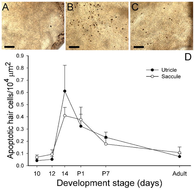
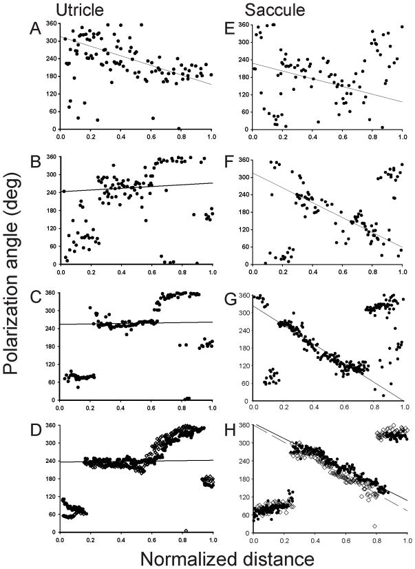
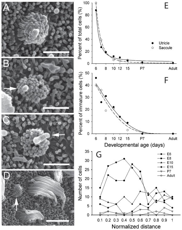
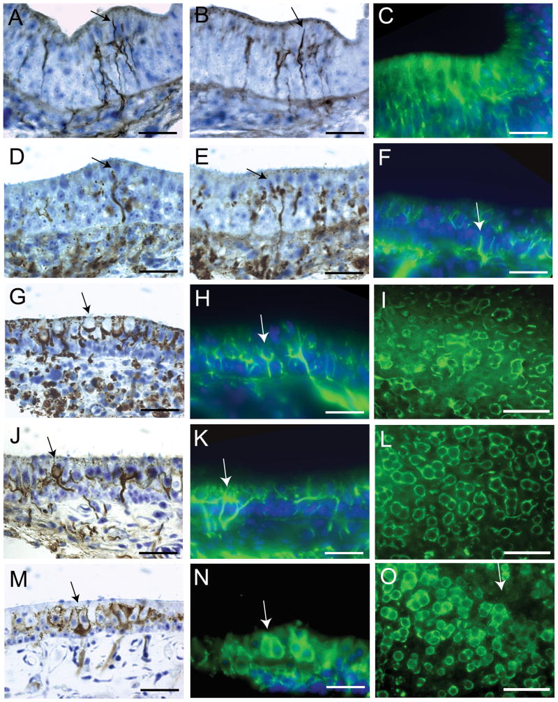
This study examined the morphological development of the otolith vestibular receptors in quail. Here, we describe epithelial growth, hair cell density, stereocilia polarization, and afferent nerve innervation during development. The otolith maculae epithelial areas increased exponentially throughout embryonic development reaching asymptotic values near posthatch day P7. Increases in hair cell density were dependent upon macular location; striolar hair cells developed first followed by hair cells in extrastriola regions. Stereocilia polarization was initiated early, with defining reversal zones forming at E8. Less than half of all immature hair cells observed had nonpolarized internal kinocilia with the remaining exhibiting planar polarity. Immunohistochemistry and neural tracing techniques were employed to examine the shape and location of the striolar regions. Initial innervation of the maculae was by small fibers with terminal growth cones at E6, followed by collateral branches with apparent bouton terminals at E8. Calyceal terminal formation began at E10; however, no mature calyces were observed until E12, when all fibers appeared to be dimorphs. Calyx afferents innervating only Type I hair cells did not develop until E14. Finally, the topographic organization of afferent macular innervation in the adult quail utricle was quantified. Calyx and dimorph afferents were primarily confined to the striolar regions, while bouton fibers were located in the extrastriola and Type II band. Calyx fibers were the least complex, followed by dimorph units. Bouton fibers had large innervation fields, with arborous branches and many terminal boutons.
Figures


Similar articles
-
Afferent innervation of the utricular macula in pigeons.J Neurophysiol. 2003 Mar;89(3):1660-77. doi: 10.1152/jn.00690.2002. J Neurophysiol. 2003. PMID: 12626631
-
Afferent innervation patterns of the saccule in pigeons.J Neurophysiol. 2003 Jan;89(1):534-50. doi: 10.1152/jn.00817.2001. J Neurophysiol. 2003. PMID: 12522200
-
Regeneration of vestibular otolith afferents after ototoxic damage.J Neurosci. 2006 Mar 15;26(11):2881-93. doi: 10.1523/JNEUROSCI.3903-05.2006. J Neurosci. 2006. PMID: 16540565 Free PMC article.
-
Morphophysiological and ultrastructural studies in the mammalian cristae ampullares.Hear Res. 1990 Nov;49(1-3):89-102. doi: 10.1016/0378-5955(90)90097-9. Hear Res. 1990. PMID: 2292511 Review.
-
Hair cells in mammalian utricles.Otolaryngol Head Neck Surg. 1998 Sep;119(3):172-81. doi: 10.1016/S0194-5998(98)70052-X. Otolaryngol Head Neck Surg. 1998. PMID: 9743073 Review.
Cited by
-
Transcription factor Emx2 controls stereociliary bundle orientation of sensory hair cells.Elife. 2017 Mar 7;6:e23661. doi: 10.7554/eLife.23661. Elife. 2017. PMID: 28266911 Free PMC article.
-
Morphology and innervation of the vestibular lagena in pigeons.Neuroscience. 2012 May 3;209:97-107. doi: 10.1016/j.neuroscience.2012.02.014. Epub 2012 Feb 15. Neuroscience. 2012. PMID: 22387112 Free PMC article.
-
Function of bidirectional sensitivity in the otolith organs established by transcription factor Emx2.Nat Commun. 2022 Oct 24;13(1):6330. doi: 10.1038/s41467-022-33819-3. Nat Commun. 2022. PMID: 36280667 Free PMC article.
-
Molecular microdomains in a sensory terminal, the vestibular calyx ending.J Neurosci. 2011 Jul 6;31(27):10101-14. doi: 10.1523/JNEUROSCI.0521-11.2011. J Neurosci. 2011. PMID: 21734302 Free PMC article.
References
-
- Baird RA, Shuff NR. Peripheral innervation patterns of vestibular nerve afferents in the bullfrog utriculus. J Comp Neurol. 1994;342:279–298. - PubMed
-
- Bever MM, Jean YY, Fekete DM. Three-dimensional morphology of inner ear development in xenopus laevis. Developmental Dynamics. 2003;227:422–430. - PubMed
-
- Blasiole B, Canfield VA, Vollrath MA, Huss D, Manzoor-Ali PKM, Dickman JD, Cheng KC, Fekete DM, Levenson R. Separate Na, K-ATPase genes are required for otolith formation and semicircular canal development in zebrafish. J Neuroscience. 2006;294:148–160. - PubMed
-
- Cajal RS. Terminación periférica del nervio acústico del las aves. Trav Lab Invest Biol Univ Mad. 1908;6:161–176.
-
- Cajal RS. Histology of the Nervous System of Man and Vertebrates. New York: Oxford; 1909. 1995, English translation by Neely and Larry Swanson from the 1909 French translation by L. Azoulay), 1899 original Spanish version.
Publication types
MeSH terms
Grants and funding
LinkOut - more resources
Full Text Sources
