Review
. 2010 Apr 26;11(6):1172-80.
doi: 10.1002/cphc.200900894.

A brief history of phytochromes

Affiliations
Review

A brief history of phytochromes

Nathan C Rockwell et al. Chemphyschem. .

Abstract

Photosensory proteins enable living things to detect the quantity and quality of the light environment and to transduce that physical signal into biochemical outputs which entrain their metabolism with the ambient light environment. Phytochromes, which photoconvert between red-absorbing P(r) and far-red-absorbing P(fr) states, are the most extensively studied of these interesting proteins. Critical regulators of a number of key adaptive processes in higher plants, including photomorphogenesis and shade avoidance, phytochromes are widespread in photosynthetic and nonphotosynthetic bacteria, and even in fungi. Cyanobacterial genomes also possess a plethora of more distant relatives of phytochromes known as cyanobacteriochromes (CBCRs). Biochemical characterization of representative CBCRs has demonstrated that this class of photosensors exhibits a broad range of wavelength sensitivities, spanning the entire visible spectrum. Distinct protein-bilin interactions are responsible for this astonishing array of wavelength sensitivities. Despite this spectral diversity, all members of the extended family of phytochrome photosensors appear to share a common photochemical mechanism for light sensing: photoisomerization of the 15/16 double bond of the bilin chromophore.


Figures
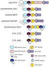
Figure 1
Figure 1
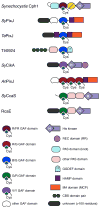
Domain structure and chromophore configuration of phytochromes and Cph2 sensors. R/FR phytochromes have a conserved PAS/GAF/PHY tridomain photosensory core. Phytochromes from different organisms have characteristic variations of this architecture. Plant and some algal phytochromes have an additional pair of PAS domains C-terminal to the core and lack a recognizable His residue that serves as a phosphoacceptor in bona fide His kinases. Bacteriophytochromes (BphPs) and related proteins from fungi (Fph) and diatoms (Dph) differ from plant and cyanobacterial phytochromes in the location of the conserved Cys residue that forms the covalent linkage to the bilin chromophore. Both plant phytochromes and Fph/Dph proteins have N-terminal extensions, while Fph/Dph proteins have C-terminal response regulator receiver domains. Cph2 sensors retain the GAF and PHY domains but lack the PAS domain and PAS/GAF knot. The family is named after Synechocystis Cph2, which has an additional GAF domain that has the hallmarks of a blue/green CBCR. Most Cph2 sensors have His kinase output domains, as is shown for two representatives from thermophilic Synechococcus species.[44]
Figure 2
Figure 2
Bilin cofactor adducts of phytochromes and related photosensors. Phytochromes utilize linear tetrapyrrole (bilin) precursors to form a variety of covalent adducts with different spectral properties. Such adducts are no longer equivalent to the parent named bilin; for example, the covalent adduct formed upon Michael addition of Cys to the C32 atom of BV is formally a (2,3)-dihydrobiliverdin. Plant and cyanobacterial phytochromes incorporate the reduced phytobilins phycocyanobilin (PCB) or phytochromobilin (PΦB) to form adducts shown at top left. A conserved Cys in the GAF domain becomes linked to the C31 carbon. This bilin adduct is shown in the (5Z)-syn (10Z)-syn (15Z)-anti configuration of the Pr state, and PCB and PΦB vary in the substituent at C18. By contrast, BphPs and Fphs utilize biliverdin IXα (BV) as chromophore precursor to form the adduct shown at top right. A conserved Cys N-terminal to the PAS/GAF knot becomes linked to the C32 carbon. This bilin is shown in the (5Z)-syn (10Z)-syn (15E)-anti configuration of the Pfr state. A subset of CBCRs and phycobiliproteins are able to isomerize PCB to PVB (bottom left), which has a saturated 4/5 bond. It is not known whether CBCRs carry out this isomerization before or after covalent attachment of the PCB precursor. This bilin is shown in the same configuration as the Pr state of phytochromes. BV can also be reduced to bilirubin IXα, which has a saturated C10 atom. The (4Z, 15E) configuration of BR is generated during phototherapy for neonatal jaundice.[–32] C10 adducts (bottom right, X = nucleophiles) of bilins disrupt the conjugation of the π system and generate blue-absorbing “rubinoid” pigments. Combining such rubinoid C10 adducts with covalent linkage to CBCR Cys residues, as illustrated, could provide an explanation for the large blueshifts observed in some CBCRs.
Figure 3
Figure 3
Structures of the phytochrome photosensory core module. Top, the PAS/GAF fragment of DrBphP in the Pr state (PDB accession 2O9C)[22] is shown with the PAS domain in blue and the GAF domain in purple. Middle, the PAS/GAF/PHY photosensory core of Cph1 in the Pr state (PDB accession 2VEA)[54] is shown with the PAS and GAF domains colored as for DrBphP and with the PHY domain in silver. Bottom, the PAS/GAF/PHY core of PaBphP in the Pfr state (chain A, PDB accession 3C2W)[57] is shown colored as for Cph1. In all cases, the knotted PAS/GAF fold is conserved. The two PAS/GAF/PHY structures exhibit a conserved, flexible ‘tongue’ reaching from the PHY domain and interacting with the chromophore-binding pocket of the GAF domain.
Figure 4
Figure 4
Stereo views of the DrBphP structure in the Pr state (top: PDB accession 2O9C)[22] and the PaBphP structure in the Pfr state (bottom: PDB accession 3C2W)[57] reveal structural changes associated with photoconversion. In DrBphP, the BV chromophore adopts a (5Z)-syn (10Z)-syn (15Z)-anti configuration. His290 is hydrogen bonded to the D-ring carbonyl oxygen (dashed line), and a water molecule is closely associated with the A-, B-, and C-rings. The sidechains of Asp207 and Tyr176 are not in close contact with chromophore. In PaBphP the BV chromophore adopts a (5Z)-syn (10Z)-syn (15E)-anti configuration. The D-ring is a-facial in both structures, consistent with CD spectroscopy.[63] The water molecule is similarly located. His277 (equivalent to His290 in DrBphP) remains in approximately the same place but no longer interacts with O19. Asp194 (equivalent to Asp207) is in approximately the same place but is now positioned to interact with the D-ring amide NH. Tyr163 (equivalent to Tyr176) adopts a different sidechain rotamer and is hydrogen bonded to the 12-propionate sidechain of BV (dashed line).
Figure 5
Figure 5
Domain structure of representative cyanobacteriochromes (CBCRs). CBCRs contain isolated bilin-binding GAF domains without PHY domains or PAS/GAF knots. Phylogenetic analyses suggest the existence of several classes of CBCR, and biochemical analyses demonstrate the existence of at least three different photoswitching behaviors.[47, 48, 76] Cyanobacteriochromes exhibit much more diverse domain organization than phytochromes or Cph2 sensors.
Figure 6
Figure 6
Schematic view of phytochrome and CBCR photochemistry. In phytochromes and CBCRs examined to date, the common element is photoisomerization of the bilin chromophore about the 15/16 double bond. In the cyanobacterial phytochrome Cph1 (red), the (15Z) Pr form converts to a (15E) lumi-R photoproduct which is then thermally converted to the (15E) Pfr form in a process requiring the 12-propionate side chain.[63] Pfr is converted to the (15Z) lumi-R photoproduct, which is then thermally converted to Pr to complete the photocycle. In the B/G photocycle of CBCR Tlr0924 (blue), we propose formation of a second covalent linkage between Cys499 and C10 of the chromophore in the (15Z) state.[46] Such a linkage at C10 would generate a species similar to bilirubin IXα or phycocyanorubin,[29, 36] explaining the blue absorbance of PbS. The (15Z) PbS form can be photoconverted to a (15E) Pb L photoproduct which is in thermal equilibrium with the Pg form.[46] Pg is generated from PbL by cleavage of the second linkage, generating a free thiol side chain at Cys499. Photoisomerization of Pg generates an as-yet unidentified (15Z) photoproduct which regenerates PbS by reformation of the second linkage at C10. This B/G cycle can occur in either PCB or PVB chromophores, because it does not involve the 4/5 bond which varies between these two bilins. Thermal relaxation of the (15E) form to the (15Z) form (dark reversion) is known in plant phytochrome, Cph1, and some BphPs. Thermal conversion of (15Z) to (15E) is also known in a subset of BphP proteins termed bathyphytochromes, including PaBphP (purple).

Similar articles

Cited by

References

    1. Butler WL, Norris KH, Seigelman HW, Hendricks SB. Proc Natl Acad Sci U S A. 1959;45:1703–1708. - PMC - PubMed
    1. Sage LC. Pigment of the Imagination : A History of Phytochrome Research. Academic Press, Inc; San Diego: 1992.
    1. Blumenstein A, Vienken K, Tasler R, Purschwitz J, Veith D, Frankenberg-Dinkel N, Fischer R. Curr Biol. 2005;15:1833–1838. - PubMed
    1. Brandt S, von Stetten D, Gunther M, Hildebrandt P, Frankenberg-Dinkel N. J Biol Chem. 2008;283:34605–34614. - PMC - PubMed
    1. De Riso V, Raniello R, Maumus F, Rogato A, Bowler C, Falciatore A. Nucl Acids Res. 2009;37:e96. - PMC - PubMed

Publication types