. 2010 Feb;26(2):173-92.
doi: 10.1089/aid.2009.0068.

Genetic characterization of HIV type 1 Nef-induced vesicle secretion

Affiliations

Genetic characterization of HIV type 1 Nef-induced vesicle secretion

Syed A Ali et al. AIDS Res Hum Retroviruses. 2010 Feb.

Abstract

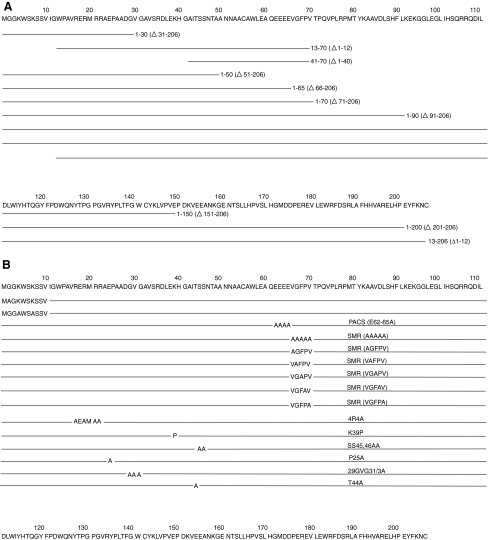
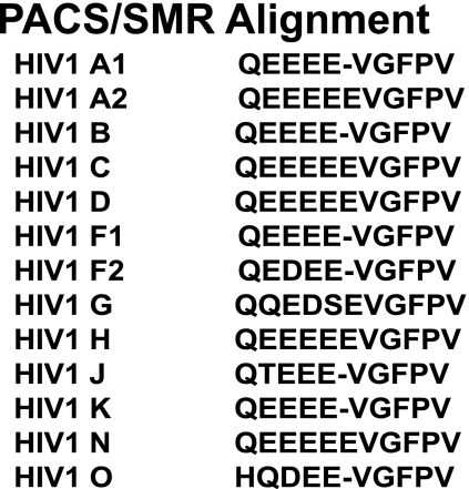
The HIV-1 Nef protein is known to be secreted, and our group has shown that Nef is secreted from nef-transfected and HIV-1-infected cells in small exosome-like vesicles (d. 40-100 nm). The role of secreted Nef remains to be fully characterized. Thus, it is important to characterize the nature of and the mechanisms regulating Nef secretion. We hypothesized that specific structural domains on the Nef protein interact with components of the endosomal trafficking machinery, sorting Nef into multivesicular bodies (MVB) and packaging it in exosome-like vesicles. To identify those domains, a series of mutants spanning the entire nef sequence were made and cloned into the expression vector pQB1, which expresses the mutants as Nef-GFP fusion proteins. These constructs were used in transient transfection assays to identify sequences necessary for secretion of the Nef-GFP fusion protein. N-terminal domains were identified as critical for Nef-induced vesicle secretion: (1) a basic cluster of four arginine residues (aa 17, 19, 21, 22), (2) the phosphofurin acidic cluster sequence (PACS; Glu62-65), and (3) a previously uncharacterized domain spanning amino acid residues 66-70 (VGFPV), which we named the secretion modification region (SMR). Additional amino acids P25, 29GVG31, and T44 were identified in HIV-1 Nef as regulating its secretion. These residues have not been associated with other reported Nef functions. The myristoylation domain, ubiquitination lysine residues, and the C-terminal portion of Nef (aa 71-206) had no effect on secretion. A minimal HIV-1 Nef sequence, comprising the identified motifs, was sufficient for Nef-induced vesicle secretion.


Figures

FIG. 1.
FIG. 1.
PCR and Site-Directed Mutagenesis Primers Used in This Study
FIG. 2.
FIG. 2.
The measurement of AChE and CD45 release from untransfected and Nef-GFP-transfected Jurkat cells. Naive or HIV-1 wtNef-GFP transfection in 1 × 106 Jurkat cells for 48 h at 37°C. (A) Cell lysates were examined for HIV-1 Nef, AChE, CD45, and tubulin by Western analysis. Columns: UT, untransfected Jurkat cell lysates; Nef, HIV-1 wtNef-GFP-transfected cell lysates. Rows: Probed with: Nef, HIV-1 Nef monoclonal antibody; AChE, AChE antibody; CD45, CD45 antibody; tubulin, tubulin antibody. (B) Differential centrifugal high-molecular-weight pellets were examined for HIV-1 Nef, AChE, and CD45 by Western analysis. Columns: lane 1, 10,000 × g pellet; lane 2, 50,000 × g pellet; lane 3, 100,000 × g pellet; lane 4, 400,000 × g pellet; lane 5, 400,000 × g spent supernatant. Rows: top two panel set: untransfected Jurkat culture pellet examined with AChE or CD45 antibodies; lower three panel set: HIV-1 NefGFP-transfected Jurkat culture pellet examined with HIV-1 Nef monoclonal, AChE, or CD45 antibodies. (C) Densitometric analysis of Western data (NIH Image J software analysis). AChE and CD45 band densities from untransfected cells and AChE, CD45, and Nef band densities from HIV-1 Nef-transfected cells were normalized against intracellular tubulin, and to report combined 100,000 × g plus 400,000 × g band density units per 1 × 106 cells. This is the combined data from multiple experiments and the data were analyzed using Student's t-test comparing untransfected values and wtNef-GFP-transfected values and displaying the p-values where p < 0.05 is significant. Transfection efficiencies for these experiments were 85% ± 2%.
FIG. 3.
FIG. 3.
Analysis of the vesicular nature of secreted Nef protein. Cells and conditioned media were collected from untransfected or wtNef-GFP-transfected Jurkat cultures. Culture media were processed via differential centrifugation, with spins at 1200 × g, 10,000 × g, 50,000 × g, 200,000 × g, and 400,000 × g. Both 200,000 × g and 400,000 × g pellets were subjected to sucrose gradient flotation. Cell lysates, culture medium, 50,000 × g pellets and 400,000 × g pellets and flotation gradient fractions were examined by Western blotting for Nef, GFP, and Alix. (A) Representative images from one experiment: Cell lysate (lane 1); culture media (supernatant; lane 2); 50,000 × g pellet (lane 3); 400,000 × g pellet (Diff. Cent.; lane 4); Gradient fractions 4–11 (lanes 5–12). Gradient fractions 1, 2, 3, and 12, which had no protein in them, are not shown. (B) Data shown are collated from multiple experiments. Bands visualized on Western blots were measured by densitometry. Data were analyzed using Student's t-test comparing Alix from untransfected cell cultures and wtNef-GFP-transfected cell cultures, with p values < 0.01 being scored as significant.
FIG. 4.
FIG. 4.
The structure and amino acid sequence of HIV-1 Nef. (A) Three-dimensional model generated by UCSF Chimera production version 1 from the Resource for Biocomputing, Visualization, and Informatics at the University of California, San Francisco. The model shows a subset of relevant, previously reported structural as well as functional motifs. (B) Sequence of HIV-1 Nef showing structural domains required for cellular interactions. The basic amino acid 1 and 2 motifs (BAA-1, BAA-2), helix-1 and helix-2, membrane targeting domain, PACS, and SMR motifs were investigated in the present study.
FIG. 5.
FIG. 5.
(A). Truncation mutagenesis to determine Nef secretion sequences. The relative fluorescence of carboxy-terminal deletion mutants of Nef compared to the wtNef-GFP is shown. Media were collected and assayed from the 48-h-old cultures of HEK293 cells transfected with the wt, NefΔ31–206 (1–30), NefΔ51–206 (1–50), NefΔ66–206 (1–65), NefΔ71–206 (1–70), NefΔ91–206 (1–90), NefΔ151–206 (1–150), NefΔ201–206 (1–200), NefΔ1–12 (13–206), NefΔ1–12 and Δ71–206 (13–70), NefΔ1–40 and Δ71–206 (41–70), and untransfected HEK293 cells (bar 12). (B) Replacement mutagenesis to fine map Nef secretion sequences. The relative fluorescence of N-terminal replacement mutants of Nef in the 1–70 aa region and compared to the wtNef-GFP is shown. Media were collected and assayed from the 48-h cultures of HEK293 cells transfected with wt Nef, NefΔ71–206 (1–70), NefG2A, NefK4K7, Nef K39P, NefK/P, Nef,S/2A, NefP25A, NefGVG/3A, NefT44A, NefVGFPV/5A, NefEEEE/4A (PACS), Nef,,, R/4A, and untransfected HEK293 cells. (C) A newly identified domain on HIV-1 Nef. The relative fluorescence of N-terminal deletion and replacement mutants of Nef in the 66–70 aa region compared to the wt Nef is shown. Media were collected and assayed from the 48-h cultures of HEK293 cells transfected with wt Nef, GFP, NefAGFPV, NefVAFPV, NefVGAPV, NefVGFAV, NefVGFPA, and untransfected HEK293 cells. (DF) The Nef-induced secretion domains function similarly in multiple cell types. Jurkats (1 × 106) transfected with HIV-1 wt Nef (bar 1), NefΔ71–206 (bar 2), NefΔ1–12 and Δ71–206 (bar 3), NefΔ1–12 (bar 4), Nef,,, R/4A (bar 5), NefEEE/4A (PACS, bar 6), NefAGFPV (SMR, bar 7), GFP (bar 8), and untransfected cells (bar 9) (D), THP-1 (E), and U937 (F) monocytes by Gene Pulser Xcell Electroporation System (Bio-Rad Laboratories, Inc., CA). Cells were incubated in RPMI 1640 medium for 48 h at 37°C and removed from the culture supernatant by centrifugation at 2000 × g for 5 min. In all experiments, the error bars show the standard errors of the measurements. Transfection efficiencies for Jurkat cells (80–86.67%), for THP-1 cells (60–65%), and for U937 cells (55–60%). These results are a compilation of at least three independent experiments.
FIG. 6.
FIG. 6.
The alignment of the PACS/SMR region of HIV-1 Nef. Amino acid consensus sequences for 13 HIV-1 subtypes were determined as described in Materials and Methods. The PACS-SMR consensus sequences were then aligned to illustrate the degree of homology in these required secretion domains of Nef. Dashes (-) indicate gaps inserted to facilitate the alignment.
FIG. 7.
FIG. 7.
HIV Nef expressed in cells is not toxic/apoptotic to transfected cells. HIV-1 Nef–GFP mutants were transfected into HEK293 cells at 37°C for 48 h. Subsequently, the cultures were stained with propidium iodide (PI) to visualize the nucleus. Finally, a comparative morphological examination of the individual cells in these cultures was performed to determine whether and how much cytotoxicity or apoptosis was observed in the transfected cells. (A) HEK293 cells were transfected with HIV-1 Nef-GFP mutants and stained by PI. Columns: bar 1, mock, untransfected HEK293 cells; bar 2, pQBI-GFP, transfected pQBI-GFP; bar 3, wtNef-GFP(1–206), transfected HIV-1 wtNef-GFP; bar 4, wtNef-GFP (1–70), transfected NefΔ71–206; bar 5, wtNef-GFP (13–70), transfected NefΔ1–12 and Δ71–206; bar 6, Nef-4R4A-GFP (1–206), transfected Nef,,,R/4A; bar 7, Nef-PACS-GFP (1–206), transfected NefEEEE/4A; bar 8, Nef-AGFPV-GFP (1–206), transfected NefAGFPV. (B) HEK293 cells were transfected with HIV-1 Nef at 37°C for 48 h and then cells were assayed by TUNEL. Columns: bar 1, pQBI-RFP, transfected pQBI-RFP in HEK293 cells; bar 2, HIV-1 wtNef-RFP, transfected HIV-1 wtNef-RFP.
FIG. 8.
FIG. 8.
Nef-induced vesicles do not display attributes of apoptotic vesicles. HIV-1 wtNef-GFP and Nef-GFP mutants were transfected into HEK293 cells. (A) Cell lysates and vesicles collected from each condition were examined for histones through Coomassie brilliant blue staining of PAGE gels. Lanes 1, 3, 5, and 7 are cell lysates from each condition; lanes 2, 4, 6, and 8 are pellets from cell lysates spun at 130,000 × g. Lanes 1 and 2 are from cells treated with 10 μM camptothecin; lanes 3 and 4 are from cells transfected with HIV-1 wtNef; lanes 5 and 6 are from cells transfected with Nef AGFPV; lanes 7 and 8 are from untransfected cells. His, denotes the region of the gel containing the histone bands. (B) Cell lysates and vesicles were analyzed by Western analysis for the presence of histones (top panel set, histone antibody), GFP (middle panel set, GFP antibody), and HIV-1 Nef (bottom panel set, Nef polyclonal antibody). Lane 1, cell lysates; lane 2, 300 × g pellet; lane 3, 1200 × g pellet; lane 4, 10,000 × g pellet; lane 5, 130,000 × g pellet. Individual panels of each panel set: top panel, Camp, cells were treated with 10 μM camptothecin; second panel, Nef, HIV-1 wtNef-GFP-transfected cells; third panel, Nef-SMR (HIV-1 Nef-VGFPV/5A-GFP)-transfected cells; bottom panel, UT, untransfected cells.
FIG. 9.
FIG. 9.
The effect of Nef mutants is not due to variable transfection/expression efficiencies. (AC) Transfected 1 μg of HIV-1 wtNef-GFP and its mutants in 1 × 106 cells of HEK293 for 48 h and then Western analysis of HEK293 cell lysates from wtNef or mutant transfections was performed. Cell cultures were transfected with pQBI-Nef-GFP (NefGFP; lane 1), pQBI-GFP (GFP; lane 2), pQBI-NefEEEE/4AGFP (PACS replacement; lane 3), pQBI-NefAGFPVGFP (SMR replacement lane 4), pQBI-Nef,,,R/4AGFP (BAA-2 replacement; lane 5), or untransfected HEK293 cells (UT; lane 6). Cell lysates were collected and analyzed by SDS–PAGE followed by Western analysis probing with anti-GFP antiserum. This is one representative image of several independent experiments. The relative positions of Nef-GFP and Nef-GFP deletion mutants' cellular protein that hybridizes to the anti-GFP antiserum are indicated. (D) Densitometry was performed and the readings from multiple independent analyses are displayed as the average densitometric units for any particular assay with standard error of measurement displayed.

References

    1. Ho DD. Neumann AU. Perelson AS, et al. Rapid turnover of plasma virions and CD4 lymphocytes in HIV-1 infection. Nature. 1995;373(6510):123–126. - PubMed
    1. Wei X. Ghosh SK. Taylor ME, et al. Viral dynamics in human immunodeficiency virus type 1 infection. Nature. 1995;373(6510):117–122. - PubMed
    1. Perelson AS. Neumann AU. Markowitz M. Leonard JM. Ho DD. HIV-1 dynamics in vivo: Virion clearance rate, infected cell life-span, and viral generation time. Science. 1996;271(5255):1582–1586. - PubMed
    1. Bagasra O. Hauptman SP. Lischner HW. Sachs M. Pomerantz RJ. Detection of human immunodeficiency virus type 1 provirus in mononuclear cells by in situ polymerase chain reaction. N Engl J Med. 1992;326(21):1385–1391. - PubMed
    1. Biberfeld P. Chayt KJ. Marselle LM, et al. HTLV-III expression in infected lymph nodes and relevance to pathogenesis of lymphadenopathy. Am J Pathol. 1986;125(3):436–442. - PMC - PubMed

Publication types

MeSH terms

Substances

LinkOut - more resources