

Decellularized rhesus monkey kidney as a three-dimensional scaffold for renal tissue engineering
- PMID: 20156112
- PMCID: PMC2947947
- DOI: 10.1089/ten.tea.2009.0602
Decellularized rhesus monkey kidney as a three-dimensional scaffold for renal tissue engineering
Abstract
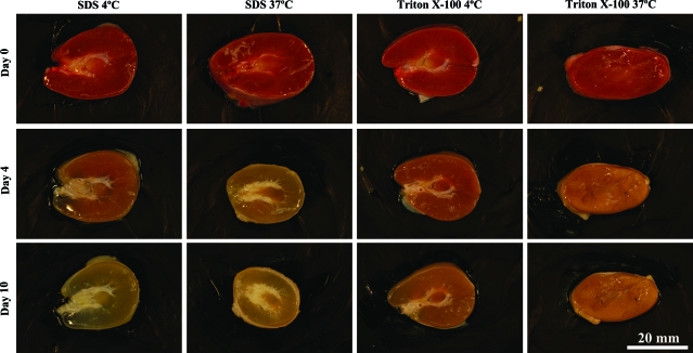
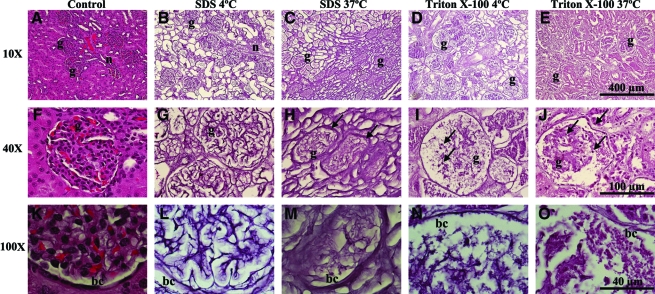
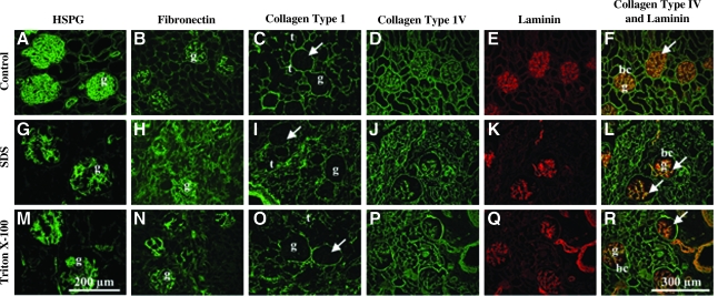
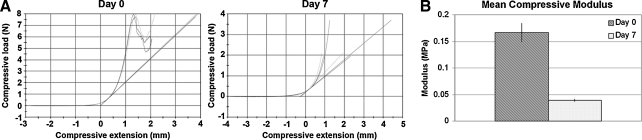
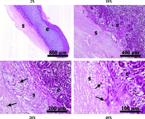
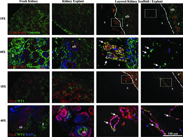
The goal of this study was the production of a decellularized kidney scaffold with structural, mechanical, and physiological properties necessary for engineering basic renal structures in vitro. Fetal, infant, juvenile, and adult rhesus monkey kidney sections were treated with either 1% (v/v) sodium dodecyl sulfate or Triton X-100 followed by quantitative and qualitative analysis. Comparison of decellularization agents and incubation temperatures demonstrated sodium dodecyl sulfate at 4 degrees C to be most effective in preserving the native architecture. Hematoxylin and eosin staining confirmed the removal of cellular material, and immunohistochemistry demonstrated preservation of native expression patterns of extracellular matrix proteins, including heparan sulfate proteoglycan, fibronectin, collagen types I and IV, and laminin. Biomechanical testing revealed a decrease in the compressive modulus of decellularized compared to fresh kidneys. Layering of fetal kidney explants on age-matched decellularized kidney scaffolds demonstrated the capacity of the scaffold to support Pax2+/vimentin+ cell attachment and migration to recellularize the scaffold. These findings demonstrate that decellularized kidney sections retain critical structural and functional properties necessary for use as a three-dimensional scaffold and promote cellular repopulation. Further, this study provides the initial steps in developing new regenerative medicine strategies for renal tissue engineering and repair.
Figures
Similar articles
-
Renal tissue engineering with decellularized rhesus monkey kidneys: age-related differences.Tissue Eng Part A. 2011 Dec;17(23-24):2891-901. doi: 10.1089/ten.TEA.2010.0714. Epub 2011 Oct 18. Tissue Eng Part A. 2011. PMID: 21902603 Free PMC article.
-
Biocompatibility and hemocompatibility of efficiently decellularized whole porcine kidney for tissue engineering.J Biomed Mater Res A. 2018 Jul;106(7):2034-2047. doi: 10.1002/jbm.a.36407. Epub 2018 Apr 17. J Biomed Mater Res A. 2018. PMID: 29569325
-
Development of decellularized aortic scaffold for regenerative medicine using Sapindus mukorossi fruit pericarp extract.Micron. 2021 Mar;142:102997. doi: 10.1016/j.micron.2020.102997. Epub 2020 Dec 24. Micron. 2021. PMID: 33388519
-
Organ reconstruction: Dream or reality for the future.Biomed Mater Eng. 2017;28(s1):S121-S127. doi: 10.3233/BME-171633. Biomed Mater Eng. 2017. PMID: 28372287 Review.
-
Decellularized kidney matrix as functional material for whole organ tissue engineering.J Appl Biomater Funct Mater. 2017 Nov 10;15(4):e326-e333. doi: 10.5301/jabfm.5000393. J Appl Biomater Funct Mater. 2017. PMID: 29131298 Review.
Cited by
-
Hydrogel-Based Skin Regeneration.Int J Mol Sci. 2024 Feb 6;25(4):1982. doi: 10.3390/ijms25041982. Int J Mol Sci. 2024. PMID: 38396661 Free PMC article. Review.
-
Repopulating Decellularized Kidney Scaffolds: An Avenue for Ex Vivo Organ Generation.Materials (Basel). 2016 Mar;9(3):190. doi: 10.3390/ma9030190. Epub 2016 Mar 11. Materials (Basel). 2016. PMID: 27375844 Free PMC article. Review.
-
Towards human organ generation using interspecies blastocyst complementation: Challenges and perspectives for therapy.Front Cell Dev Biol. 2023 Jan 19;11:1070560. doi: 10.3389/fcell.2023.1070560. eCollection 2023. Front Cell Dev Biol. 2023. PMID: 36743411 Free PMC article. Review.
-
Fabricating a Kidney Cortex Extracellular Matrix-Derived Hydrogel.J Vis Exp. 2018 Oct 13;(140):58314. doi: 10.3791/58314. J Vis Exp. 2018. PMID: 30371659 Free PMC article.
-
Mimicking the Kidney: A Key Role in Organ-on-Chip Development.Micromachines (Basel). 2016 Jul 20;7(7):126. doi: 10.3390/mi7070126. Micromachines (Basel). 2016. PMID: 30404298 Free PMC article. Review.
References
-
- National transplant statistics. Kidney transplants. Scientific Registry for Transplant Recipients. 2008. www.ustransplant.org/csr/current/nats.aspx. [Sep 6;2009 ]. www.ustransplant.org/csr/current/nats.aspx
-
- Gilbert T.W. Sellaro T.L. Badylak S.F. Decellularization of tissues and organs. Biomaterials. 2006;27:3675. - PubMed
-
- Kolker A.R. Brown D.J. Redstone J.S. Scarpinato V.M. Wallack M.K. Multilayer reconstruction of abdominal wall defects with acellular dermal allograft (AlloDerm) and component separation. Ann Plast Surg. 2005;55:36. - PubMed
-
- Wainwright D.J. Use of an acellular allograft dermal matrix (AlloDerm) in the management of full-thickness burns. Burns. 1995;21:243. - PubMed
-
- Ferguson R.E., Jr. Pu L.L. Repair of the abdominal donor-site fascial defect with small intestinal submucosa (Sugrisis) after TRAM flap breast reconstruction. Ann Plast Surg. 2007;58:95. - PubMed
Publication types
MeSH terms
Substances
Grants and funding
LinkOut - more resources
Full Text Sources
Other Literature Sources
