

Molecular mechanisms of adrenergic stimulation in the heart
- PMID: 20156590
- PMCID: PMC2891219
- DOI: 10.1016/j.hrthm.2010.02.013
Molecular mechanisms of adrenergic stimulation in the heart
Abstract
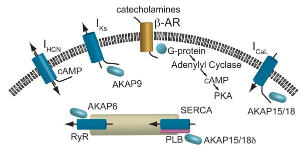
Increased cardiac output in response to beta-adrenergic receptor stimulation is achieved by rapid alteration of the activity of cardiac ion channels, pumps, and exchangers. Over the past decade, the discovery of macromolecular complexes, which include the ion channels and pumps and the kinases that control their level of phosphorylation, has led to an increased understanding of the molecular mechanisms behind the cardiac adrenergic response. The increased understanding has led to the discovery of a new long QT gene encoding an accessory protein in one of these macromolecular complexes. This article briefly reviews the major components of the beta-adrenergic pathway in the heart and discusses the direction of current and future research.
Copyright 2010 Heart Rhythm Society. Published by Elsevier Inc. All rights reserved.
Figures
Similar articles
-
Regulation of cardiac L-type Ca²⁺ channel CaV1.2 via the β-adrenergic-cAMP-protein kinase A pathway: old dogmas, advances, and new uncertainties.Circ Res. 2013 Aug 16;113(5):617-31. doi: 10.1161/CIRCRESAHA.113.301781. Circ Res. 2013. PMID: 23948586
-
Tyrosine phosphatase inhibitors selectively antagonize beta-adrenergic receptor-dependent regulation of cardiac ion channels.Mol Pharmacol. 2000 Dec;58(6):1213-21. doi: 10.1124/mol.58.6.1213. Mol Pharmacol. 2000. PMID: 11093756
-
Beta-adrenergic pathways in nonfailing and failing human ventricular myocardium.Circulation. 1990 Aug;82(2 Suppl):I12-25. Circulation. 1990. PMID: 2164894 Review.
-
Distinct beta-adrenergic receptor subtype signaling in the heart and their pathophysiological relevance.Sheng Li Xue Bao. 2004 Feb 25;56(1):1-15. Sheng Li Xue Bao. 2004. PMID: 14985822 Review.
-
Deletion of the C-terminal phosphorylation sites in the cardiac β-subunit does not affect the basic β-adrenergic response of the heart and the Ca(v)1.2 channel.J Biol Chem. 2012 Jun 29;287(27):22584-92. doi: 10.1074/jbc.M112.366484. Epub 2012 May 15. J Biol Chem. 2012. PMID: 22589548 Free PMC article.
Cited by
-
Molecular Pathophysiology of Congenital Long QT Syndrome.Physiol Rev. 2017 Jan;97(1):89-134. doi: 10.1152/physrev.00008.2016. Physiol Rev. 2017. PMID: 27807201 Free PMC article. Review.
-
Sex-Gender Disparities in Cardiovascular Diseases: The Effects of Estrogen on eNOS, Lipid Profile, and NFATs During Catecholamine Stress.Front Cardiovasc Med. 2021 Feb 12;8:639946. doi: 10.3389/fcvm.2021.639946. eCollection 2021. Front Cardiovasc Med. 2021. PMID: 33644139 Free PMC article. Review.
-
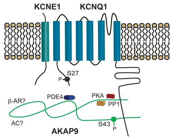
The A-kinase anchoring protein Yotiao facilitates complex formation between adenylyl cyclase type 9 and the IKs potassium channel in heart.J Biol Chem. 2012 Aug 24;287(35):29815-24. doi: 10.1074/jbc.M112.380568. Epub 2012 Jul 9. J Biol Chem. 2012. PMID: 22778270 Free PMC article.
-
Dual role of Tropomyosin-R160 in thin filament regulation: Insights into phosphorylation-dependent cardiac relaxation and cardiomyopathy mechanisms.Arch Biochem Biophys. 2025 Jun;768:110380. doi: 10.1016/j.abb.2025.110380. Epub 2025 Mar 6. Arch Biochem Biophys. 2025. PMID: 40057222
-
Polymorphisms in the GNAS Gene as Predictors of Ventricular Tachyarrhythmias and Sudden Cardiac Death: Results From the DISCOVERY Trial and Oregon Sudden Unexpected Death Study.J Am Heart Assoc. 2016 Nov 28;5(12):e003905. doi: 10.1161/JAHA.116.003905. J Am Heart Assoc. 2016. PMID: 27895044 Free PMC article.
References
-
- Gray PC, Scott JD, Catterall WA. Regulation of ion channels by cAMP-dependent protein kinase and A- kinase anchoring proteins. Curr.Opin.Neurobiol. 1998;8(3):330–334. - PubMed
-
- Marx SO, Kurokawa J, Reiken S, Motoike H, D'Armiento J, Marks AR, Kass RS. Requirement of a macromolecular signaling complex for beta adrenergic receptor modulation of the KCNQ1-KCNE1 potassium channel. Science. 2002;295(5554):496–499. - PubMed
Publication types
MeSH terms
Substances
Grants and funding
LinkOut - more resources
Full Text Sources
Miscellaneous
