

Case Reports
doi: 10.1016/j.hrthm.2009.12.019.
Epub 2010 Jan 4.
Pathologic examination after epicardial ablation of ventricular tachycardia in cardiac sarcoidosis
Affiliations
- PMID: 20156617
- PMCID: PMC4140187
- DOI: 10.1016/j.hrthm.2009.12.019
Item in Clipboard
Case Reports
Pathologic examination after epicardial ablation of ventricular tachycardia in cardiac sarcoidosis
Heart Rhythm.
2010 May.
No abstract available
Figures
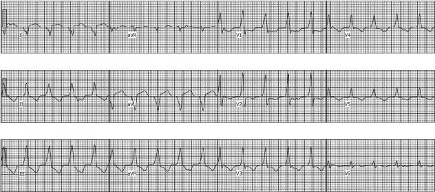
Ventricular tachycardia with a RBBB morphology and right superior axis and VA dissociation. Note the “pseudo-delta” wave in lead V1 is suggesting an epicardial site of origin.

ECGI isochrones and potential maps localizing the VT origin to the basal antero-lateral surface of the LV. Note the Q-wave morphology of the ECGI-reconstructed epicardial electrogram (EGM)at that site [inset], indicating an epicardial source. The asterisk in the isochrone map marks the site of earliest activation; the asterisk in the potential map is the local potential minimum associated with earliest activation.

CARTO electroanatomic activation maps in Left Anterior Oblique Cranial orientation. The radiofrequency energy applications are represented by the red spheres. Lesions 4–7 (labeled) affected the ventricular tachycardia. (Two points were collected during the 4th application).
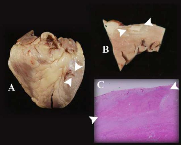
Gross, sectioned, and microscopic examination of the heart displays the epicardial ablation lesions. In each panel, the ablation lesions are indicated by the two white arrowheads. A: The lesions are seen at the basal anterolateral aspect of the whole heart. The lesions to the left of the arrowheads were superficial, as demonstrated in panel B. B: Section through the area ablation, revealing dense scar of ablation surrounded by scar from sarcoidosis. The depth of the lesion is 2 mm. C: The microscopic cross sectional examination shows that the ablation lesion is in an area affected by sarcoidosis. There is normal myocardium at the lower border of the panel.
References
-
- Jefic D, Joel B, Good E, et al. Role of radiofrequency catheter ablation of ventricular tachycardia in cardiac sarcoidosis: report from a multicenter registry. Heart Rhythm. 2009;6:189–195. - PubMed
-
- Berruezo A, Mont L, Nava S, Chueca E, Barholomay E, Brugada J. Electrocardiographic recognition of the epicardial origin of ventricular tachycardias. Circulation. 2004;109:1842–1847. - PubMed
-
- Sosa E, Scanavacca M. Percutaneous pericardial access for mapping and ablation of epicardial ventricular tachycardia. Circulation. 2007;115:e542–44. - PubMed
Publication types
MeSH terms
Grants and funding
LinkOut - more resources
Full Text Sources
Medical
