

Serotoninergic modulation of cortical and respiratory responses to episodic hypoxia
- PMID: 20156721
- PMCID: PMC3521363
- DOI: 10.1186/2047-783x-14-s4-32
Serotoninergic modulation of cortical and respiratory responses to episodic hypoxia
Abstract
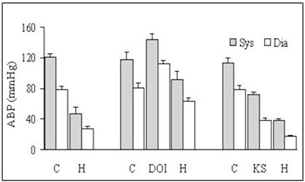
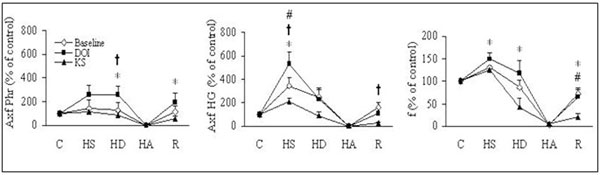
Biphasic respiratory response to hypoxia in anesthetized animals is accompanied by changes in the EEG mostly in the low EEG frequency bands. Serotonin is a potent modulator of cortical and respiratory activity through 5-HT(2) receptors. Present study investigated whether 5-HT(2) receptors might be involved in the EEG and respiratory relationship during normoxic and hypoxic respiration assessed from integrated phrenic (Phr) and hypoglossal (HG) nerve activities. EEG signal recorded from the frontal cortex was subjected to power spectral analysis in delta, theta, alpha, and beta frequency bands. Systemic administration of 5-HT(2) agonist DOI (1-(2,5-dimethoxy-4-iodophenyl)-2-aminopropane) enhanced tonic and lowered peak phasic respiratory activity, and increased frequency of bursts of Phr and HG activity. At the same time, EEG activity became desynchronized and arterial blood pressure (ABP) increased. Following DOI pretreatment, 11% hypoxia induced an augmented respiratory response in comparison with the response in the baseline condition. ABP fell less then in the control hypoxia. EEG pattern changed less than in the baseline state. Subsequent administration of ketanserin, a 5-HT(2) antagonist increased respiratory activity, elicited a synchronization of EEG activity and hypotension. The respiratory response to hypoxia was attenuated and cortical response was more potent in comparison with that after DOI injection. Arterial blood pressure decreased more then during baseline hypoxic response. The results suggest that modulation of cortical synchronization and desynchronization through 5-HT(2) receptor active agents may impact to hypoxic respiratory response.
Figures


Similar articles
-
Electroencephalographic and respiratory activities during acute intermittent hypoxia in anesthetized rats.J Physiol Pharmacol. 2007 Nov;58 Suppl 5(Pt 1):85-93. J Physiol Pharmacol. 2007. PMID: 18204119
-
Intracerebroventricular serotonin reduces the degree of acute hypoxic ventilatory depression in peripherally chemodenervated rabbits.Chin J Physiol. 2008 Jun 30;51(3):136-45. Chin J Physiol. 2008. PMID: 18935908
-
Serotonergic system in hypoxic ventilatory response in unilateral rat model of Parkinson's disease.J Biomed Sci. 2017 Mar 27;24(1):24. doi: 10.1186/s12929-017-0331-2. J Biomed Sci. 2017. PMID: 28347345 Free PMC article.
-
Time-dependent hypoxic ventilatory responses in rats: effects of ketanserin and 5-carboxamidotryptamine.Am J Physiol. 1999 Sep;277(3):R658-66. doi: 10.1152/ajpregu.1999.277.3.R658. Am J Physiol. 1999. PMID: 10484481
-
[Serotonin and blood pressure regulation--antihypertensive mechanism of ketanserin].Nihon Yakurigaku Zasshi. 1989 Oct;94(4):207-22. doi: 10.1254/fpj.94.207. Nihon Yakurigaku Zasshi. 1989. PMID: 2575564 Review. Japanese.
Cited by
-
Comodulation of dopamine and serotonin on prefrontal cortical rhythms: a theoretical study.Front Integr Neurosci. 2013 Aug 5;7:54. doi: 10.3389/fnint.2013.00054. eCollection 2013. Front Integr Neurosci. 2013. PMID: 23935568 Free PMC article.
-
Toward a multiscale modeling framework for understanding serotonergic function.J Psychopharmacol. 2017 Sep;31(9):1121-1136. doi: 10.1177/0269881117699612. Epub 2017 Apr 18. J Psychopharmacol. 2017. PMID: 28417684 Free PMC article. Review.
References
-
- Gonzalez C, Dinger BG, Fidone SJ. In: Regulation of breathing. Dempsey JA, Pack AI, editor. Vol. 79. 1995. Mechanisms of carotid body chemoreception; pp. 391–471.
Publication types
MeSH terms
Substances
LinkOut - more resources
Full Text Sources