. 2010 Nov;20(11):2684-93.
doi: 10.1093/cercor/bhq016. Epub 2010 Feb 15.

Perisomatic GABAergic innervation in prefrontal cortex is regulated by ankyrin interaction with the L1 cell adhesion molecule

Affiliations

Perisomatic GABAergic innervation in prefrontal cortex is regulated by ankyrin interaction with the L1 cell adhesion molecule

Hanjun Guan et al. Cereb Cortex. 2010 Nov.

Abstract

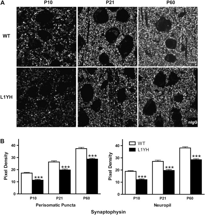
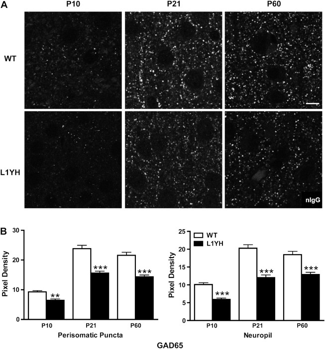
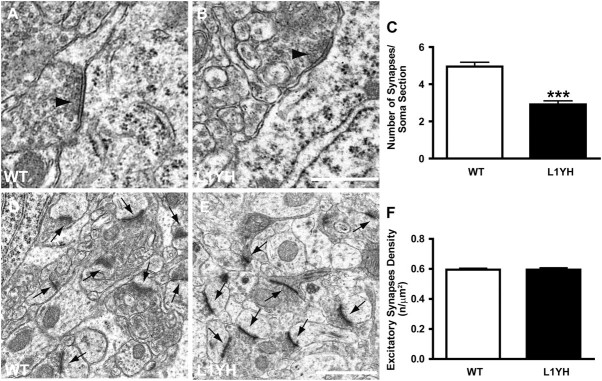
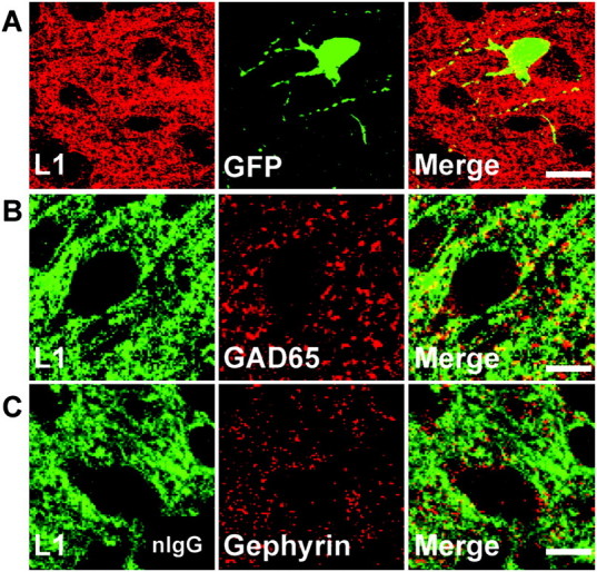
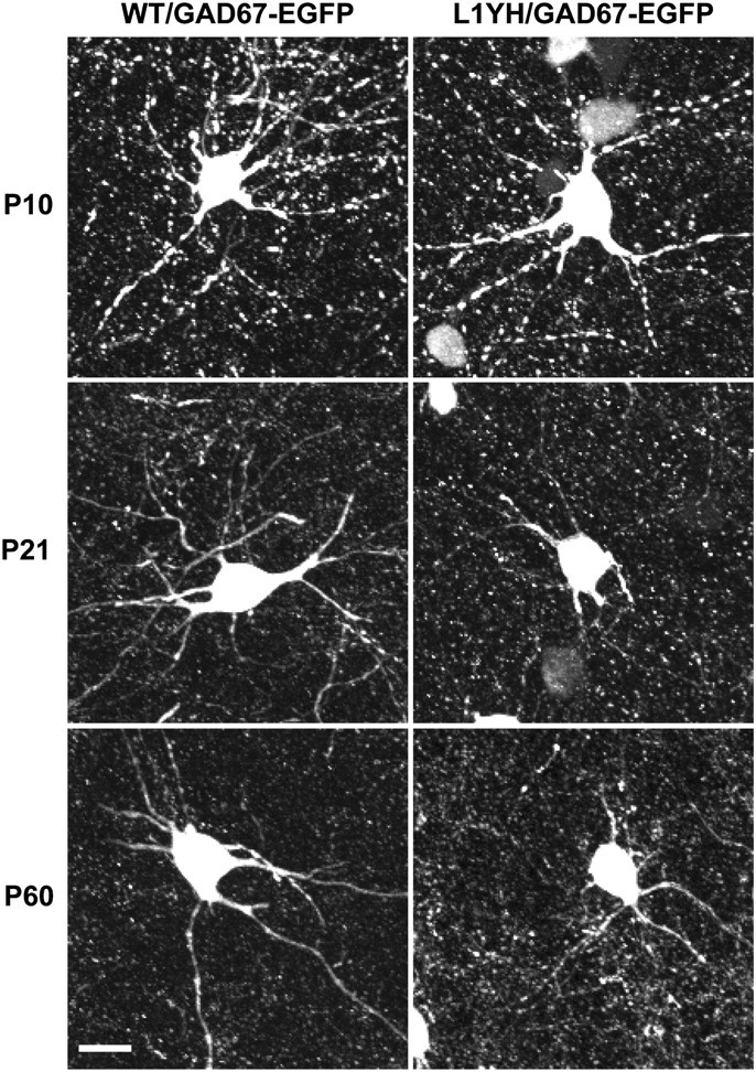
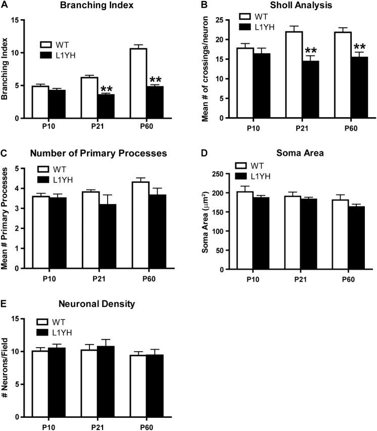
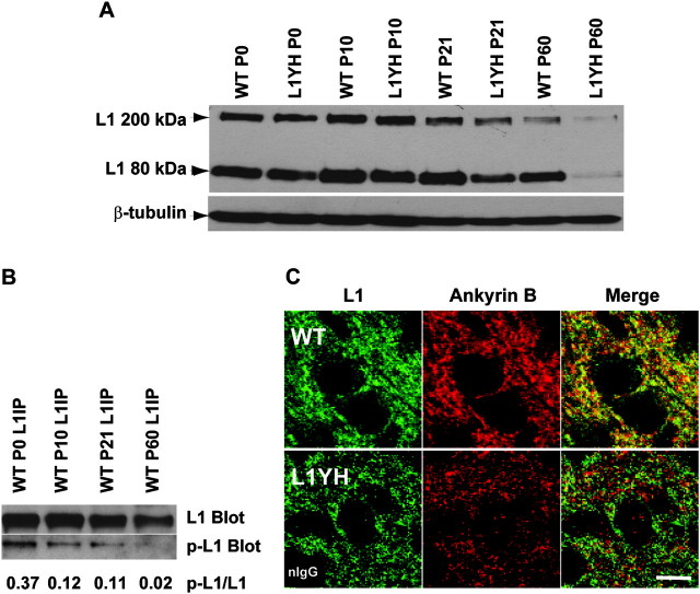
The L1 adhesion molecule functions in axon growth and guidance, but a role in synaptic development of cortical inhibitory interneurons is largely unexplored. L1 mediates adhesion by engaging the actin cytoskeleton through binding the actin/spectrin adapter protein ankyrin. Loss of L1-ankyrin interaction impaired process elaboration/branching by GABAergic interneurons, including basket cells, and reduced the number of perisomatic synapses in the cingulate cortex as shown in L1 mutant mice (L1YH) with a mutation in the ankyrin-binding site, either alone or intercrossed with GAD67-enhanced green fluorescence protein reporter mice. Electron microscopy revealed that perisomatic inhibitory synapses but not excitatory synapses in the neuropil were specifically affected. In wild-type cingulate cortex, L1 colocalized with perisomatic synaptic markers, whereas L1 phosphorylation on Tyr(1229) decreased postnatally, correlating with increased ankyrin binding and synaptic development. These results suggest a novel role for L1 engagement with the actin cytoskeleton in development of inhibitory connectivity within the cingulate cortex.


Figures

Figure 1.
Figure 1.
Synaptic puncta in the mouse prefrontal cortex increase postnatally during normal development but are reduced in L1YH mutant mice. (A) Representative confocal images of synaptophysin immunoreactivity from WT and L1YH cingulate cortex (layers II/III) at postnatal ages. Scale bar: 10 μm. (B) Pixel densities of synaptophysin immunoreactivity in perisomatic puncta and neuropil. In L1YH mice, synaptophysin immunofluorescence intensities in perisomatic puncta rings and neuropil were significantly lower than those of age-matched WT mice at all ages examined. Error bars represent SEM; ***P < 0.001 relative to WT mice. Four animals per genotype per stage were analyzed.
Figure 2.
Figure 2.
Developmental increase in GAD65-postive perisomatic puncta and neuropil is decreased in L1YH mutant prefrontal cortex. (A) Representative confocal images of GAD65 immunoreactivity from WT and L1YH cingulate cortex (layers II/III) at postnatal ages. Scale bar: 10 μm. (B) Pixel densities of GAD65 immunoreactivity in perisomatic puncta and neuropil. In L1YH mice, GAD65 immunofluorescence intensities in perisomatic puncta rings and neuropil were significantly lower than those of age-matched WT mice at all ages examined. Error bars represent SEM; **P < 0.01 and ***P < 0.001 relative to WT mice. Four animals per genotype per stage were analyzed.
Figure 3.
Figure 3.
Reduced perisomatic inhibitory synapses but not excitatory synapses in L1YH mice revealed by electron microscopy analysis. Electron microscopy was performed in layers II/III cingulate cortex of L1YH mice and WT littermates at P60. At least 20 randomly selected pyramidal neurons and 6 neuropil areas per animal (n = 4 per genotype) were analyzed. (A,B) Ultrastructural analysis showed perisomatic inhibitory synapses (arrow heads in A, B) along pyramidal cells in cingulate cortex of L1YH mice (B), with largely normal morphology and organelle organization compared with WT (A). Scale bar: 0.5 μm. (C). Quantification of perisomatic inhibitory synapses along the somal perimeter of pyramidal cells in cingulate cortex of L1YH mice revealed an approximately 40% reduction compared with WT. Error bars represent SEM; ***P < 0.001. (D,E) Representative images of excitatory synapses (arrows in D, E) in neuropil areas in cingulate cortex of L1YH mice (E) and WT littermates (D). Scale bar: 1μm. (F) Quantification of excitatory synapses density in neuropil areas in cingulate cortex of L1YH mice and WT littermates. No difference was observed between WT and L1YH mice. n/μm2 = number of synapses per square micron of somal perimeter. Error bars represent SEM; ***P < 0.001.
Figure 4.
Figure 4.
L1 localizes to GABAergic inhibitory synaptic puncta in postnatal mouse cingulate cortex. (A) Double immunofluorescence staining of L1 and GFP in cingulate cortex (layers II/III) of WT/GAD67-EGFP mice at P10 showed L1 localized along processes and somal membranes of EGFP-positive basket interneurons. Scale bar: 20 μm. (B) Double staining of L1 and GAD65, a presynaptic inhibitory synapse marker, in cingulate cortex (layers II/III) of WT mice at P10 revealed L1 colocalized in part with GAD65. Scale bar: 10 μm. (C) Double staining of L1 and gephyrin, a postsynaptic inhibitory synapse marker, in cingulate cortex (layers II/III) of WT mice at P10 showed less colocalization of L1 with gephyrin. Scale bar: 10 μm.
Figure 5.
Figure 5.
Reduced process elaboration by basket interneurons in cingulate cortex in L1YH/GAD67-EGFP mice. Individual EGFP-labeled basket interneurons from layers II/III of the cingulate cortex of L1YH/GAD67-EGFP and WT/GAD67-EGFP mice were imaged and reconstructed from confocal z-stacks. Representative images of basket interneurons at each stage are shown for each time point (P10, P21, and P60). EGFP-positive basket interneurons initiated primary processes (axons and dendrites exiting the soma) in both WT and L1YH mice at P10, and no morphological difference was observed between WT and L1YH mice (upper panel). WT basket interneurons developed extensively branched arbors from P21 to P60, whereas L1YH basket interneurons show much less branching (middle and lower panels). Scale bar: 20μm.
Figure 6.
Figure 6.
Quantification of basket interneuron arborization in the cingulate cortex of WT and L1YH/GAD67-EGFP mice. Individual EGFP-labeled basket interneurons from layers II/III of the cingulate cortex of L1YH/GAD67-EGFP and WT/GAD67-EGFP mice were imaged and reconstructed from confocal z-stacks, then analyzed using Neurolucida software. At least 60 EGFP-labeled basket interneurons from 6 random selected fields per animal were analyzed, using 4 mice per stage and genotype. (A) In L1YH/GAD67-EGFP mice, the branching index (the total number of ends/total number of primary processes for each neuron evident in the z-stack images) of basket interneurons did not increase with age and was significantly lower than that of WT at P21 and P60. (B) Sholl analysis revealed that in L1YH/GAD67-EGFP mice, the numbers of crossings by basket interneurons processes at a given distance from the soma were similar at P10 but significantly less than that of WT basket interneurons at P21 and P60 (mean number of crossings per neuron at Sholl distance of 30 μm was shown). (C–E) The mean numbers of primary processes per neuron (C), mean soma area (D), and neuronal density (E) were not affected in L1YH mice. Error bars represent SEM; **P < 0.01.
Figure 7.
Figure 7.
Regulation of L1 expression and phosphorylation in postnatal brain development. (A) Developmental regulation of L1 expression in mouse forebrain. Lysates of mouse forebrain (75 μg) were analyzed at ages of P0, P10, P21, and P60 for L1 expression by SDS-PAGE and immunoblotting. The expression levels of L1 in WT and L1YH forebrain were similar from P0 to P21; however, the L1 level in L1YH brain was lower than WT at P60 (adult). The data shown were representative of three independent brain lysates. Blots were reprobed for β-tubulin as an internal control. (B) Developmental regulation of L1 phosphorylation on Tyr1229 in forebrain. L1 was immunoprecipitated from lysates of P0, P10, P21, and P60 WT forebrain and subjected to SDS-PAGE and immunoblotting with antibody specific for L1 phosphorylated at Tyr1229 (p-L1 Blot), then reprobed for total L1 with antibody specific for L1 protein (L1 Blot). Blots were densitometrically scanned, and band densities were analyzed using NIH Image J software. The ratios of phospho-L1 to L1 intensities are indicated below. (C) L1 colocalizes with ankyrin B in WT but not L1YH cingulate cortex. Double immunofluorescence staining of L1 and ankyrin B in layers II/III of cingulate cortex of P10 WT and L1YH mice shows colocalization in WT but not L1YH cortex in merged confocal images. Scale bar: 10 μm. Insert in lower left panel shows control staining of L1YH cortex with nonimmune IgG (nIgG).

Similar articles

Cited by

References

    1. Ango F, di Cristo G, Higashiyama H, Bennett V, Wu P, Huang ZJ. Ankyrin-based subcellular gradient of neurofascin, an immunoglobulin family protein, directs GABAergic innervation at purkinje axon initial segment. Cell. 2004;119:257–272. - PubMed
    1. Ango F, Wu C, Van der Want JJ, Wu P, Schachner M, Huang ZJ. Bergmann glia and the recognition molecule CHL1 organize GABAergic axons and direct innervation of Purkinje cell dendrites. PLoS Biol. 2008;6:e103. - PMC - PubMed
    1. Benes FM, Lim B, Matzilevich D, Subburaju S, Walsh JP. Circuitry-based gene expression profiles in GABA cells of the trisynaptic pathway in schizophrenics versus bipolars. Proc Natl Acad Sci U S A. 2008;105:20935–20940. - PMC - PubMed
    1. Bennett V, Baines AJ. Spectrin and ankyrin-based pathways: metazoan inventions for integrating cells into tissues. Physiol Rev. 2001;81:1353–1392. - PubMed
    1. Bennett V, Chen L. Ankyrins and cellular targeting of diverse membrane proteins to physiological sites. Curr Opin Cell Biol. 2001;13:61–67. - PubMed

Publication types

MeSH terms