. 2010 Apr 1;588(Pt 7):1097-115.
doi: 10.1113/jphysiol.2009.184838. Epub 2010 Feb 15.

Mixed GABA-glycine synapses delineate a specific topography in the nucleus tractus solitarii of adult rat

Affiliations

Mixed GABA-glycine synapses delineate a specific topography in the nucleus tractus solitarii of adult rat

Amandine Dufour et al. J Physiol. .

Abstract

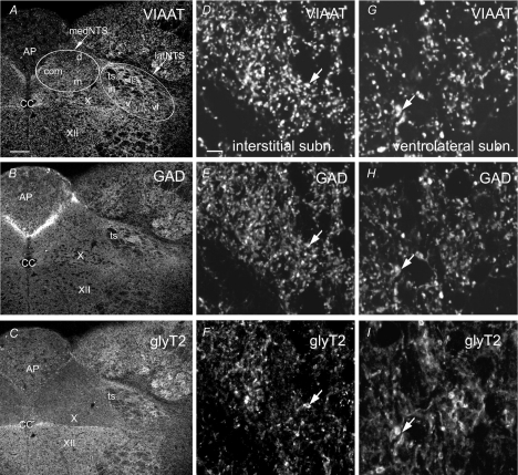
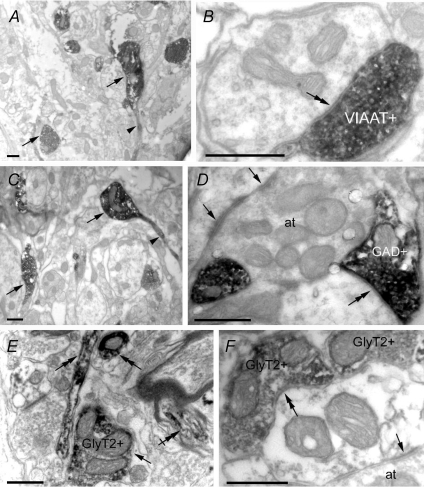
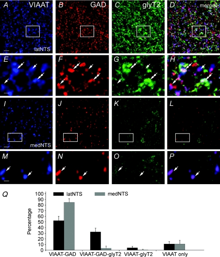
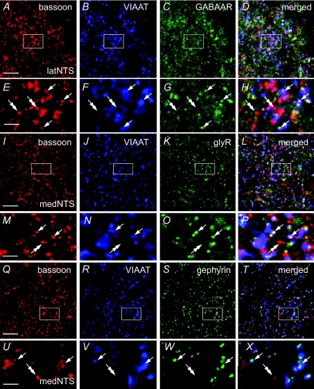
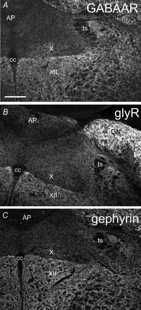
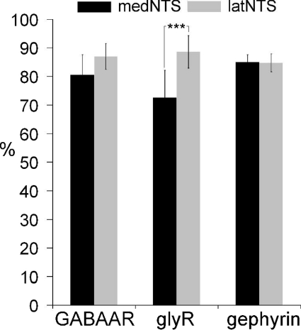
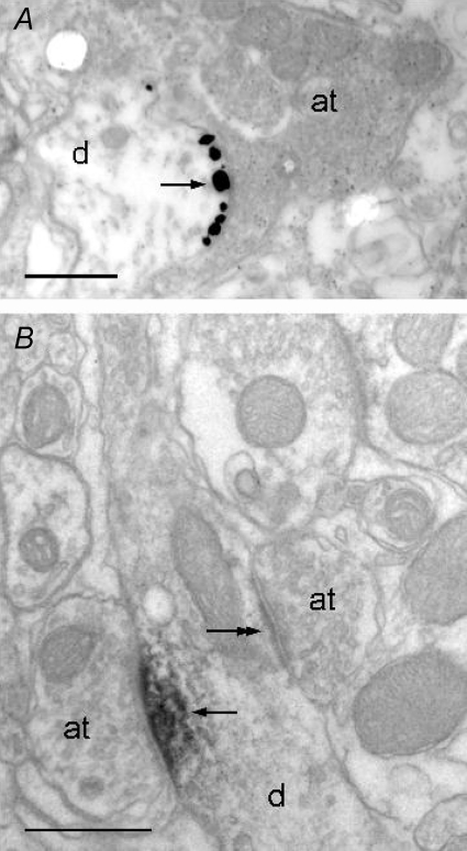
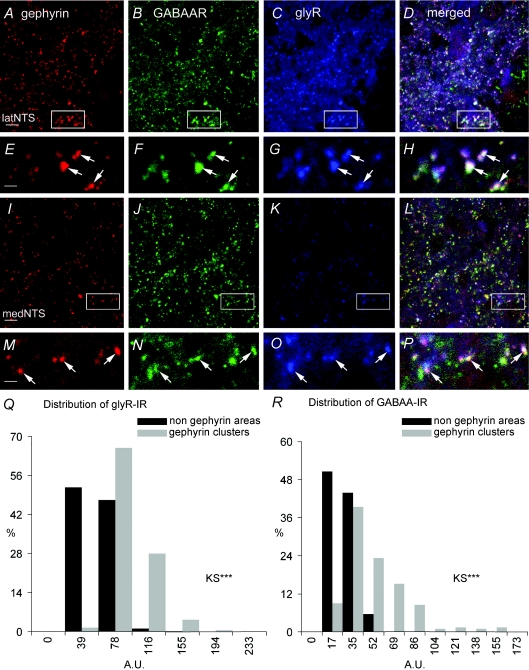
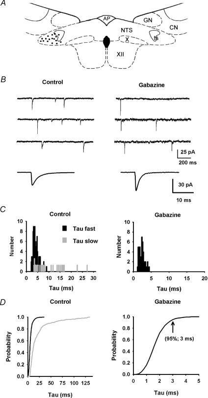
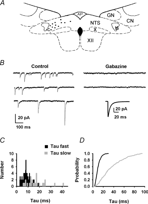
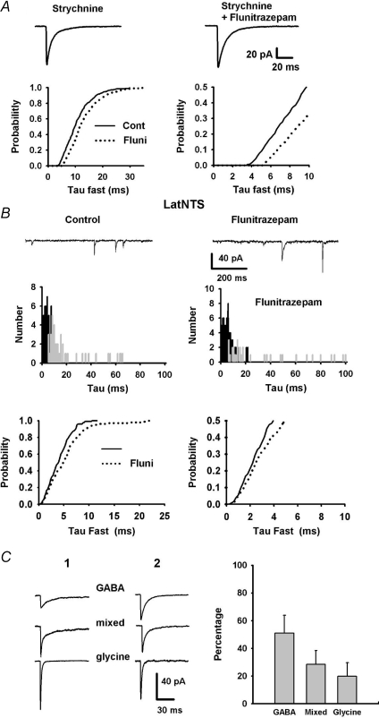
Using combined morphological and electrophysiological approaches, we have determined the composition of inhibitory synapses of the nucleus tractus solitarii (NTS), a brainstem structure that is a gateway for many visceral sensory afferent fibres. Immunohistochemical experiments demonstrate that, in adult rat, GABA axon terminals are present throughout the NTS while mixed GABA-glycine axon terminals are strictly located to the lateral part of the NTS within subnuclei surrounding the tractus solitarius. Purely glycine axon terminals are rare in the lateral part of the NTS and hardly detected in its medial part. Electrophysiological experiments confirm the predominance of GABA inhibition throughout the NTS and demonstrate the existence of a dual inhibition involving the co-release of GABA and glycine restricted to the lateral part of NTS. Since GABA(A) and glycine receptors are co-expressed postsynaptically in virtually all the inhibitory axon terminals throughout the NTS, it suggests that the inhibition phenotype relies on the characteristics of the axon terminals. Our results also demonstrate that glycine is mostly associated with GABA within axon terminals and raise the possibility of a dynamic regulation of GABA/glycine release at the presynaptic level. Our data provide new information for understanding the mechanisms involved in the processing of visceral information by the central nervous system in adult animals.


Figures

Figure 1
Figure 1. Confocal views showing immunofluorescence for VIAAT, GAD65/67 (GAD) and glyT2 within adult rat NTS
A–C, two subregions are distinguished: the medial part of the NTS (medNTS) is located medial to the tractus solitarius (ts); the lateral part of the NTS (latNTS) regroups subnuclei surrounding the tractus solitarius. DI, higher magnifications of the interstitial (D–F) and the ventrolateral (G–I) subnuclei showing numerous putative immunoreactive axon terminals (e.g. arrows). AP, area postrema; cc, central canal; com, commissural subnucleus; d, dorsal subnucleus; in, intermediate subnucleus; is, interstitial subnucleus; m, medial subnucleu; v, ventral subnucleus; vl, ventrolateral subnucleus; X, dorsal motor nucleus of the vagus; XII, hypoglossal nucleus. Scale bars = 146 μm for A–C, 5.8 μm for D–I.
Figure 2
Figure 2. Electronic micrographs of VIAAT (A and B), GAD65/67 (GAD; C and D) and glyT2 (E and F) within adult rat NTS after immunoperoxidase
Labelling for VIAAT, GAD or glyT2 is present in axon terminals (arrows in A, C and E, respectively). GAD and VIAAT labelling can also be found present in pre-terminal axonal processes (arrowhead in A and C). GlyT2 labelling is present in axons (double arrows in E) that can be myelinated (crossed double arrow in E). Axon terminals positive for VIAAT, GAD or glyT2 establish symmetric synapses (double arrow in B, D and F). Asymmetric synapses (arrows in D and F) are visible and associated to negative axon terminals (at). Scale bars = 0.5 μm.
Figure 3
Figure 3. Relative distribution of axon terminals immunoreactive for VIAAT, GAD65/67 (GAD) and glyT2 in rat NTS
A–D, numerous axon terminals are immunoreactive for VIAAT (A), GAD (B) and glyT2 (C) in latNTS. E–H, enlarged views of areas framed in AD show that VIAAT positive terminals (arrows and double arrows) co-express immunoreactivity for GAD only (arrows) or for both GAD and glyT2 (double arrows). I–L, in medNTS numerous terminals are immunoreactive for VIAAT and GAD, but glyT2 labelling is almost absent. M–P, enlarged views of framed areas in IL show that VIAAT positive terminals are immunoreactive only for GAD (arrows). Scales bar = 5.8 μm for A–D and I–L; 1.45 μm for E–H and M–P. Q, percentages of axon terminals immunoreactive for VIAAT and GAD65/67, or immunoreactive for VIAAT and glyT2, or immunoreactive for VIAAT, GAD65/67 and glyT2 in the lateral (black) and medial (grey) NTS. Error bars show s.d., n= 6 rats.
Figure 5
Figure 5. GABAAR, glyR and gephyrin immunoreactivity in inhibitory synapses of rat NTS
A–X, simultaneous detection of the presynaptic active zones (bassoon, red), inhibitory axon terminals (VIAAT, blue) and GABAAR, glyR or gephyrin (green). Immunoreactivity is visible as numerous fluorescent clusters throughout the NTS. Images in E–H, M–P and U–X represent enlarged views of framed areas in A–D, I–L and Q–T, respectively. Bassoon immunoreactive puncta colocalized with VIAAT labelling (inhibitory synapses, arrows) are associated with GABAAR (E–H), glyR (M–P) or gephyrin (U–X) positive clusters within latNTS and medNTS. Inversely, bassoon immunoreactive puncta that are not colocalized with VIAAT labelling (non-inhibitory synapses, double arrows) are not associated to GABAAR, glyR or gephyrin positive clusters. Scale bars = 11.6 μm for A–D, I–L and Q–T; 2.9 μm for E–H, M–P and U–X.
Figure 4
Figure 4. Confocal views showing immunofluorescence for the β2/3 subunits of GABAAR, the α subunits of glyR and gephyrin within adult rat NTS
AP, area postrema; cc, central canal; GN, gracilis nucleus; ts, tractus solitarius; X, dorsal motor nucleus of the vagus; XII, hypoglossal nucleus. Scale bar = 224 μm.
Figure 6
Figure 6. Distribution of inhibitory synapses expressing GABAAR, glyR and gephyrin in rat NTS
Percentages of inhibitory synapses containing GABAAR or glyR in medNTS and latNTS. Note that the percentage of glyR containing synapses is significantly lower in medNTS (Mann–Whitney test, P < 0.005).
Figure 7
Figure 7. Electronic micrographs of gephyrin labelling in NTS after pre-embedding immunogold (A) or immunoperoxidase (B)
Labelling is present at synapses (arrows) between dendrites (d) and axon terminals (at). Symmetrical synapses are labelled (arrows in A), while no labelling is found at asymmetrical synapses (e.g. double arrows in B). Scale bars = 500 nm.
Figure 8
Figure 8. GABAAR and glyR are co-expressed with gephyrin at inhibitory synapses in the NTS
A–P, immunofluorescence for gephyrin, GABAAR and glyR is colocalised in latNTS (A–D and arrows in enlarged views E–H), and also in medNTS although immunoreactivity for glyR is weaker (I–L and arrows in enlarged views M–P). Q–R, distributions of fluorescence intensities (A.U., arbitrary unit) for glyR immunoreactivity (glyR-IR) and GABAAR immunoreactivity (GABAAR-IR). Fluorescence intensities in gephyrin clusters are significantly different from those in non-gephyrin areas (KS, P < 0.01). Scale bars = 5.8 μm for A–D and I–L; 1.39 μm for E–H and M–P.
Figure 10
Figure 10. Characterization of mIPSCs in lateral NTS neurons
A, schematic drawing of a coronal section of brainstem showing the location of recorded cells in the latNTS (black dots). AP, area postrema; CN, cuneatus nucleus; GN, gracilis nucleus; NTS, nucleus tractus solitarii; ts, tractus solitarius; X, dorsal motor nucleus of the vagus; XII, hypoglossal nucleus. B, in control conditions, inward inhibitory current exhibited either mono- or bi-exponential decay. Bottom trace: average mIPSCs obtained from 150 events. Addition of 5 μm gabazine decreased synaptic activity frequency and revealed fast inward currents with mono-exponential decay. Bottom trace: average mIPSCs obtained from 50 events. C, distribution of τfast (black) and τslow (grey) for the neuron shown in A in control condition (117 mIPSCs) or after addition of gabazine (42 mIPSCs). D, normalized cumulative distribution of τfast and τslow for 8 latNTS neurons (values computed from 1504 mIPSCs) in control conditions and after addition of gabazine (800 mIPSCs). Note that under gabazine, all mIPSC decays were best fitted to a single exponential.
Figure 9
Figure 9. Characterization of mIPSCs in medial NTS neurons
A, schematic drawing of a coronal section of brainstem showing the location of recorded cells in the medNTS (black dots). AP, area postrema; CN, cuneatus nucleus; GN, gracilis nucleus; NTS, nucleus tractus solitarii; ts, tractus solitarius; X, dorsal motor nucleus of the vagus; XII, hypoglossal nucleus. B, in control conditions (bath added 2 mm kynurenate, TTX, holding potential: −70 mV), inward inhibitory current exhibited either mono- or bi-exponential decay. Addition of 5 μm gabazine abolished synaptic activity (same scale as control). Inset: average mIPSC obtained from 50 events. C, distribution of τfast (black) and τslow (grey) for the neuron shown in B (132 mIPSCs). D, normalized cumulative distribution of τfast and τslow for 7 medial NTS neurons (values computed from 821 IPSCs).
Figure 11
Figure 11. Evidence for co-release of GABA and glycine in NTS neurons
A, addition of Flunitrazepam into the saline increased GABA averaged mIPSCs duration as seen by a prolonged decay times (upper traces). Normalized cumulative distribution of τfast for isolated GABA events (TTX and strychnine) before (plain lines) and after addition of Flunitrazepam (dashed lines). Note the rightward shift of the distributions after Flunitrazepam addition (right, expanded axes for clarity). B, addition of Flunitrazepam into the saline increased mIPSC duration in latNTS neurons (upper traces). Distribution of τfast (black) and τslow (grey) for the same neuron in control condition or after addition of Flunitrazepam (middle panel). Normalized cumulative distribution of τfast the same neuron before (plain lines) and after addition of Flunitrazepam (dashed lines). Note that the fastest decays (<3 ms, right, expanded axes) were not affected by Flunitrazepam (bottom panel). C, left, examples of averaged mIPSCs for two different latNTS neurons (1, 2) obtained from mIPSCs separated according to the fitting procedure. C, right, bar graph of the relative proportion of the GABA-, mixed and glyR-mediated mIPSCs estimated in 5 latNTS neurons. Error bars show s.e.m.

Similar articles

Cited by

References

    1. Andresen MC, Mendelowitz D. Sensory afferent neurotransmission in caudal nucleus tractus solitarius: common denominators. Chem Senses. 1996;21:387–395. - PubMed
    1. Aubrey KR, Rossi FM, Ruivo R, Alboni S, Bellenchi GC, Le Goff A, Gasnier B, Supplisson S. The Transporters GlyT2 and VIAAT cooperate to determine the vesicular glycinergic phenotype. J Neurosci. 2007;27:6273–6281. - PMC - PubMed
    1. Baccei ML, Fitzgerald M. Development of GABAergic and glycinergic transmission in the neonatal rat dorsal horn. J Neurosci. 2004;24:4749–4757. - PMC - PubMed
    1. Bailey TW, Appleyard SM, Jin Y-H, Andresen MC. Organization and properties of GABAergic neurons in solitary tract nucleus (NTS) J Neurophysiol. 2008;99:1712–1722. - PubMed
    1. Bailey TW, Hermes SM, Andresen MC, Aicher SA. Cranial visceral afferent pathways through the nucleus of the solitary tract to caudal ventrolateral medulla or paraventricular hypothalamus: target-specific synaptic reliability and convergence patterns. J Neurosci. 2006;26:11893–11902. - PMC - PubMed

Publication types

LinkOut - more resources