Review
. 2010 Jun 1;588(Pt 11):1887-95.
doi: 10.1113/jphysiol.2010.186874. Epub 2010 Feb 15.

Voltage-sensor mutations in channelopathies of skeletal muscle

Affiliations
Review

Voltage-sensor mutations in channelopathies of skeletal muscle

Stephen C Cannon. J Physiol. .

Abstract

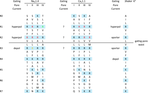
Mutations of voltage-gated ion channels cause several channelopathies of skeletal muscle, which present clinically with myotonia, periodic paralysis, or a combination of both. Expression studies have revealed both loss-of-function and gain-of-function defects for the currents passed by mutant channels. In many cases, these functional changes could be mechanistically linked to the defects of fibre excitability underlying myotonia or periodic paralysis. One remaining enigma was the basis for depolarization-induced weakness in hypokalaemic periodic paralysis (HypoPP) arising from mutations in either sodium or calcium channels. Curiously, 14 of 15 HypoPP mutations are at arginines in S4 voltage sensors, and recent observations show that these substitutions support an alternative pathway for ion conduction, the gating pore, that may be the source of the aberrant depolarization during an attack of paralysis.


Figures

Figure 1
Figure 1. Alignment of S4 sequences for NaV1.4, CaV1.1 and Shaker K+ channels
Positively charged residues are delineated with a shaded background (blue) for the R0 to R7 position. Mutations of these basic residues may produce a gating pore current activated by hyperpolarization (hyperpol), by depolarization (depol), or give rise to a proton transporter (xporter). The gating pore waist lies between R2 and R3, and voltage-dependent translocation of substituted R residues on S4 through this region regulates the flow of gating pore current. HypoPP mutations (red) are clustered at arginines in the R1 or R2 position of NaV1.4 and CaV1.1. Mutations of NaV1.4 at IV-R1 (green) are associated with PMC, not HypoPP, and do not produce gating pore current. A mixed variant of periodic paralysis is associated with mutations at NaV1.4 II-R3 (yellow) which causes a depolarization-activated gating pore.
Figure 2
Figure 2. Hyperpolarization-activated gating pore current in the R1H HypoPP mutation of NaV1.4
A, currents elicited by 300 ms step depolarizations between −140 mV and +30 mV from a holding potential of −100 mV in oocytes expressing wild-type (WT) or R1H channels. Saturating concentration of TTX has been added to suppress INa. B, steady-state I–V relation, after subtraction of linear background leak: WT, open circles; R1H, filled circles. Modified from Struyk & Cannon (2007) with permission.
Figure 3
Figure 3. The gating pore current increases the susceptibility to paradoxical depolarization in low extracellular [K+]
The steady-state I–V relation for mammalian skeletal muscle was simulated by the combination of an inward rectifier K+ current, IKir, a delayed rectifier K+ current, IKDR, and a leakage current with a reversal potential of 0 mV, ILeak, as in Struyk & Cannon (2008). A, in 4 mm[K+]o the resting potential of −91.3 mV is determined primarily from the balance of an inward ILeak and outward IKir. Addition of a gating pore current, Igp, to simulate HypoPP (dashed red line), shifts the I–V relation downward (dashed black line) but results in only a small depolarization of Vrest to −87.3 mV. B, reduction of [K+]o to 2.5 mm shifts EK and IKir to more negative potentials, with a predicted hyperpolarization of Vrest to −101.6 mV in WT fibres. For HypoPP fibres, however, Vrest depolarizes to −65.3 mV (arrow) because the combination of inward currents (ILeak+Igp) exceeds the outward current from IKir. Under these conditions, which would result in paralysis from inactivation of sodium channels, Vrest is now set by the balance of IKDR and the inward currents. Because KDR channels are active only for >−55 mV, Vrest is strongly dependent on KDR gating, which results in an apparent decoupling of Vrest from EK. C, phase plot of Vrest as a function of [K+]o for simulated WT and HypoPP fibres. The inward gating pore current in HypoPP fibres causes only a small depolarization of Vrest from −91.3 mV to −87.3 mV in 4 mm[K+]o (arrow). More importantly, the catastrophic depolarization of Vrest shifts leftward from 1.5 mm[K+]o for WT to 3 mm for HypoPP. The Nernst potential for K+, EK, is shown for comparison (blue line).

Similar articles

Cited by

References

    1. Adrian RH, Bryant SH. On the repetitive discharge in myotonic muscle fibres. J Physiol. 1974;240:505–515. - PMC - PubMed
    1. Ashcroft FM. Ion Channels and Disease. Oxford: Elsevier; 2000.
    1. Bulman DE, Scoggan KA, van Oene MD, Nicolle MW, Hahn AF, Tollar LL, Ebers GC. A novel sodium channel mutation in a family with hypokalemic periodic paralysis. Neurology. 1999;53:1932–1936. - PubMed
    1. Cannon SC. Pathomechanisms in channelopathies of skeletal muscle and brain. Annu Rev Neurosci. 2006;29:387–415. - PubMed
    1. Cannon SC, Brown RH, Jr, Corey DP. A sodium channel defect in hyperkalemic periodic paralysis: potassium-induced failure of inactivation. Neuron. 1991;6:619–626. - PubMed

Publication types