. 2010 Mar 15;207(3):651-67.
doi: 10.1084/jem.20091921. Epub 2010 Feb 15.

Naive tumor-specific CD4(+) T cells differentiated in vivo eradicate established melanoma

Affiliations

Naive tumor-specific CD4(+) T cells differentiated in vivo eradicate established melanoma

Ying Xie et al. J Exp Med. .

Abstract

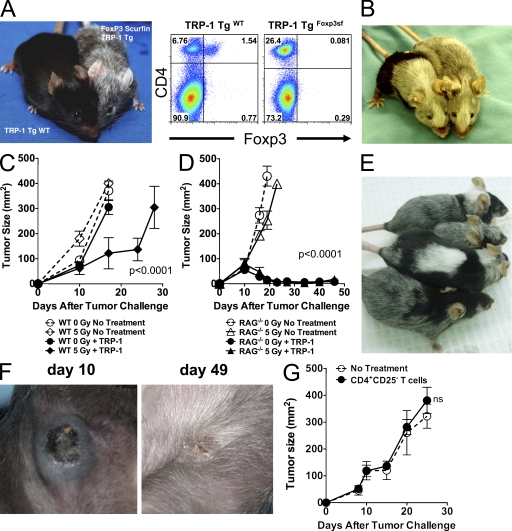
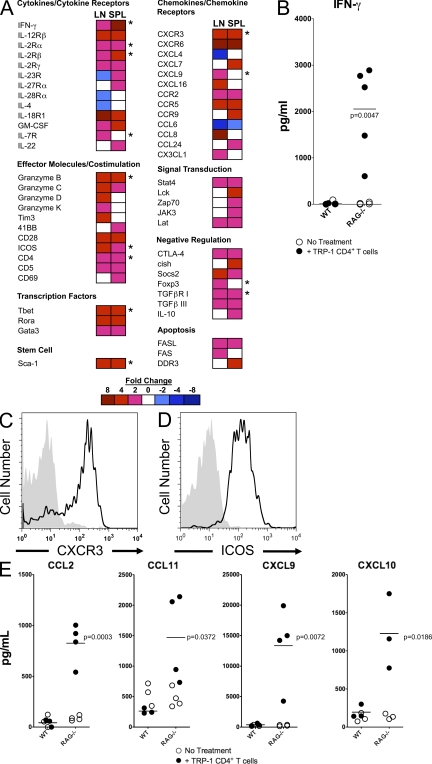
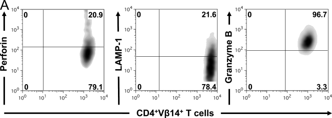
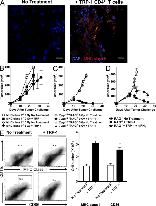
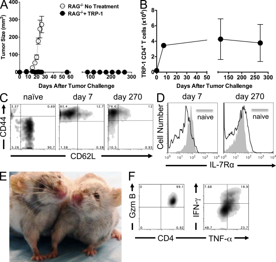
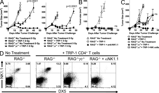
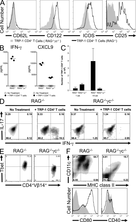
In vitro differentiated CD8(+) T cells have been the primary focus of immunotherapy of cancer with little focus on CD4(+) T cells. Immunotherapy involving in vitro differentiated T cells given after lymphodepleting regimens significantly augments antitumor immunity in animals and human patients with cancer. However, the mechanisms by which lymphopenia augments adoptive cell therapy and the means of properly differentiating T cells in vitro are still emerging. We demonstrate that naive tumor/self-specific CD4(+) T cells naturally differentiated into T helper type 1 cytotoxic T cells in vivo and caused the regression of established tumors and depigmentation in lymphopenic hosts. Therapy was independent of vaccination, exogenous cytokine support, CD8(+), B, natural killer (NK), and NKT cells. Proper activation of CD4(+) T cells in vivo was important for tumor clearance, as naive tumor-specific CD4(+) T cells could not completely treat tumor in lymphopenic common gamma chain (gamma(c))-deficient hosts. gamma(c) signaling in the tumor-bearing host was important for survival and proper differentiation of adoptively transferred tumor-specific CD4(+) T cells. Thus, these data provide a platform for designing immunotherapies that incorporate tumor/self-reactive CD4(+) T cells.


Figures

Figure 1.
Figure 1.
Treatment of established melanoma with adoptive transfer of TRP-1 CD4+ T cells into lymphopenic mice is specific. (A) Enhanced specific autoimmune disease in TRP-1 CD4+ Foxp3sf (Foxp3 negative) mice. TRP-1 CD4+ WT Tg mice (left; n = 13) and TRP-1 CD4+ Foxp3sf mice (right; n = 7) were compared for incidence of depigmentation over time. 1-mo-old littermates are shown. TRP-1 CD4+ Foxp3sf mice have no Foxp3+ T cells as shown by flow cytometry. (B) Depigmentation (vitiligo) can be adoptively transferred to lymphopenic hosts through TRP-1–specific CD4+ T cells. 2 × 10⁵ TRP-1 CD4+ T cells from Tyrp1B-wRAG−/− mice were transferred to nontumor-bearing RAG−/− hosts. Mice (n = 20) developed depigmentation after 35–45 d. A representative picture is shown. (C) Tumor-bearing WT mice were irradiated with 500 rads (5 Gy) or not irradiated (0 Gy) on day 7 after tumor challenge, and 2 × 10⁵ naive TRP-1 CD4+ T cells were adoptively transferred by i.v. tail vein injection. Experiments were repeated two times. P < 0.0001 for WT mice receiving 5 Gy and TRP-1 CD4+ T cells versus no treatment. (D) Tumor-bearing RAG−/− mice were irradiated with 500 rads (5 Gy) or not irradiated (0 Gy) on day 7 after tumor challenge, and 2 × 10⁵ naive TRP-1 CD4+ T cells were adoptively transferred by i.v. tail vein injection. Experiments were repeated nine times. P < 0.0001 for RAG−/− no treatment versus RAG−/− + TRP-1 CD4+ T cells (0 Gy or 5 Gy). (E) Whole body vitiligo in tumor-bearing mice treated with TRP-1 CD4+ T cells (n = 40 mice). (F) Tumor regression in lymphopenic mice treated with TRP-1 CD4+ T cells at days 10 and 49. The picture is representative of 40 mice over eight different experiments. (G) Adoptive transfer of 106 open repertoire CD4+CD25 T cells into lymphopenic tumor-bearing RAG−/− mice on day 7 after tumor challenge does not affect tumor growth. Data represent three independent experiments. n = 5 mice/group; P = NS. Error bars indicate SEM.
Figure 2.
Figure 2.
TRP-1 CD4+ T cells treating established tumors differentiate into Th1 CD4+ cytotoxic T cells in lymphopenic mice. (A) Gene expression analysis of TRP-1 CD4+ T cells from spleen and LNs of lymphopenic mice undergoing tumor regression. Heat maps represent fold changes in mRNA expression between naive TRP-1 CD4+ T cells and TRP-1 CD4+ T cells differentiated in vivo for 1 wk. The gene array is representative of one experiment. * indicates data confirmed by flow cytometry or multiplex assay. (B) IFN-γ levels in the serum of tumor-bearing WT and RAG−/− mice with and without adoptive transfer of 2 × 10⁵ TRP-1 CD4+ T cells 1 wk after transfer. Open circles represent individual mice receiving no treatment and closed circles represent individual mice receiving TRP-1 CD4+ T cells. Horizontal bars indicate means for treated groups only (n = 3–5 mice/group). Data are representative of three experiments (no treatment vs. treatment; P = 0.0047). (C) CXCR3 expression on naive TRP-1 CD4+ T cells before transfer (gray histogram) and 1 wk after in vivo differentiation (solid line). (D) ICOS expression on naive and 1wk in vivo differentiated TRP-1 CD4+ T cells. (E) Serum chemokines levels in WT and RAG−/− mice treated or not (n = 3–5 mice/group). Open circles represent individual mice receiving no treatment and closed circles represent individual mice receiving TRP-1 CD4+ T cells. Horizontal bars indicate means for treated groups only. Data are representative of three experiments.
Figure 3.
Figure 3.
TRP-1 CD4+ T cells become cytotoxic T cells in vivo. (A) Spleens from tumor-bearing RAG−/− mice treated with TRP-1 CD4+ T cells on day 7 after tumor challenge were stained ex vivo with antibodies to CD4, Vβ14, perforin, LAMP-1 (CD107a), and granzyme B 1 wk after adoptive cell transfer. Intracellular staining was performed as indicated in Materials and methods. Flow cytometry shows gated TRP-1 CD4+Vβ14+ cells. Data represent two independent experiments (n = 5 mice/group).
Figure 4.
Figure 4.
Mechanism of treatment by TRP-1 CD4+ T cells. (A) Confocal microscopy of day-14 tumors, 1 wk after adoptive cell transfer with 2 × 10⁵ naive TRP-1 CD4+ T cells. Tumors were frozen in OCT, cut, and stained with DAPI (blue) and MHC class II (red). Samples were analyzed by confocal microscopy (Olympus) with a 20× oil immersion objective. Bars, 50 µm. (B) 7-d tumor-bearing MHC class II−/− mice were irradiated with 5 Gy or not irradiated and treated with 2 × 10⁵ naive TRP-1 CD4+ T cells. (C) 7-d tumor-bearing non-TCR Tg Tyrp1B-wRAG−/− mice were irradiated with 5 Gy or not irradiated and treated with 2 × 10⁵ naive TRP-1 CD4+ T cells. (D) 11-d tumor-bearing RAG−/− mice were treated with naive TRP-1 CD4+ T cells or not and, in addition, some treated mice received one injection of 500 µg of neutralizing anti–IFN-γ antibodies on day 7 of ACT. Data are representative of four independent experiments with five to eight mice per group. (E) DC activation in lymphopenic mice after ACT with TRP-1 CD4+ T cells. Flow cytometry of CD11chighMHC class IIhigh and CD86high DCs from tumor-bearing RAG−/− mice undergoing treatment or not. Bar graphs indicate absolute numbers of CD11chigh MHC class II+ CD86high cells. Values represent SEM (n = 3 mice/group; **, P < 0.05). Data are representative of four experiments.
Figure 5.
Figure 5.
Activation, persistence, and memory formation of TRP-1 CD4+ T cells. (A) Long-term tumor regression. Lymphopenic mice were treated on day 7 after tumor challenge with adoptive transfer of naive TRP-1 CD4+ T cells and followed for 270 d. (B) TRP-1 CD4+ T cells persist for long periods in lymphopenic mice. Error bars indicate SEM. (C) Flow cytometry of CD44 and CD62L expression on naive TRP-1 CD4+ T cells before transfer, after 1 wk of in vivo differentiation, and at 270 d. (D) IL-7Rα expression on naive (shaded) and 1-wk in vivo differentiated and 270-d persisting TRP-1 CD4+ T cells (solid line). (E) Phenotype of 270-d-old treated RAG−/− mice. (F) Granzyme B, IFN-γ, and TNF expression in TRP-1 CD4+ T cells isolated on day 120 after treatment from tumor-free mice. Data are representative of two experiments (n = 5 mice/group).
Figure 6.
Figure 6.
NK cells are not required for tumor therapy with TRP-1 CD4+ T cells. (A) Tumor-bearing RAG−/− and RAG−/−γc−/− mice were treated on day 7 without or with irradiation (5 Gy) and with and without ACT with 2 × 10⁵ naive TRP-1 CD4+ T cells. Data are representative of three experiments (P < 0.0001). The p-value indicated is for RAG−/− + TRP-1 versus RAG−/−γc−/− + TRP-1 CD4+ T cells for both 0 and 5 Gy. (B) Tumor-bearing RAG−/− mice received 1 mg of anti-NK1.1 antibodies weekly starting 1 d before ACT and were compared with tumor-bearing RAG−/− mice receiving no treatment or 2 × 10⁵ naive TRP-1 CD4+ T cells. Data represent three experiments with five to eight mice per group. P = NS for RAG−/− + TRP-1 versus RAG−/− + TRP-1 + anti-NK1.1. (C) 7-d tumor-bearing RAG−/− and RAG−/−γc−/− mice received naive TRP-1 CD4+ T cells plus sorted NK cells from WT mice where indicated (P = NS for addition of NK cells). Data represent two independent experiments. Error bars indicate SEM. (D) Flow cytometry of NK cells (NK1.1+DX5+ cells) in the LNs of RAG−/−, RAG−/− + anti-NK1.1 antibodies, and RAG−/−γc−/− mice as indicated 3–4 wk after adoptive T cell transfer. Data are representative of three independent experiments (n = 2–3 mice/group).
Figure 7.
Figure 7.
γc signaling on host DC is required for survival and differentiation of TRP-1 T cells in vivo. (A) Differential activation of TRP-1 CD4+ T cells in RAG−/− and RAG−/−γc−/− hosts. Spleens from RAG−/− or RAG−/−γc−/− mice were isolated and stained for TRP-1 CD4+ T cells. Shown are CD62L, CD122, ICOS, and CD25 expression on gated TRP-1 CD4+ T cells from indicated host. (B) IFN-γ and CXCL9 are differentially expressed in the serum at 1 wk in RAG−/− and RAG−/−γc−/− hosts after TRP-1 CD4+ T cell transfer. Horizontal bars represent mean. (C) TRP-1 CD4+ T cells expand in RAG−/−γc−/− hosts initially but fail to survive after 4 wk. Error bars indicate SEM. (D) Flow cytometry of IFN-γ and IL-17 expression in TRP-1 CD4+ T cells isolated from tumor bearing RAG−/− and RAG−/−γc−/− mice 4 wk after transfer. T cells were activated with PMA and ionomycin for 4 h and then fixed and permeabilized and stained with anti–IFN-γ and IL-17 antibodies. (E) Tbet expression in TRP-1 CD4+ T cells 4 wk after transfer. (F) MHC class II, CD80, and CD40 expression in RAG−/−γc−/− hosts. Top flow diagram indicates CD11chigh MHC class II+ cells; bottom flow histograms show CD80 and CD40 expression on gated CD11chigh MHC class II+ cells. Solid line represents RAG−/− mice treated with TRP-1 CD4+ T cells. Shaded histogram represents RAG−/−γc−/− mice treated with TRP-1 CD4+ T cells. Data are representative of three independent experiments (n = 5 mice/group).

Similar articles

Cited by

References

    1. Antony P.A., Piccirillo C.A., Akpinarli A., Finkelstein S.E., Speiss P.J., Surman D.R., Palmer D.C., Chan C.C., Klebanoff C.A., Overwijk W.W., et al. 2005. CD8+ T cell immunity against a tumor/self-antigen is augmented by CD4+ T helper cells and hindered by naturally occurring T regulatory cells. J. Immunol. 174:2591–2601 - PMC - PubMed
    1. Antony P.A., Paulos C.M., Ahmadzadeh M., Akpinarli A., Palmer D.C., Sato N., Kaiser A., Hinrichs C.S., Heinrichs C., Klebanoff C.A., et al. 2006. Interleukin-2-dependent mechanisms of tolerance and immunity in vivo. J. Immunol. 176:5255–5266 - PMC - PubMed
    1. Banchereau J., Klechevsky E., Schmitt N., Morita R., Palucka K., Ueno H. 2009. Harnessing human dendritic cell subsets to design novel vaccines. Ann. N. Y. Acad. Sci. 1174:24–32 10.1111/j.1749-6632.2009.04999.x - DOI - PMC - PubMed
    1. Behrens G., Li M., Smith C.M., Belz G.T., Mintern J., Carbone F.R., Heath W.R. 2004a. Helper T cells, dendritic cells and CTL Immunity. Immunol. Cell Biol. 82:84–90 10.1111/j.1440-1711.2004.01211.x - DOI - PubMed
    1. Behrens G.M., Li M., Davey G.M., Allison J., Flavell R.A., Carbone F.R., Heath W.R. 2004b. Helper requirements for generation of effector CTL to islet beta cell antigens. J. Immunol. 172:5420–5426 - PubMed

Publication types

MeSH terms